Journal of Otolaryngology and Rhinology
An Unusual Complication of Maxillary Subperiosteal Implant Removal: Dislocation of the Parotid Gland Duct
Marciante Giulia Anna1*, Saibene Alberto Maria1, Pisani Antonia1, Biglioli Federico2 and Giovanni Felisati1
1Department of Health Sciences, Maxillo-facial Surgery Unit, Università degli Studi di Milano, San Paolo Hospital, Milan, Italy
2Department of Health Sciences, Otorhinolaryngology Unit, Università degli Studi di Milano, San Paolo Hospital, Milan, Italy
*Corresponding author:
Marciante Giulia Anna, Department of Health Sciences, Maxillo-facial Surgery Unit, Università degli Studi di Milano, San Paolo Hospital, Milan, Italy, E-mail:
giuliaanna.marciante@gmail.com
J Otolaryngol Rhinol, JOR-2-018, (Volume 2, Issue 2), Case Report
Received: January 28, 2016: Accepted: April 27, 2016: Published: April 30, 2016
Citation: Anna MG, Maria SA, Antonia P, Federico B, Felisati G (2016) An Unusual Complication of Maxillary Subperiosteal Implant Removal: Dislocation of the Parotid Gland
Duct. J Otolaryngol Rhinol 2:018.
Copyright: © 2016 Anna MG, et al. This is an open-access article distributed under the terms of the Creative Commons Attribution License, which permits unrestricted use, distribution, and reproduction in any medium, provided the original author and source are credited.
Abstract
A 73-year-old man, treated with a maxillary subperiosteal implant in 1985, was referred to our ENT clinic by his dental practitioner due to the onset of sudden edema, pain and purulent nasal discharge. Although the patient had experienced painful recurrent mobilization of the implant from 1995, he did not approach his dentist until 2006, when edema and purulent nasal discharge occurred. The radiologic imaging revealed a maxillary sinusitis with severe maxillary bone resorption and an oro-nasal/oro-antral communication, for which the patient underwent removal of the implant, functional endoscopic sinus surgery and repair of the oro-nasal/oro-antral communication with multiple local flaps. Two months later, the patient started complaining of recurrent nasal watery discharge, however, no signs of intranasal lesions or infections had been found at fiberoptic endoscopy. The lasting of this symptom and its peculiar presentation during meals were reported at the successive follow up evaluation, leading to the suspect of parotid duct's dislocation into the maxillary sinus due to the scarring of the vestibular flap. An accurate nasal and oral examination and the onset of the nasal discharge while eating confirmed the hypothesis. The patient underwent duct ligation with complete resolution of symptoms.
Keywords
Subperiosteal implant, Nasal watery discharge, Parotid duct dislocation, Maxillary sinusitis, Functional endoscopic sinus surgery
Introduction
Subperiosteal implants (SI) are prosthetic devices consisting of a metallic framework fixed beneath the mucoperiostium of atrophied maxillas in edentulous patients. The value of SI, first described by Dahl in the '40s [1], progressively decreasedin the last 30 years, thanks to the widespread resort to endosseous implants. The greater efficacy of these implants in rehabilitation and their lower failure rate have already been discussed in literature [2-4]. At the best of our knowledge, little is known about failure rates of SI, which is reported to be around 34-50% [5], as only one large case series and a few case reports or small case series concerning survival rates and complications of SI are available in literature [6,7]. However, none of these studiesdescribed possible nasal complications. In 2013, Felisati et al. presented their own experience about nasal complications of different types of dental implants [8]: in this study, early oro-nasal communication (ONC), oro-antral communication (OAC) and important infections were observed in all the patients with SI complications. The treatment of these conditions was based on the association of functional endoscopic sinus surgery (FESS) to local flaps (buccal Rehrmann/Moczair flaps, palatal flaps and/or buccal fat pad), as already proposed for displaced oral implants [9]. Management of dental treatment complication is an area of expertise for ENT and oral/maxillofacial surgeons. This manuscript describes a case of failed subperiosteal maxillary implant, its treatment and the unexpected duct complication with its atypical presentation. Furthermore, a short review of the literature will be presented.
Case Report
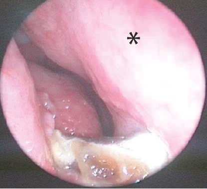
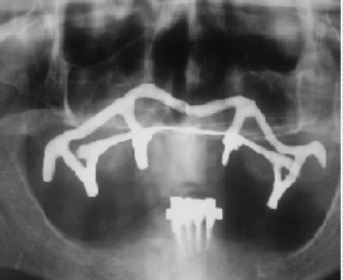
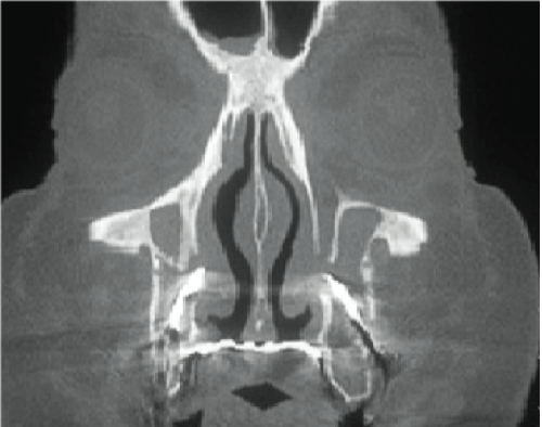
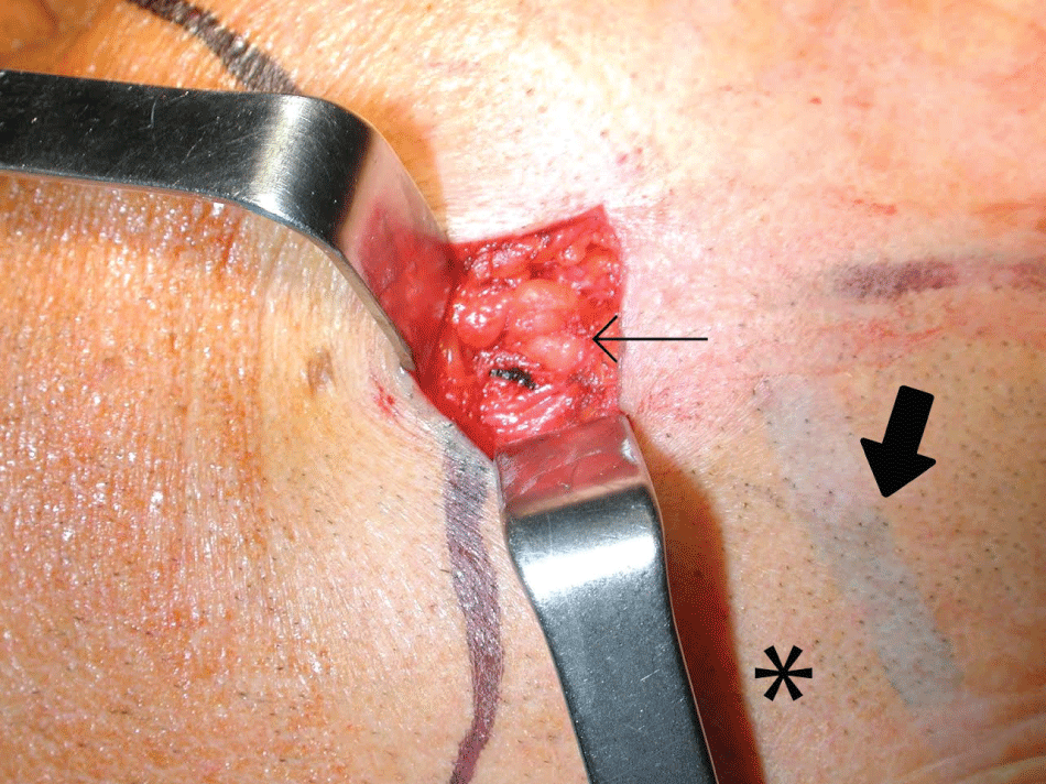
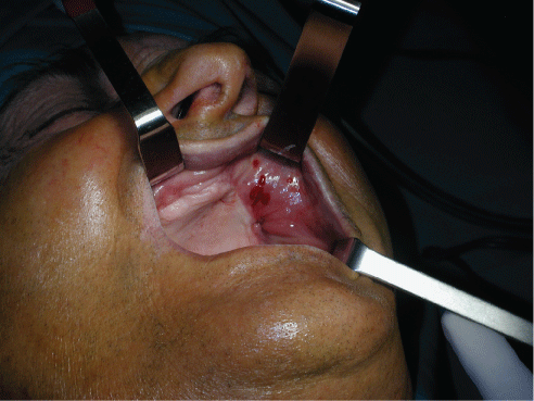
A 73-year-old man, implanted with a maxillary subperiosteal device in 1985, started referring to his dentist several times after surgery because of painful mobilization of the implant. In 1990, considering the persistence of symptoms without signs of infection, the patient's dental practitioner performed an unspecified dental procedure in order to further fix the implant to the maxillary bone. In spite of that treatment, although painful mobilization still recurred after surgery, the patient decided not to approach his medical practitioner until 2006, when painful facial edema and purulent nasal discharge occurred. Considering the patient's past medical history and symptoms, he was promptly referred to our ENT Clinic, where he underwent a complete ENT evaluation, comprehensive of a nasal flexible fiberoptic endoscopy (Figure 1), which showed bilateral mucopurulent secretions from both the osteomeatal complexes with a greater discharge in the left nasal fossa and the presence of a metallic foreign body under the inferior turbinates. Therefore, in order to study the maxillary arch, a Panoramic Radiograph (Figure 2) was first performed, showing a maxillary full arch subperiosteal implant with surrounding bone resorption and four additional endosseous implants in the mandibular bone. A maxillofacial Computed Tomography (CT) Scan (Figure 3) was then carried out,revealing the nasal dislocation of the subperiosteal implant and bilateral maxillary sinus infection. The patient underwent a broad-spectrum antibiotic, steroidal and anti-inflammatory intravenous therapy to treat the infection; however, since the presence of the implant represented the main causal factor, its removal was mandatory. A combined approach of nasal endoscopic and oral surgery was therefore proposed for implant removal and the concurrent treatment of the sinonasal infection. A first incision at the gum fornix level was needed to expose the meshwork and the underlying OAC/ONC, and endoscopic sinonasal surgery was then performed in order to widen the maxillary sinus ostium with uncinectomy and middle antrostomy and to eradicate the maxillary sinus' infection by means of an antibiotic toilette. Lastly, a palatal flap was raised to cover the central palatal area exposed after the implant removal, while a mucoperiosteal Rehrmann flap was positioned over its left side and a Bichat fat flap over its right side to close the oro-antral and oro-nasal fistulas. Two months after surgery, the patient started complaining of a watery discharge from the left nasal fossa and xerostomia. A complete ENT and maxillofacial evaluation were performed: no intranasal lesions or signs of sinusitis responsible for rhinorrhea were found at nasal fiberoptic endoscopy, and both the flaps were regularly healing. A more accurate recollection of signs and symptoms revealed the existence of a strict correlation between meal consumption and onset of the nasal watery discharge, and so an iatrogenic dislocation of the parotid duct into the maxillary sinus due to the lifting and repositioning of the flap was suggested. The patient was therefore asked to eat during the clinical examination, and the production of watery discharge from the left nasal fossa was immediately observed, confirming the hypothesis. In order to perform a minimally-invasive treatment of the discharge, a transoral ligation of the left parotid duct was firstattempted, without success, since the ductal in cannulation failed because of the conspicuous scarring of the site and the surgically altered local anatomy (Figure 4). As a consequence, the parotid duct was reached by means of a transfacial approach during which a rerouting of the duct was firstly attempted, however without success. Resolution of the complication was finally gained with an incision at the crossing point between the anterior margin of the left parotid gland and the natural course of the parotid duct along the masseter and buccinator muscles, ductal identification and its ligation at the origin (Figure 5).
.
Figure 1: Nasal flexible fiberoptic endoscopy of the left nasal fossa: mucopurulent secretions from the osteomeatal complex with the metallic subperiosteal implant under the inferior turbinate (*).
View Figure 1
.
Figure 2: Panoramic Radiograph shows the presence of the subperiosteal implant with surrounding bone resorption and four additional mandibular endosseous implants in the mandibular bone.
View Figure 2
.
Figure 3: Coronal CT Scan reveals implant's migration into the nasal fossae with bone resorption and bilateral maxillary chronic sinusitis.
View Figure 3
Resolution of the nasal discharge was immediately observed. The patient underwent a strict follow up program, which is currently in its 7th year. No recurrence of OAC/ONC, sinusitis or meal-related discharge have been reported. Anyway, the wide maxillary bone's resorption still represents a severe contraindication for implantologic rehabilitation (Figure 6).
.
Figure 4: Intraoperative parotid duct's targeting considering the surface anatomical reperi represented by the anterior margin of the left parotid gland (*), masseter (➞) and natural course of the duct itself (➨).
View Figure 4
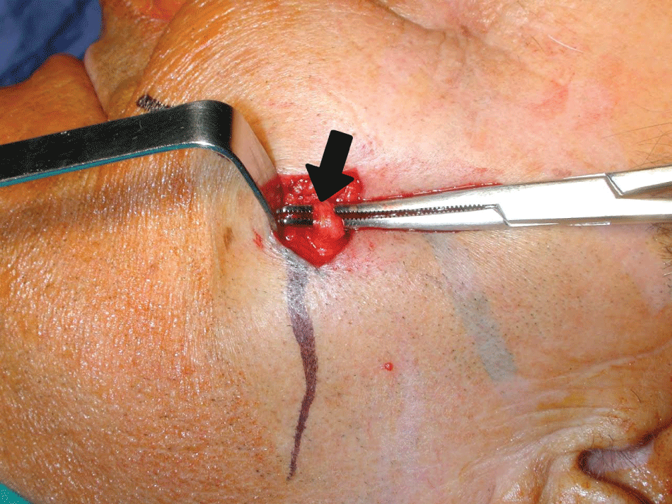
.
Figure 5: Intraoperative parotid duct's identification and ligation at the origin (➨).
View Figure 5
.
Figure 6: Follow up evaluation: both the flaps are regularly healed and no OAC/ONC can be found, but a wide maxillary bone's resorption can be appreciated.
View Figure 6
Discussion
Subperiosteal implants, that firstly represented a great advancement in implantologic surgery, had progressively revealed to determine the development of important complications such as pain, paraesthesia, implant exposure, fistulae and infections [10-12]. In literature, the main survival rate of subperiosteal implants (the majority of which is mandibular implants) is about 95-96% at 5 years, 67-96% at 10 years and 50%-60% at 15-20 years after surgery, with a statistically significant rise in failure rate if considering a longer follow up period [4,13,14]. Only few still recommend SI in specific cases [15]. On the basis of our experience, extensive infections of the maxillary sinus and the nasal cavities have to be considered as important complications as the frequent bone resorption and infection of the maxillary bone for which implant removal is necessary. In addition, maxillary bone height after implant removal is almost always insufficient for subsequent endosseous implantation, so that maxillary augmentation has to be performed (if possible) before prosthetic rehabilitation [5]. In our opinion, removal of SI should be therefore considered in case of decreased implant stability, especially in patients with maxillary bone atrophy. Treatment of the two most severe SI complications (OAC and sinonasal infections) is based on different approaches, depending on the site and extension of the infection. Sinonasal infectious involvement requires sinonasal endoscopic surgery, which allows to perform an accurate toilet of the nasal fossae and the maxillary sinus, and additional ethmoidal or frontal opening in case of a local spread of the infection [16]. Since these two conditions often coexist, a combined approach is mandatory in order to treat them and simultaneously restore the sinonasal homeostasis [17,18], especially if considering that dental treatment complications are gaining more and more importance because of the increasing diffusion of dental treatments. OAC are often closed with an endoral approach by means of Rehrmann flap, Mochzair flap or Bichat fat flap, whose most common complications include infraorbital anaesthesia, reduction of the vestibular sulcus, wound dehiscence, granulation tissue and partial necrosis of the flap [19,20]. To date, the iatrogenic transposition of the parotid duct into the maxillary sinus and the onset of meal-related nasal watery discharge as a complication of a Rehrmann flap has been described only by Neuschl et al. in 2010 [21]. Our personal experience strengthens the evidences of the role of SI as a source of sinonasal infection in patients complaining of nasal symptoms after implantologic surgery, and also underlines the importance of a multidisciplinary approach in treating oral reconstructive surgery complications, of which meal-related watery nasal discharge due to parotid duct dislocation represents a rare manifestation.
References
-
Dahl G (1943) Om mojligheten or inplantation I kaken avmetallskelett som bas elle retention or fasta eller avtagbara protester. Odontologisk Tidskrift 51: 440-449.
-
Linkow LI, Ghalili R (1998) Critical design errors in maxillary subperiosteal implants. J Oral Implantol 24: 198-205.
-
Schou S, Pallesen L, Hjørting-Hansen E, Pedersen CS, Fibaek B (2000) A 41-year history of a mandibular subperiosteal implant. Clin Oral Implants Res 11: 171-178.
-
Moore DJ, Hansen PA (2004) A descriptive 18-year retrospective review of subperiosteal implants for patients with severely atrophied edentulous mandibles. J Prosthet Dent 92: 145-150.
-
Bodine RL, Yanase RT, Bodine A (1996) Forty years of experience with subperiosteal implant dentures in 41 edentulous patients. J Prosthet Dent 75: 33-44.
-
Zwerger S, Abu-Id MH, Kreusch T (2007) Long-term results of fittig subperiosteal implants: report of twelve patient cases. Mund Kiefer Gesichtschir 11: 359-362.
-
Kreutz RW, Carr SJ (1986) Bilateral oronasal fistulas secondary to an infected maxillary subperiosteal implant. Report of a case. Oral Surg Oral Med Oral Pathol 61: 230-232.
-
Felisati G, Chiapasco M, Lozza P, Saibene AM, Pipolo C, et al. (2013) Sinonasal complications resulting from dental treatment: outcame-oriented proposal of classification and surgical protocol. Am J Rhinol Allergy 27: e101-106.
-
Chiapasco M, Felisati G, Maccari A, Borloni R, Gatti F, et al. (2009) The management of complications following displacement of oral implants in the paranasal sinuses: a multicenter clinical report and proposed treatment protocols. Int J Oral Maxillofac Surg 38: 1273-1278.
-
Mercier P, Cholewa J, Djokovic S (1981) Mandibular subperiosteal implants (a retrospective analysis in light of the Harvard Consensus). J Can Dent Assoc 47: 46-51.
-
Young L Jr, Michel JD, Moore DJ (1983) A twenty-year evaluation of subperiosteal implants. J Prosthet Dent 49: 690-694.
-
Bailey JH, Yanase RT, Bodine RL (1988) The mandibular subperiosteal implant denture: a fourteen-year study. J Prosthet Dent 60: 358-361.
-
Golec TS (1989) The mandibular full subperiosteal implant--a ten-year review of 202 cases. J Oral Implantol 15: 179-185.
-
Bodine RL (1974) Evaluation of 27 mandibular subperiosteal implant dentures after 15 to 22 years. J Prosthet Dent 32: 188-197.
-
Kusek ER (2009) The use of laser technology (Er;Cr:YSGG) and stereolithography to aid in the placement of a subperiosteal implant: case study. J Oral Implantol 35: 5-11.
-
Saibene AM, PipoloGC, Lozza P, Maccari A, Portaleone SM, Scotti A, et al. (2014) Redefining boundaries in odontogenic sinusitis: a retrospective evaluation of extramaxillary involvement in 315 patients. Int Forum All Rhinology 4:1020-1023.
-
Giovannetti F, Priore P, Raponi I, Valentini V (2014) Endoscopic sinus surgery in sinus-oral pathology. J Craniofac Surg 25: 991-994.
-
Saibene AM, Lozza P (2015) Endoscopic sinus surgery and intraoral approaches in sinus oral pathology. J Craniofac Surg 26: 322-323.
-
Martin-Granizo R, Naval L, Costas A, Goizueta C, Rodriguez F, et al. (1997) Use of buccal fat pad to repair intraoral defects: review of 30 cases. Br J Oral Maxillofac Surg 35: 81-84.
-
Guven O (1998) A clinical study on oroantral fistulae. J Craniomaxillofac Surg 26: 267-271.
-
Neuschl M, Kluba S, Krimmel M, Reinert S (2010) Iatrogenic transposition of the parotid duct into the maxillary sinus after tooth extraction and closure of an oroantral fistula. A case report. J Craniomaxillofac Surg 38: 538-540.


