

International Journal of Oral and Dental Health
Effectiveness of Shade Measurements Using a Scanning and Computer Software System: a Pilot Study
Gotfredsen K1*, Gram M1, Ben Brahem E1, Hosseini M1, Petkov M2 and Sitorovic M2
1Department of Odontology, Oral Rehabilitation, University of Copenhagen, Denmark
2University Clinic for Dental Sciences, University SS. Cyril and Methodius, Skopje, Macedonia
*Corresponding author: Klaus Gotfredsen, DDS, Ph.D, Department of Oral Rehabilitation, School of Dentistry,
Faculty of Health and Medical Sciences, University of Copenhagen, Denmark, E-mail: klg@sund.ku.dk
Int J Oral Dent Health, IJODH-1-008, (Volume 1, Issue 2), Research Article; ISSN: 2469-5734
Received: March 06, 2015 | Accepted: April 22, 2015 | Published: April 25, 2015
Citation: Gotfredsen K, Gram M, Ben Brahem E, Hosseini M, Petkov M, et al. (2015) Effectiveness of Shade Measurements Using a Scanning and Computer Software System: a Pilot Study. Int J Oral Dent Health 1:008. 10.23937/2469-5734/1510008
Copyright: © 2015 Gotfredsen K, et al. This is an open-access article distributed under the terms of the Creative Commons Attribution License, which permits unrestricted use, distribution, and reproduction in any medium, provided the original author and source are credited.
Abstract
Objectives: The aim of the present study was to evaluate a digital scanning and color measuring system in vivo in relation to the subjective, visual shade determination and an objective, well-known digital spectrophotometric system.
Methods: Shade determination with the 3D-Master system was performed at 29 patient including 87 teeth using a new scanning and computer-based system (3Shape TRIOS® Color system), a well-known spectrophotometric computer-based system (MHT SpectroShade™) and the subjective visual shade assessment. The reliability of the two objectives and the subjective color assessment methods was calculated as an inter-individual comparison of the shade determination obtain by two dentists. Thereafter the validity of the objective methods was compared with the subjective methods by a pairwise comparison of color tabs selected by two dentists with each method. Each dentist determined which color tap was the best match.
Results: The reliability of the objective, computer-based systems was high compared with the subjective, visual method for color determination. The validation test demonstrated no significant differences between the new Trios® Color System and the conventional, visual method and between the MHT SpectroShade™ spectrophotometric system and the conventional, visual method.
Conclusion: The effectiveness of the new digital scanning and computer software system for color determination was as good as an earlier tested and validated digital colorimetric system and as the conventional, visual method for color determination of teeth.
Introduction
The esthetic result has become more attention and one important aspect to obtain a good esthetic result is to obtain the most optimal shade of the prosthetic reconstruction [1,2]. The shade is of greatest importance when patients judge the quality of the restoration especially in the anterior region. The ideal shade is the shade from the natural, neighboring teeth. Shade selection is, however only the first step to duplicate the color of the natural teeth, and the next important step is to communicate the shade to the technician at the dental laboratory [3]. Thereafter the most challenging part is the shade reproduction in the dental laboratory, where the individual technician and his ability to use the information he receives from the dentist.
There is no golden standard for specific tooth shade evaluation and most dentist and dental technicians uses color tabs for the initial ceramic work. Thereafter an individual coloring by the dental technician using digital images or with the patient in front of the technician is used for reproducing the optimal restoration.
The color tabs used for color determination in the dental clinics covers most frequent tooth shades occuring in nature and is designed to provide systematic coverage of the tooth color spectrum. Some systems are using color classification with values of lightness, color saturation described as chroma and hue referring to the property by which the dominant wavelength is expressed [4]. The successful use of color tabs depends on accurate color assessment by the person choosing the shade, and by the person producing the restoration. Visual color determination with color tabs is highly subjective, and general variables such as age, experience by the examiner, light source, room conditions, and physiological variables lead to inconsistencies [5,6]. Thus, a number of in vitro experiments have demonstrated a low reliability of conventional visual compared to spectrophotometric methods [3,7,8]. Despite the limitations for visual assessment, the human eye is very efficient in detecting even small differences in the color between two objects, and when the best shade match of two colors has to be selected the visual assessment is important [5]. Thus, for an in vivo validation of a new shade-determining device a comparison to the conventional, visual method for the best shade match could be used.
To make shade selection easier and more accurate, allowing the dentist to provide a better esthetic result of the prosthetic reconstructions, a high number of shade-determining devices has been introduced [9,10]. During the last two decades various spectrophotometric devices using digital photographs combined with a computer-based shade determination systems have been constructed and tested [11-13]. Recently, a new system developed for digital impression taking has added a tool for teeth shade measurement using a high definition camera included into a single handheld digital scanner. This direct in vivo scanning of the teeth with LED light and computer software calculate the best shade for the restoration and this shade can be directly transferred to the dental technician together with the digital impression of the tooth. Thus, a CAD-CAM crown can be fabricated with the most optimal shade. However, no studies have yet evaluated the use of this new technology in clinical settings.
Aim
The purpose of the present study was to evaluate a digital scanning and color measuring system in vivo in relation to a conventional visual shade determination and a well-known digital spectrophotometric system.
The null hypotheses was: H0: The effectiveness of a new digital scanning system combined with a computer software system for color determination is as good as previous used methods for color determination.
Material and Methods
Subjects and teeth
The two maxillary incisors and the canine from 29 patients (19 women and 10 men) with an average age of 37 years (range 22-62 years) were included, resulting in a total of 87 teeth for color determination. All the test persons had received a professional tooth cleaning not longer than 6 month ago. Eight patients with 24 teeth were included for the reliability testing of the new digital system and a well-known spectrophotometric system and 21 patients with 63 teeth were included for testing the reliability of the conventional visual shade assessment and the validity of all the shade measurements.
Shade guide systems
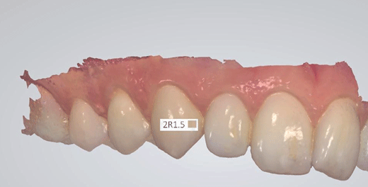
The new, tested shade guide system was based on a digital scanning and a computer software system, 3Shape TRIOS® Color (3Shape, Holmens Kanal, Copenhagen, DK). The light source for the tooth scanning is a LED covering the visual spectrum. The shade measurement tool is added to the TRIOS scanning system. TRIOS® Color automatically measures the shade while scanning the teeth by combining color information recorded in 3D images obtained from multiple angles. This color information is processed intelligently, using knowledge of the tooth's 3D geometry and the angle of scanning. The final color was in the present experiment translated into the Vita 3D-Master shade system by selecting the best matching shade in the selected shade system (Figure 1).
.
Figure 1: The TRIOS® Color System, where teeth are scanned and a computer automatically measures the shape of the selected tooth using a well-known shade guide
View Figure 1
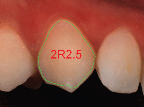
The MHT SpectroShade™ (MHT Optic Research AG, Mandachstrasse 50, CH-8155 Niederhasli) was used as the well-known, spectrophotometric device. A digital photograph is transfer to a computer system, where a software program determines the color. The measuring head was positioned with its mouthpiece on the alignment. The central position of the tooth to be measured was controlled and the measurements were carried out twice in a row on the slightly moistened teeth. The Vita 3D-Master scale was selected for the tooth color determination at the central part of the tooth (Figure 2).
The traditional, visual, subjective method using the Vita 3D-MASTER® system (Vident, Brea, CA, USA) was used as control for the shade evaluation and for the validation procedure. The Vita 3D-MASTER® system used included a total of 28 color tabs covered by 5 value levels, 3 chroma levels and 4 hue values, which were available, and the shade closest to the chromatic standard for the tooth shade was selected.
Shade definition
The shade was measured in the central region of the tooth e.g. the best match to the middle third of the patients tooth, and this was referred to as the tooth's shade.
As the shade of a tooth becomes lighter as the tooth dries out each teeth was slightly moistened with water just before the measurements was performed. The subjective measurements were performed within 1/2 minute from moistening.
Exclusion criteria
Dentist with a positive history of visual color deficiency e.g. reduced chromatic perception and teeth with composite fillings or crowns were excluded.
Prerequisites
The color determination was performed in natural daylight, but away of all windows e.g. no directs light. Patients were sitting in the same unit-chair and with the dental lamp turned off. The angle of the view for MHT Spectroshade, 3Shape Trios® Color and subjective VITA 3D-master Vitapan was the same. Lipstick or other effects that may affect color assessment was removed and patients with strong colored dress were covered with a white-grayish cloth.
Design
The experiment is divided into three steps, (1) Reliability testing of the (a) 3Shape TRIOS® Color scan and (b) MHT SpectroShade™; (2) Measuring step and (3) Validation step.
Reliability of the objective methods
Calibrations were performed with the examiners and the consistency of their evaluations was confirmed using Kappa statistic. The reliability of the 3Shape Trios® Color and MHT SpectroShade™ were performed as an interexaminer, pre-study including two measurements with each of the two devices on 24 natural teeth performed by two dentists (1 female and 1 male). The same two dentists evaluated 63 teeth with by subjective, visual shade determination using the same VITA 3D-master guide as used for the objective methods.
Measuring step
The conventional, human shade assessment comparing visually the central region of the shade tab with the central region of the natural tooth and selecting the shade tabs that matches the best was done. Two dentists performed the measurements blindly e.g. all shade numbers were hidden and the dentist performed the measurement independent of one another. Secondly the 3Shape Trios® scanner with the Trios® Color measurement tools and the MHT SpectroShade™ were calibrated and assessed the color at the same teeth. Two dentists (one women and one man) measured the shade with the 3D Vitapan Master shade guide, and two dentist measured with the Trios® Color and the MHT Spectroshade equipment. Two other dentists (1 female and 1 man) were validating the measurements. With 60 natural teeth for the measurements there was a total of 480 validating measurement, 240 for MHT Spectroshade™ versus the conventional subjective method and 240 for Trios® Color versus the conventional, subjective method.
Validation step
The true shade of the sample teeth is not known, which means the accuracy of the color determination needs to be quantified indirectly. The validation step consisted of a pair-wise comparison of two measurements for each tooth. The shade, color tap in the Vita 3D-Master system for each selected method were chosen and placed beside the assessed tooth. The question: "Which shade tap is the best match" were answered and registered for each of the two validating dentist. For each comparison the method yielding the best match was marked and given the score 1. If all methods were equally good, they should have the same number of best matches. If one method was better than the other method, the number of best-match would exceed the average. In the situation where two methods have yielded the same shade, there was no need to perform the visual comparison. The two methods then shared the mark, which means it counted as 0.5 for each.
To minimize bias in the visual comparison was performed blind as the evaluating dentist could not see the color tab number and the two color tabs for comparison was placed at random at the right and left side of the evaluated tooth.
Statistical analysis
The interexaminer reliability of the three methods was tested using unweighted and weighted Kappa values calculated using R [14]. The validity of the measurements was evaluated by calculating the frequency of best-match for each method. If one method consistently yielded better shade matches than the other, this method would account for more than 50% of the "best-match". A t-test was used for comparison of shade matching performed by the 3Shape Trios® Color versus the traditional, visual method and for the MHT SpectroShade system, respectively. The significance level was p< 0.05.
Results
The reliability of the objective, computer-based systems was high compared with the subjective, visual method for color determination. The MHT SpectroShade™ demonstrated the best agreement for the color value, whereas the 3Shape Trios® Color had the best agreement of all included methods for the color chroma and hue. An almost perfect agreement was found using the Kappa test for the color value measurements performed with the MHT SpectroShade™ equipment, and a substantial agreement was found for the color hue measurements with the same device as well as with the 3Shape Trios® Color system, whereas the conventional visual, subjective method demonstrated a moderate agreement for the color value and a fair agreement for the color hue (Table 1).
![]()
Table 1: Reliability of objective and subjective methods
View Table 1
The weighted kappa values demonstrated a substantial agreement between the two objective methods (3Shape Trios® Color and MHT SpectroShade™) and the conventional subjective, visual method, whereas an almost perfect agreement was found between the Trios® Color and the MHT Spectroshade™ methods (Table 2).
![]()
Table 2: Agreement between shade determination methods
View Table 2
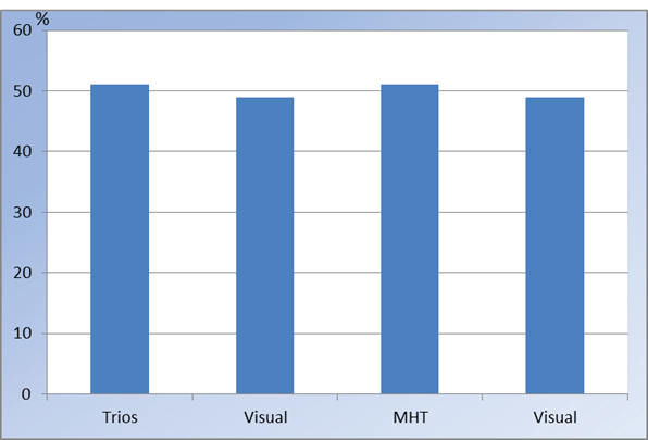
When the methods were validated by a determination of the best shade match no significant difference was found between Trios® Color and the conventional, visual method and between the MHT SpectroShade™ and the conventional, visual method (Figure 3).
.
Figure 3: Best shade match by comparing 3Shape Trios® Color (Trios) with traditional Visual method and MHT Spectroshade™ (MHT) with traditional visual method
View Figure 3
The frequency distribution of color value determinations showed that the computer-based systems had a broader use of all the 5 values with more bright and dark values compared to the visual methods, which concentrated on value 2 and 3. For all three methods the tooth 11 was the brightest and the canine the darkest tooth (Table 3).
![]()
Table 3: Frequency distribution of color value determinations
View Table 3
Discussion
The results support the use of a scanning and color measuring computer-based system in dentistry. The further development of such systems for clinical use would be warranted and could serve as a valuable tool for material selection and restoration design, particularly in the area of aesthetic, restorative dentistry.
The efficacy of the new digital scanning and computer software system for color determination was as good as an earlier validated digital colorimetric system and as the conventional, visual method for color determination in restorative dentistry.
In the present study the reliability of the computer-based systems were higher than for the conventional, visual system. This is in accordance with a number of other studies [7-9,15,16]. Most of these studies were performed in vitro, but a study by Gehrke and co-workers were performed in vivo. Three examiners looked at teeth from 40 subjects using two digital, computer-based systems and a visual determination and used color tabs from the Vita Classical Shade Guide for all included methods [9]. Different types of shade guides have been used for shade determination [9,15,17]. In the present study the 3D Vitapan Master tooth guide was chosen as it is the most commonly used color tab system by clinicians, and has demonstrated an acceptable reproducibility [18]. This color determination system has 28 color tabs, whereas the Vita Classical Shade Guide only has 14. Even though an increased number of color tabs should decrease the reliability of a system both the Trios Color scan system as well as the MHT Spectroshade system demonstrated a high reliability using the 3D Vitapan Masterguide color tabs. Another advantages of this color tab system should be that it includes the color elements, value, chroma and hue, which has been claimed to be the ideal order that best matches the capabilities of the human eye [19].
The study by Gehrke and co-workers [9] used three regions (incisal, middle and cervical) across the tooth for color determination. The Trios® Color Shade system as well as the MHT SpectroShade™ colorimetric system was able to measure all the various shades appearing all over the tooth surface, thus given a very detailed shade determination at the tested tooth. The reliability dropped, however, when the shade determination was extended to the periphery of the teeth. The incisal area was frequently translucent and affected by its background, and the scattered light from the gingiva frequently modified the cervical color [20]. Thus, the middle third of the tooth was selected for shade determination, and this has also shown to be the site that best represents the color of the tooth in a study including a high number of teeth [21].
The agreement between the new scanning and color determination method and the other computer-based colorimetric measure was almost perfect, and slightly better than the subjective methods. This is in contrast to a study by Hugo and co-workers [10], where 3 colorimetric systems and the visual method were compared in vivo and very low agreement between the 3 instruments (MHT SpectroShade™, X-Rite Shade Vision®, Rieth DSG4) was found. As in the present study no true color of the in vivo teeth could be determined with certainty. In the study by Hugo and co-workers they defined the "true" color as the color of every tooth that was determined by the majority of the methods. This was different from the present study, and it can be argued that the method used for testing agreement between methods and validations of the devices are essential for the outcome.
The validation of all three methods in the present study was performed by a pairwise, visual comparison of selected color tabs of one of the computer-based methods with the conventional visual method and two dentists had to assess the best shade match of the two. This visual pairwise comparison with the conventional, visual method was chosen in the present study, as the human eye and perception is the most important factor in color evaluation. This visual pairwise comparison demonstrated no significant differences between the computer-based methods including the new scanning method and the golden standard. Even though visual shade selection is inconsistent in nature the complexity of tooth color makes it difficult for computer-based systems to perform ideal color determinations under all conditions, which also has been highlighted in other publications [9]. However in the present study the computer-based systems were comparable to the human color perception, when tested in vivo. The newly developed color determination scanner used in the present study was easy to handle and more convenient to the patient than the colorimetric camera system used. The system was developed for digital impressions, but a color determination program was added, and in the present study, it seemed to be quite reliable and valid. However, the present study was relatively limited in number of tested teeth and can be looked as a pilot study. On the other hand the difference between the methods in the validation part was so small, that it would not be possible to have significant differences even though the number of examined teeth was tripled.
In the present study the frequency distribution of color determination of the anterior teeth in the upper jaw was only performed for the most important color dimension, value, caused by the limited number of included teeth. Furthermore the color value mainly depends of the incident light, surface texture and shape [4]. It was not surprisingly that the canine was darker that the incisors measured using all 3 methods. On the other hand it was a new observation that the central incisors was assessed brighter with the computer-based methods, than with the visual method, which concentrated on fewer values.
Conclusion
The effectiveness of the new digital scanning and computer software system for color determination was as good as an earlier tested and validated digital colorimetric system and as the conventional, visual method for color determination of teeth.
Acknowledgment
The investigators express their gratitude to 3Shape A/S (Holmens Kanal 7, 1060 Copenhagen, Denmark) for lending us the TRIOS equipment.
The authors acknowledge the assistance of the statistician, Ulla Mogensen.
References
-
Dueled E, Gotfredsen K, Trab Damsgaard M, Hede B (2009) Professional and patient-based evaluation of oral rehabilitation in patients with tooth agenesis. Clin Oral Implants Res 20: 729-736.
-
Peng M, Fei W, Hosseini M, Gotfredsen K (2014) Crown color match of implant-supported zirconia and porcelain-fused-to-metal restorations: a spectrophotometric comparison. Hua Xi Kou Qiang Yi Xue Za Zhi 32: 62-65.
-
Schropp L (2009) Shade matching assisted by digital photography and computer software. J Prosthodont 18: 235-241.
-
Molin MTVS PV (2013) Cosmetic and Esthetic Considerations. In: Nilner K, Karlsson S, Dahl BL (eds) A Textbook of fixed prosthodontics. The scandinavian approach 7: 344-357.
-
Paul S, Peter A, Pietrobon N, Hämmerle CH (2002) Visual and spectrophotometric shade analysis of human teeth. J Dent Res 81: 578-582.
-
Hassel AJ, Koke U, Schmitter M, Beck J, Rammelsberg P (2005) Clinical effect of different shade guide systems on the tooth shades of ceramic-veneered restorations. Int J Prosthodont 18: 422-426.
-
Bahannan SA (2014) Shade matching quality among dental students using visual and instrumental methods. J Dent 42: 48-52.
-
Judeh A, Al-Wahadni A (2009) A comparison between conventional visual and spectrophotometric methods for shade selection. Quintessence Int 40: 69-79.
-
Gehrke P, Riekeberg U, Fackler O, Dhom G (2009) Comparison of in vivo visual, spectrophotometric and colorimetric shade determination of teeth and implant-supported crowns. Int J Comput Dent 12: 247-263.
-
Hugo B, Witzel T, Klaiber B (2005) Comparison of in vivo visual and computer-aided tooth shade determination. Clin Oral Investig 9: 244-250.
-
Al-Dwairi Z, Shaweesh A, Kamkarfar S, Kamkarfar S, Borzabadi-Farahani A, et al. (2014) Tooth shade measurements under standard and nonstandard illumination and their agreement with skin color. Int J Prosthodont 27: 458-460.
-
Guan YH, Lath DL, Lilley TH, Willmot DR, Marlow I, et al. (2005) The measurement of tooth whiteness by image analysis and spectrophotometry: a comparison. J Oral Rehabil 32: 7-15.
-
Paravina RD (2002) Evaluation of a newly developed visual shade-matching apparatus. Int J Prosthodont 15: 528-534.
-
Team RC (2014) The R Project for Statistical Computing. R Journal 6: 6.
-
Derdilopoulou FV, Zantner C, Neumann K, Kielbassa AM (2007) Evaluation of visual and spectrophotometric shade analyses: a clinical comparison of 3758 teeth. Int J Prosthodont 20: 414-416.
-
Horn DJ, Bulan-Brady J, Hicks ML (1998) Sphere spectrophotometer versus human evaluation of tooth shade. J Endod 24: 786-790.
-
Joiner A (2004) Tooth colour: a review of the literature. J Dent 32 Suppl 1: 3-12.
-
Hammad IA (2003) Intrarater repeatability of shade selections with two shade guides. J Prosthet Dent 89: 50-53.
-
Gómez-Polo C, Gómez-Polo M, Celemin-Viñuela A, Martínez Vazquez De Parga JA (2014) Differences between the human eye and the spectrophotometer in the shade matching of tooth colour. J Dent 42: 742-745.
-
O'Brien WJ, Hemmendinger H, Boenke KM, Linger JB, Groh CL (1997) Color distribution of three regions of extracted human teeth. Dental Materials: Official Publication of the Academy of Dental Materials 13: 179-185.
-
Goodkind RJ, Schwabacher WB (1987) Use of a fiber-optic colorimeter for in vivo color measurements of 2830 anterior teeth. J Prosthet Dent 58: 535-542.
