

International Journal of Brain Disorders and Treatment
Diagnostic Challenge of Ovarian Teratoma Related Anti N-Methyl-D-Aspartate Receptor Encephalitis in a Patient with Giant Arachnoid Cyst
Kilinc O1*, Gulatar B1, Gonul O1, Yildizhan B2 and Midi I1
1Marmara University Hospital, Department of Neurology, Istanbul, Turkey
2Marmara University Hospital, Department of Obstetrics and Gynecology, Istanbul, Turkey
*Corresponding author:
Ozden Kilinc, MD, Department of Neurology, Marmara University Hospital, Fevzi Cakmak Mah., Mimar Sinan Cad., Ust Kaynarca, Pendik, 34899, Istanbul, Turkey, Tel: 90-216-6570606/8035, Fax: +90-216-6570695, E-mail: ozdenkilinc@gmail.com
Int J Brain Disord Treat, IJBDT-2-009, (Volume 2, Issue 1), Case Report; ISSN: 2469-5866
Received: January 08, 2016 | Accepted: January 23, 2016 | Published: January 26, 2016
Citation: Kilinc O, Gulatar B, Gonul O, Yildizhan B, Midi I (2016) Diagnostic Challenge of Ovarian Teratoma Related Anti N-Methyl-D-Aspartate Receptor Encephalitis in a Patient with Giant Arachnoid Cyst. Int J Brain Disord Treat 2:009. 10.23937/2469-5866/1510009
Copyright: © 2016 Kilinc O, et al. This is an open-access article distributed under the terms of the Creative Commons Attribution License, which permits unrestricted use, distribution, and reproduction in any medium, provided the original author and source are credited.
Abstract
Anti-N-methyl-D-aspartate receptor (NMDAr) encephalitis is well-characterized and treatable subtype of inflammatory encephalitis. This type of encephalitis is often associated with ovarian teratoma in young women and characterized by memory deficits, seizures, confusion and psychological disturbances. In this report, we presented a case of anti-NMDAr encephalitis in a woman with a previously asymptomatic, giant posterior fossa arachnoid cyst (AC). With our report, we present our clinical approach anti-NMDAr encephalitis with challenging magnetic resonance imaging (MRI) findings of giant AC. Also, we aim to emphasize the issue of the consideration of surgical indications and management of ACs on the basis of the presence of neuropsychiatric symptoms, misleading results of addressing to the diagnosis with only imaging studies and importance of elaboration of patient’s history and laboratory investigations.
Keywords
Anti N-methyl-D-aspartate receptor, Encephalitis, Arachnoid cyst, Magnetic resonance imaging
Introduction
Anti-N-methyl-D-aspartate receptor (NMDAr) encephalitis has been identified as a significant subtype of autoimmune encephalitis [1]. The disorder is known to be associated with ovarian teratoma in young women, also frequently affects men and children of all ages [2]. Available literature guides requirement of early resection of the teratoma for better outcomes in the treatment process [1,2]. Herein, we report a case of anti-NMDAr encephalitis in a previously healthy 22-year old woman with giant posterior fossa arachnoid cyst (AC). Symptomatic adult ACs are extremely rare but over the years, many reports have been published that describe neuropsychiatric symptoms in patients with AC, as well as asymptomatic reported cases [3]. There is a great deal of controversy regarding the changes in sizes over time, clinical manifestations and management of ACs [3,4]. In this case report, we aim to present our clinical approach in a patient with anti-NMDAr encephalitis with challenging magnetic resonance imaging (MRI) findings of giant AC. To the best of our knowledge, an engrossing coincidence of anti-NMDAr encephalitis and AC has not been reported so far.
Case Report
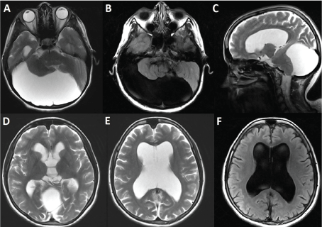
A 22-year-old gravida-0, para-0 sexually inactive female patient with no history of neurological disorder was admitted to our emergency service with complaints of bizarre behavior, irritability and visual hallucinations continuing beyond two weeks. Initial symptoms also included echolalia and inability to recognize her family members. In the emergency service, patient was assessed by both neurology and neurosurgery physicians. Her neurological examination revealed impaired memory, agitation, echolalia and orofacial dyskinesias. She had a temperature of 36.6 °C without meningeal signs. Brain computerized tomography (CT) and MRI demonstrated giant posterior fossa AC and tetraventricular hydrocephalus (Figure 1). First, she was hospitalized into neurosurgery clinic in order to assess the necessity for surgical intervention with the thought that the AC might be a source of patient’s subacute onset symptoms due to compression to the adjacent brain areas. Treatment with acyclovir was started immediately for possible viral encephalitis. Cerebrospinal fluid (CSF) analysis revealed normal glucose and protein levels. Viral and bacterial cultures and polymerase chain reaction analysis of CSF for herpes simplex virus also showed negative results and acyclovir treatment was stopped. At that time, involuntary movements on the right arm were added to her orofacial dyskinetic movements, which were thought to be in epileptic nature. Electroencephalography (EEG) monitoring demonstrated slowing of the background activity without epileptiform discharges. Due to the absence of any neurological symptoms previously, giant AC was considered to be chronic and asymptomatic and patient’s current complaints were not found to be related to the AC. Therefore, surgical intervention was not performed. She was transferred to neurology clinic. Anti-thyroid antibodies were found elevated in the serum and intravenous methylprednisolone treatment was started for possible Hashimoto's encephalopathy, but no clinical improvement was observed. On the 10th day of her admission, consecutive complex partial seizures were observed. Based on the combination of behavior, mood and cognitive changes with seizures and dyskinesia, the clinical presentation was found to be suggestive for anti NMDAr encephalitis. Autoimmune encephalitis and paraneoplastic antibody panels were sent and autoantibodies against to the NMDAr were detected in CSF. The patient was treated with intravenous immunoglobulin (IVIG) 400 mg/kg per day for five days. Abdominal MRI scan was performed, which demonstrated a right ovarian 7 cm in size mature cystic teratoma with normal left ovary. Surgical intervention for teratoma resection was performed promptly as well as immunotherapy. Monthly courses of IVIG were continued for additional two months and three months after initial presentation, the patient showed gradual improvement in cognitive functions without apparent deficits. Six months after the initial symptoms, the neuropsychological test results demonstrated normal cognitive functions except mild memory deficits.
.
Figure 1: T2-weighted axial (A-D-E) and sagittal (C) magnetic resonance images and fluid attenuated inversion recovery (FLAIR) sequence axial magnetic resonance images (B-F) show the giant posterior fossa arachnoid cyst and tetraventricular hydrocephalus.
View Figure 1
Discussion
Anti-NMDAr encephalitis is increasingly well-recognized inflammatory encephalitis with autoantibodies against to the extracellular epitopes of NMDAr [1]. It is characterized by subacute memory impairment, seizures, confusion and psychological disturbances, also often associated with a variety of different neurological symptoms [1,2]. Brain MRI is often normal or shows transient fluid attenuated inversion recovery or contrast-enhancing abnormalities in some patients [1]. Antibodies in CSF combined with a characteristic clinical picture have 100% sensitivity and specificity [5].
This type of encephalitis is frequently associated with ovarian teratomas in young women, but association with ovarian teratomas is rare in pediatric populations [2]. It has been suggested that as part of an anti-tumour response and/or with immune system stimulation after an infection, autoimmune antibodies against NMDAr are produced, which are ectopically expressed in ectodermic teratoma tissue derived from totipotent stem cells [6]. If there is an identified ovarian teratoma, early resection of the teratoma is considered to be essential for the treatment of anti-NMDAr encephalitis as well as initiation of immunotherapy [1,2]. In a recent study, it has been shown that greater than 80% of the patients with anti-NMDAr encephalitis had full or substantial recovery after immunotherapy and tumor resection [7].
The differential diagnosis of anti-NMDAr encephalitis is broad and includes neuroleptic malignant syndrome, viral encephalitis, metabolic and toxic encephalopathies [2]. With detailed evaluation, we excluded all of these etiological factors in our patient. The main difference of our case from the previously published reports was the challenging MRI findings. Initially, the subacute onset of symptoms of our patient enabled us to evaluate the possibility of a causal relationship between the AC and the genesis of the symptoms. However, serological investigations showed positive results for anti-NMDAr encephalitis.
The ACs are benign, non-tumorous, space-occupying congenital collections of CSF, though can be located all along the craniospinal axis. They are most commonly seen in the middle fossa and the second most frequent localization is the posterior fossa [8]. Symptomatic adult ACs are extremely rare, but over the years, many reports have been published that describe cognitive impairment, psychiatric symptoms and epileptic seizures in patients with AC [3,8]. The clinical signs of the ACs depend on the cyst's location and size and patient's age [4].
Selecting patients with AC for surgical treatment is still controversial [3]. In general, conservative management is recommended if the patient is asymptomatic and the size of the cyst is stable on repeated assessments [8]. However, if neuropsychiatric symptoms can be attributed to the cyst or if the cyst is enlarging significantly, decompression of the cyst is recommended [8]. In our case, there was much debate about the necessity of decompressing of the cyst, but the patient had not experienced any neurological symptoms previously, so her current complaints were not found to be related to the AC and surgical intervention was not performed. However, as the patient did not have a baseline cranial imaging before her clinical complaints, changes in cyst size over time was not known.
To the best of our knowledge, an engrossing coincidence of anti-NMDAr encephalitis and AC has not been reported so far. This case raises the issue of the consideration of surgical indications and management of ACs on the basis of the presence of neuropsychiatric symptoms. More importantly, it is a reminder of misleading results of addressing to the diagnosis with only imaging studies, importance of elaboration of patient’s history and laboratory investigations. Anti-NMDAr encephalitis represents a new category of immune-mediated disorder that is often paraneoplastic. Any reported case is implemental in increasing the awareness of this treatable disorder.
References
-
Dalmau J, Gleichman AJ, Hughes EG, Rossi JE, Peng X, et al. (2008) Anti-NMDA-receptor encephalitis: case series and analysis of the effects of antibodies. Lancet Neurol 7: 1091-1098.
-
Florance NR, Davis RL, Lam C, Szperka C, Zhou L, et al. (2009) Anti-N-methyl-D-aspartate receptor (NMDAR) encephalitis in children and adolescents. Ann Neurol 66: 11-18.
-
Wester K (2008) Intracranial arachnoid cysts--do they impair mental functions? J Neurol 255: 1113-1120.
-
Baquero GA, Molero P, Pla J, Ortuno F (2014) A schizophrenia-like psychotic disorder secondary to an arachnoid cyst remitted with neurosurgical treatment of the cyst. Open Neuroimag J 8: 1-4.
-
Wandinger KP, Saschenbrecker S, Stoecker W, Dalmau J (2011) Anti-NMDA-receptor encephalitis: a severe, multistage, treatable disorder presenting with psychosis. J Neuroimmunol 231: 86-91.
-
Mangler M, Trebesch de Perez I, Teegen B, Stocker W, Pruss H, et al. (2013) Seroprevalence of anti-N-methyl-D aspartate receptor antibodies in women with ovarian teratoma. J Neurol 260: 2831-2835.
-
Kayser MS, Titulaer MJ, Gresa-Arribas N, Dalmau J (2013) Frequency and characteristics of isolated psychiatric episodes in anti-N-methyl-d-aspartate receptor encephalitis. JAMA Neurol 70: 1133-1139.
-
Gan YC, Connolly MB, Steinbok P (2008) Epilepsy associated with a cerebellar arachnoid cyst: seizure control following fenestration of the cyst. Childs Nerv Syst 24: 125-134.
