

Clinical Medical
Reviews and Case Reports
Bilateral Clavicle Fracture: A Rare Presentation of a Common Form of Orthopedic Birth Injury
Joana Oliveira1*, Andreia Abrantes1, Raque1 Gouveia1,2 and Graça Oliveira1,2
1Paediatrics Department, Hospital de Santa Maria, Portugal
2Neonatology Unit, Hospital de Santa Maria, Portugal
*Corresponding author: Joana Oliveira, Paediatrics Department, Hospital de Santa Maria, Centro Hospitalar Lisboa Norte Av. Prof. Egas Moniz, 1649-028 Lisbon, Portugal, Tel: +351217805202, E-mail: joana.a.oliveira@hotmail.com
Clin Med Rev Case Rep, CMRCR-3-119, (Volume 3, Issue 7), Case Report; ISSN: 2378-3656
Received: May 09, 2016 | Accepted: July 11, 2016 | Published: July 14, 2016
Citation: Oliveira J, Abrantes A, Gouveia R, Oliveira G (2016) Bilateral Clavicle Fracture: A Rare Presentation of a Common Form of Orthopedic Birth Injury. Clin Med Rev Case Rep 3:119. 10.23937/2378-3656/1410119
Copyright: © 2016 Oliveira J, et al. This is an open-access article distributed under the terms of the Creative Commons Attribution License, which permits unrestricted use, distribution, and reproduction in any medium, provided the original author and source are credited.
Abstract
A female newborn presented at day-1 with isolated right clavicle crepitus after vaginal delivery by certified nurse midwives. Shoulder dystocia was suspected at birth but delivery was successful with the sole aid of suprapubic pressure. The adequate weight-for-gestational-age female newborn (3605 gr. at 39 Wk.) was vigorous at birth and no resuscitation was needed. Bilateral clavicle fracture was later confirmed by radiographic studies and associated birth injuries like brachial plexus paralysis were excluded during newborn physical exam screening. Outcome was favorable with complete recovery after three weeks of conservative treatment under orthopedic guidance.
Keywords
Birth injuries, Bone fractures, Clavicle, Neonate
Abbreviations
Gr: Grams, Wk: Weeks (postconceptional age)
Introduction
Bilateral clavicle fracture (CF) is a rare presentation of one of the most common forms of orthopedic birth injury [1,2]. Bilateral CF has been reported [3]. Unilateral CF incidence estimates vary greatly [1,4-7], especially if we consider undetected fractures at initial hospital discharge (up to 40% of all live births in some settings) [8]. Recent data point to an incidence of 0.6-1.6% of unilateral CF complicating vaginal delivery births [4-6].
CF is a benign and usually self-limiting injury resulting from mechanical stress forces applied to the fetus during the passage through the birth canal [7]. It is commonly associated with shoulder dystocia and to a lesser extent with brachial plexus injury, although the interrelation between these different entities has been difficult to interpret and is still under investigation [6].
CF may present with clavicular crepitus, palpable bony irregularity and sternocleidomastoid muscle spasm. Pain or discomfort may occur with variable degree of upper limb movement limitation. Moro reflex is usually absent or diminished but may be entirely normal. Peripheral nerve damage exclusion is mandatory as it has the potential to complicate an otherwise favorable prognosis. Radiographic studies confirm the fracture [9].
Incomplete diaphyseal fractures of the clavicle are the most common and frequently missed [3]. When misalignment occurs the medial fragment is pulled upwards by the action of the sternocleidomastoid muscle whereas the lateral fragment is pulled downward as a result of upper limb traction. Treatment is conservative. Fracture stabilization is not indicated in bilateral CF and/or thoracic trauma [9,10]. Associated nerve injury may require specific rehabilitation exercises and nerve reconstruction or nerve graft surgery in severe cases (avulsion) [11].
Other predisposing factors that contribute independently to increasing the risk of CF include fetal birth weight greater than 4250 Gr., advanced gestational age (> 42 Wk.), protracted labor, multiparity and short second stage of labor [6]. Prophylactic use of the McRobert's maneuver with or without the application of suprapubic pressure has not proved superior to therapeutic obstetric maneuvers in preventing birth trauma and unfavorable outcomes in cases of shoulder dystocia [12]. Recent data suggest that clinicians should use the therapeutic maneuver most likely to result in a successful delivery [13].
Case Report
A 24 h year-old female newborn presented with isolated right clavicle crepitus on physical examination and displaced bilateral clavicle fractures on radiograph studies after vaginal delivery by certified nurse midwives. Although shoulder dystocia was suspected at the time no instrumentation was needed: delivery was successful after resorting to suprapubic pressure measures. The adequate weight-for-gestational-age newborn (3605 gr at 39 wk) was vigorous at birth with no criteria for asphyxia (Apgar index: 9 at 1 minute; 10 at 5 minutes) and no need for resuscitation. A complete physical exam was performed at day-1 with exclusion of associated birth injuries including peripheral nerve injury: active and passive limb movements were unaffected on both sides and did not elicit any pain or cause discomfort, Moro reflex and grasp reflex were both present and symmetrical. Gestation was uneventful.
The mother's obstetric history included a vacuum delivery of a healthy female baby (2700 gr at 38 wk) four years earlier with no documented birth injuries. There was no other relevant family history.
Investigations
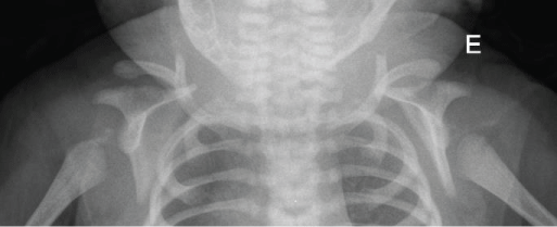
Clavicle radiographic studies confirmed a diagnosis of bilateral clavicle fracture. Bilateral misaligned diaphyseal clavicle fractures are visible on figure 1.
.
Figure 1: Newborn upper chest radiography at day 1 showing completely misaligned diaphyseal fractures on both clavicles.
View Figure 1
Treatment
After orthopedic consultation conservative measures were proposed to the mother: minimal handling and avoidance of tight clothing until further clinical follow-up within three weeks. The newborn was discharged at day three.
Outcome and follow-up
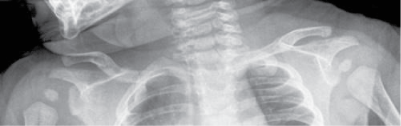
Complete resolution of bone fracture was documented at three-weeks of age during orthopedic consultation (Figure 2). Neither bone distortion nor peripheral nerve damage were found. The baby was discharged from orthopedic consultation and neurodevelopment is to be assessed during regular paediatric follow-up.
.
Figure 2: Newborn upper chest radiography at day 21 showing consolidated diaphyseal fractures on both clavicles (bony callus).
View Figure 2
Discussion
Bilateral CF fracture is a rare presentation of a common orthopedic birth injury, commonly associated with shoulder dystocia that is secondary to mechanical stress forces applied to the fetus in utero and most commonly during passage through birth canal [9,10]. Many fractures are undiagnosed at first hospital discharge as newborns are asymptomatic especially if there is no peripheral nerve damage as we verified in the reported case [8].
To our knowledge bilateral CF birth injuries reports are scarce and all report additional injuries. Kanik A, et al. [3] report a series of two macrosomic newborn babies with bilateral CF and associated brachial plexus palsy: the first male newborn (38 wk, 4200 g - 97th percentile) presented with bilateral Erb-Duchenne paralysis with complete recovery after two months of conservative treatment; the second male newborn (34 wk, 3200 g - < 97th percentile) presented with bilateral Erb-Duchenne paralysis and a small pneumomediastinum with complete recovery after two months of conservative treatment as well. Shoulder dystocia and normal birth-weight-for-age in our case are in accordance with the reported series; female gender and absence of associated birth injuries, especially brachial plexus paralysis, make our report rather unusual.
Published evidence of bilateral CF seems to suggest that fetuses may have been exposed to greater mechanical forces that predisposed them to additional birth injuries. We suspect similar cases of isolated bilateral CF may be under diagnosed as isolated unilateral CF has been well documented [6], or there may be some degree of report bias as prognosis is generally favorable for both unilateral and bilateral fractures [2].
References
-
Roberts SW, Hernandez C, Maberry MC, Adams MD, Leveno KJ, et al. (1995) Obstetric clavicular fracture: the enigma of normal birth. Obstet Gynecol 86: 978-981.
-
Ahn ES, Jung MS, Lee YK, Ko SY, Shin SM, et al. (2015) Neonatal clavicular fracture: recent 10 year study. Pediatr Int 57: 60-63.
-
Kanik A, Sutcuoglu S, Aydinlioglu H, Erdemir A, Arun Ozer E (2011) Bilateral clavicle fracture in two newborn infants. Iran J Pediatr 21: 553-555.
-
Kaplan B, Rabinerson D, Avrech OM, Carmi N, Steinberg DM, et al. (1998) Fracture of the clavicle in the newborn following normal labor and delivery. Int J Gynaecol Obstet 63: 15-20.
-
Lam MH, Wong GY, Lao TT (2002) Reappraisal of neonatal clavicular fracture: relationship between infant size and neonatal morbidity. Obstet Gynecol 100: 115-119.
-
Parantainen J, Palomaki O, Talola N, Uotila J (2014) Clinical and sonographic risk factors and complications of shoulder dystocia - a case-control study with parity and gestational age matched controls. Eur J Obstet Gynecol Reprod Biol 177: 110-114.
-
Dajani NK, Magann EF (2014) Complications of shoulder dystocia. Semin Perinatol 38: 201-204.
-
Paul SP, Heaton PA, Patel K (2013) Breaking it to them gently: fractured clavicle in the newborn. Pract Midwife 16: 31-34.
-
http://emedicine.medscape.com/article/980112-overview.
-
Mangurten HH (2015) Birth injuries. In: Martin RJ, Fanaroff AA, Walsh MC, Fanaroff and Mantin's Neonatal and Perinatal Medicine. Disease of the fetuses and infant. (10th edn), Mosby Elsevier, Philadelphia, e1-5.
-
Coroneos CJ, Voineskos SH, Coroneos MK, Alolabi N, Goekjian SR, et al. (2015) Primary Nerve Repair for Obstetrical Brachial Plexus Injury: A Meta-Analysis. Plast Reconstr Surg 136: 765-779.
-
Athukorala C, Middleton P, Crowther CA (2006) Intrapartum interventions for preventing shoulder dystocia. Cochrane Database of Systematic Reviews, CD005543.
-
Spain JE, Frey HA, Tuuli MG, Colvin R, Macones GA, et al. (2015) Neonatal morbidity associated with shoulder dystocia maneuvers. Am J Obstet Gynecol 212: 353.
