Clinical Medical
Reviews and Case Reports
Multiple Myeloma and Amyloidosis Presenting as a Restrictive Lung Disease with Respiratory Failure
Marta Pereira1*, Luís Afonso1, Gonçalo Fernandes2 and Rui Araújo3
1Internal Medicine Resident, Medical Department, Hospital Pedro Hispano, Matosinhos, Portugal
2Anesthesiology Consultant, Intensive Medicine Unit, Hospital Pedro Hispano, Matosinhos, Portugal
3Anesthesiology Consultant, Director of Intensive Medicine Unit, Hospital Pedro Hispano, Matosinhos, Portugal
*Corresponding author: Marta Pereira, Internal Medicine Resident, Medical Department, Hospital Pedro Hispano, Rua Dr. Eduardo Torres, 4454-509 Senhora da Hora, Matosinhos, Portugal, Tel: 229391000, E-mail: martabarpereira@gmail.com
Clin Med Rev Case Rep, CMRCR-3-091, (Volume 3, Issue 2), Case Report; ISSN: 2378-3656
Received: January 12, 2016 | Accepted: February 18, 2016 | Published: February 20, 2016
Citation: Pereira M, Afonso L, Fernandes G, Araújo R (2016) Multiple Myeloma and Amyloidosis Presenting as a Restrictive Lung Disease with Respiratory Failure. Clin Med Rev Case Rep 3:091. 10.23937/2378-3656/1410091
Copyright: © 2016 Pereira M, et al. This is an open-access article distributed under the terms of the Creative Commons Attribution License, which permits unrestricted use, distribution, and reproduction in any medium, provided the original author and source are credited.
Abstract
Amyloidosis refers to the extracellular tissue deposition of fibrils resulting from abnormal folding of proteins. It may affect multiple organs, causing a broad range of symptoms, thus making the diagnosis particularly challenging. Polyneuropathy is one of the recognized manifestations of Amyloid Light-chain (AL) amyloidosis.
We present a case of Multiple Myeloma and AL amyloidosis with associated motor axon neuropathy and thickened chest wall leading to restrictive ventilatory pattern and respiratory failure as presenting symptoms.
Polyneuropathy and respiratory failure may be a prominent or presenting feature of AL amyloidosis, and this diagnosis should be considered in all older patients who seem to have a neuromuscular basis for the respiratory failure.
Keywords
Amyloidosis, Multiple myeloma, Respiratory failure, Polyneuropathy
Introduction
Light chain amyloidosis is a disorder characterized by deposition of insoluble monoclonal immunoglobulin light chain fragments in various tissues. It is associated with various B cell lymphoproliferative disorders encompassing the multiple myeloma (MM) - plasma cell dyscrasia spectrum [1]. Clinical features depend on organs involved but can include restrictive cardiomyopathy, nephrotic syndrome, hepatic failure and peripheral/autonomic neuropathy. Peripheral neuropathy occurs in 17% of patients with Amyloid Light-chain (AL) amyloidosis, making it the most common type of acquired amyloid polyneuropathy. Due to varying clinical symptoms, the diagnosis of amyloid neuropathy is often a challenge [2,3].
We present a case of Multiple Myeloma AL amyloidosis with associated motor axon neuropathy and thickened chest wall leading to restrictive ventilatory pattern and respiratory failure as presenting symptoms.
Case Report
A 61-year-old female with no previously known diseases was admitted to the Emergency Room with dyspnoea and peripheral oedema. She had been asymptomatic until 9 months before this episode, when she fractured her right femur after a minor fall. She underwent surgery, but nevertheless became progressively more dependent, in part due to concurrent global muscle weakness. She lost about 10-15 kg since then. Around that time, a bilateral carpal tunnel syndrome was documented and she also had surgery with clinical improvement. More recently, for the previous 3 weeks, she had been complaining of exertional dyspnoea with increasing intensity (symptomatic with minor efforts), peripheral oedema, dysphonia and dysphagia, frequently choking with liquid food. On admission, there were signs of severe respiratory distress with concurrent respiratory acidemia (pH 7.21 | pO2 124 mmHg | pCO2 93 mmHg, on 40% FiO2), and orotracheal intubation and mechanical ventilation (MV) were needed after an unsuccessful trial of non-invasive ventilation (NIV). She was hypotensive (BP 95/47 mmHg), needing vasopressor support. Mild rales were audible on both pulmonary bases. Peripheral oedema was evident at admission along with diffuse cutaneous thickening, mainly of the thoracic wall and limbs, and macroglossia. She was apyrexial. Neurological examination showed bulbar dysfunction (hypophonia and dysphagia), horizontal nystagmus and proximal reduced motor strength in all four limbs (2/5 proximal; 4/5 distal). There was no sensitive dysfunction noted.
After admission in Intensive Care, respiratory gas exchange quickly improved, but weaning from MV was very difficult - she was extubated twice when all conditions were optimized and support was minimal, but showed poor ventilatory dynamics and subsequent respiratory failure, culminating in tracheostomy by day 6. There was the impression that this was a restrictive disorder due to respiratory muscle weakness and some restraining effect from the thickened skin of the chest wall. From the extensive study that took place, we highlight:
- Normochromic normocytic anemia, with normal renal function, and no electrolyte disturbances (particularly no hypercalcemia); increased β2 microglobulin; Serum protein electrophoresis with immunofixation showed a monoclonal protein IgA/kappa and Urinary protein electrophoresis confirmed Bence-Jones proteinuria.
- Electromyography was suggestive of motor axon polyneuropathy, with reduced motor amplitude in all motor nerves evaluated and normal results in every sensitive nerve evaluated. - ECG with right bundle branch block and low voltage.
- Transthoracic Echocardiogram with left ventricular (LV) wall thickening (12 mm) and dyastolic dysfunction.
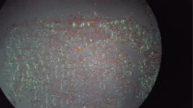
- Skin biopsy specimen collected from the chest wall (where the skin appeared to be more thickened) showed amyloid deposition – apple-green birefringence with Congo red staining under polarized light (Figure 1).

.
Figure 1: Apple-green birefringence on Congo red histological staining under polarized light, suggestive of amyloid deposits (10x).
View Figure 1
According to these results, the diagnosis of AL amyloidosis associated with MM was considered as the main hypothesis. Immunohistochemical typing of amyloid was not possible due to the characteristics of the sample available (it is rarely possible from a fat specimen and renal and/or gastrointestinal biopsies are preferable for this purpose). Nevertheless, the deposition of amyloid and the diagnosis of MM made the diagnosis of AL amyloidosis the most probable. Furthermore, the clinical status of the patient required an early treatment, and so, performing another biopsy, although ideal, was not considered a priority for starting treatment (which would always be necessary for MM alone). There was no evidence of kidney failure but motor polyneuropathy and possibly cutaneous thickening by amyloid deposition were responsible for this severe and atypical presentation with respiratory failure and dependence of ventilatory support. Furthermore, there was evidence suggestive of cardiac infiltration, which might justify symptoms of heart failure previously described.
The patient remained in hospital for three months, being decannulated about one month after tracheostomy, but always dependent on positive pressure for adequate gas exchange. Chemotherapy was started with dexamethasone and bortezomib, with very good results - almost two years after diagnosis, and still on chemotherapy, she is almost completely independent and is slowly being able to tolerate increasing periods without NIV, although currently still needing this ventilator support overnight. Her strength improved and electromyography was repeated with complete normalization of the initial results, documenting total recovery of motor neuropathy. Dyspnoea also improved (currently class II of the New York Heart Association), echocardiogram showed reduction of LV wall thickness and brain natriuretic peptide (BNP) is currently normal. Serum and urinary free light chains decreased more than 50%, which corresponds to a good partial response.
![]()
Table 1: Diagnostic tests.
View Table 1
Discussion
Amyloidosis refers to a collection of conditions in which abnormal protein folding results in insoluble fibril deposition in tissues [3,4]. Histochemically, this abnormally folded protein binds to Congo red, revealing green birefringence under polarized light [2]. There are about twenty described biochemical forms of amyloidosis, and one of the major types is AL, in which case the precursor proteins are Kappa or Lambda immunoglobulin light chains derived from plasma cells. MM coexists with AL amyloidosis in around 10-15% of cases and this scenario is associated with poor prognosis [1]. AL amyloidosis may affect multiple organs and diagnosis is often a challenge due to multiple and apparently unrelated symptoms [1-4]. They can include haemorrhage, skin and soft tissue thickening, painful seronegative arthropathy, hoarse voice lymphadenopathy and signs of hypoadrenalism or hypothyroidism, paired with more frequent presentations like nephrotic syndrome, heart failure, sensorimotor peripheral neuropathy and hepatomegaly.
In fact, the case presented is a major example of this challenge - a patient admitted with respiratory failure and recent symptoms suggesting heart failure, although completely disproportionate to the severity of respiratory acidemia, and with difficult ventilatory weaning even after clinical improvement. A complete neurological exam and an electromyography led to the diagnosis of a motor polyneuropathy with respiratory muscle weakness that was interpreted as the major cause for an inability to breathe without the help of a minor positive pressure. Aetiology was established afterwards with the diagnosis of AL amyloidosis associated with MM (monoclonal immunoglobulin IgA/kappa with related organ impairment - Anemia and possible pathological fracture). Electromyography results were suggestive of a motor axon polyneuropathy - amyloid polyneuropathy versus possible Guillain-Barré Syndrome (AMAN type), which can be associated to MM by itself. Absence of sensitive neuron lesion was less in favour of amyloid neuropathy. Nerve biopsy was not performed to distinguish between both aetiologies.
Peripheral neuropathy can present as a para-neoplastic manifestation of MM (25-30% cases), usually mimicking polyradiculoneuropathy, which could be what happened in the present case. In can also be a common complication of many of the systemic amyloidosis, occurring in 17% of patients with AL amyloidosis and making it the most common type of acquired amyloid polyneuropathy [3]. Although the cause of neuropathy is not entirely clear, it is likely related to amyloid deposition within the nerve [2-4]. This may lead to focal, multifocal or diffuse neuropathies involving sensory, motor and/or autonomic fibres. As the disease progresses, it can affect larger nerve fibres and patients may complain of major motor weakness. The presenting symptoms depend on the distribution of nerves affected and might be something as atypical as a restrictive pulmonary disorder like this patient experienced [2,4]. Carpal tunnel syndrome, previously diagnosed in this case, is also one of the most common types of neuropathy associated with AL amyloidosis [3,4]. It is also possible that muscle deposits of amyloid contributed to this scenario, although no histological study was done to support this. In fact, amyloid myopathy, a rare condition in which amyloid is deposited in the muscle, is usually characterized by muscle weakness, pseudohypertrophy and macroglossia and has been progressively recognized as a cause of respiratory failure [5].
Curiously, and even with important proteinuria, this patient did not have signs of renal disease, one of the most prevalent in MM and amyloidosis. However, she also presented with recent signs and symptoms of congestive heart failure, thickening of ventricular walls on echocardiography and low voltage QRS complexes on ECG suggesting, in this particular context, myocardial deposition of amyloid. In fact, cardiac involvement is described as either a positive cardiac biopsy demonstrating amyloid infiltration or as an increased LV wall thickness > 12 mm in the absence of arterial hypertension or other potential causes of true LV hypertrophy with a positive non-cardiac biopsy. The presence and severity of amyloid cardiomyopathy is the major factor influencing prognosis of affected subjects [1,4].
Treatment options for MM associated AL amyloidosis include chemotherapy and autologous peripheral blood stem cell transplantation [4]. Peripheral neuropathy often persists even in treated patients, although some recovery based on electrophysiologic tests and histopathological evidence of AL amyloid regression may occur [3]. This patient had a positive response to treatment with clinical improvement showed by progressive reduction of the need for NIV and complete normalization of electromyography findings.
Learning Points
- Increasing medical alertness of the natural history of AL amyloidosis might reduce the time to achieve the diagnosis and therapeutic decisions, also improving the prognosis.
- Some symptoms seemingly unrelated to each other (asthenia, weight loss, oedema, neuropathy, macroglossia) are actually early and specific red flags of the amyloid process.
- Polyneuropathy and respiratory failure may be a prominent or presenting feature, and consequently the diagnosis should be considered in all older patients who seem to have a neuromuscular basis for the respiratory problem.
Acknowledgments
The authors would like to thank Dr. Herberto Oliveira, from the Anatomopathology Department, for his precious help in this case.
References
-
Fikrle M, Palecek T, Kuchynka P, Nemecek E, Bauerová L, et al. (2013) Cardiac amyloidosis: A comprehensive review. Cor et vasa 55: 60-75.
-
Adams D, Lozeron P, Lacroix C (2012) Amyloid neuropathies. Curr Opin Neurol 25: 564-572.
-
Streeten EA, de la Monte SM, Kennedy TP (1986) Amyloid infiltration of the diaphragm as a cause of respiratory failure. Chest 89: 760-762.
-
Rajkumar SV (2015) Clinical presentation, laboratory manifestations and diagnosis of immunoglobulin light chain (AL) amyloidosis (primary amyloidosis). UpToDate, Waltham, MA.
-
Ashe J, Borel CO, Hart G, Humphrey RL, Derrick DA, et al. (1992) Amyloid myopathy presenting with respiratory failure. J Neurol Neurosurg Psychiatry 55: 162-165.


