

Clinical Medical
Reviews and Case Reports
A Rare Case of Bilaterale Ureterohydronephrosis due to Cystitis Cystica Et Glandularis and Review of the Literature
Zafer Demirer1*, Ali Guragac2, Burak Kopru2, Bilal Firat Alp2 and Ibrahim Yildirim2
1Department of Urology, Eskisehir Military Hospital, Eskisehir, Turkey
2Department of Urology, Gülhane Military Medical Academy, School of Medicine, Ankara, Turkey
*Corresponding author:
Zafer Demirer, Department of Urology, Eskisehir Military Hospital, Eskisehir, Turkey, Tel: +90 222 2204530-4250, Fax: +90 222 2303433, E-mail: zaferdemirer@mynet.com, zaferdemirer1903@gmail.com
Clin Med Rev Case Rep, CMRCR-2-061, (Volume 2, Issue 10), Case Report; ISSN: 2378-3656
Received: June 22, 2015 | Accepted: October 02, 2015 | Published: October 05, 2015
Citation: Demirer Z, Guragac A, Kopru B, Alp BF, Yildirim I (2015) A Rare Case of Bilaterale Ureterohydronephrosis due to Cystitis Cystica Et Glandularis and Review of the Literature. Clin Med Rev Case Rep 2:061. 10.23937/2378-3656/1410061
Copyright: © 2015 Demirer Z, et al. This is an open-access article distributed under the terms of the Creative Commons Attribution License, which permits unrestricted use, distribution, and reproduction in any medium, provided the original author and source are credited.
Abstract
Cystitis Cystica Et Glandularis (CCEG) is a metaplastic lesion of the bladder mucosa with a characteristic histopathologic appearance. CCEG are commonly found in specimens taken from the bladder wall of patients with cancer and other diseases or at autopsy. In most cases the course of CCEG is asymptomatic. Some patients complain of hematuria, symptoms associated with the obstruction of the upper urinary tract, or lower urinary tract symptoms. The main symptoms of CCEG accompanied with upper urinary tract obstruction are renal colic and abdominal pain, and the symptoms in most CCEG without upper urinary tract obstruction are frequency, urgency, dysuria, hematuria and fever. The primary treatment for CCEG comprises cause elimination and transurethral resection (TUR). We present a rare case of CCEG, intestinal type, which caused bilaterale ureterovesical obstruction leading to renal colic, bilaterale ureterohydronephrosis and haematuria. The case was treated by TUR.
Keywords
Bladder, Cystitis glandularis, Cystitis cystica, Ureterohydronephrosis, Transurethral resection
Introduction
The urothelium shows intermediate histologic features between squamous and glandular epithelium, forming the transitional epithelium. CCEG is a metaplastic lesion of the bladder mucosa with a characteristic histopathologic appearance [1]. These 2 terms are often used interchangeably. Cystitis Cystica (CC) occurs with cystic dilatation of von Brunn's nests, where the nests acquire a luminal space and may become markedly dilated [1,2]. Cysistitis Glandularis (CG) is a common benign proliferative lesion of the bladder which is characterized with mucus producing glands within the mucosa and submucosa of urinary bladder epithelium [1-3].
CCEG is a metaplastic alteration of the urothelium in the urinary bladder due to persistent infection, outlet obstruction, bladder exstrophy, bladder stones, indwelling catheterization, or even tumors [4]. In most cases the course of CCEG is asymptomatic. However, some patients complain of hematuria, symptoms associated with the obstruction of the upper urinary tract, or lower urinary tract symptoms (LUTS) [5].
We present a rare case of CCEG, intestinal type, causing bilaterale ureterovesical obstruction and leading to renal colic, bilaterale ureterohydronephrosis and haematuria, which was managed by TUR.
Case Report
A 27-year-old man presented with recurrent macroscopic haematuria and bilaterale loin pain. Urological history and physical examination were otherwise unremarkable. Urine analysis was significant only for more than 300 red blood cells per high power field. The level of serum creatinine was 1.40 mg/dl. Urine culture and cytology as well as routine blood examinations, serum biochemistry profiles were all unremarkable.
Bilaterale hydroureteronephrosis and bladder mass in the bladder floor were also demonstrated by ultrasonographic evaluation (Figure 1). Then a non contrast computed tomography scan of the abdomen and pelvis was performed in order to show that no renal or ureteric calculi accounted for hydronephrosis. A rigit cystoscopy revealed a large mass arising from the bladder neck, extending along the base and covering the bilaterale orifices; neither of the orifices was visible while on his cystoscopic evaluation. Then TUR of the mass was performed down to the base and area was fulgurated, ureteral orifices couldn't be visualized after the mass resection either (Figure 2).
.
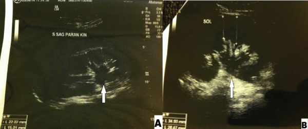
Figure 1: Ultrasonographic evaluation revealed bilateral (A-right, B-left) hydronephrosis, and proximal hydroureter.
View Figure 1
.
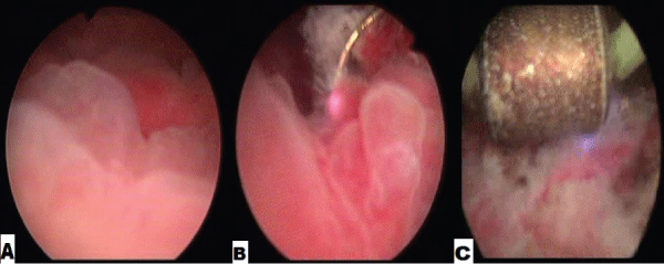
Figure 2: Cystoscopic evaluation demonstrated large mass arising from the bladder floor (A), TUR (B) and fulguration (C) was performed for the treatment.
View Figure 2
Because of his increasing bilaterale hydronephrosis and loin pain, bilaterale percutaneous nephrostomy was performed and a week later bilaterale JJ stents were placed via antegrade approach (Figure 3).
.
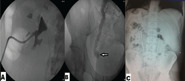
Figure 3: Bilaterale ureterohydronephrosis due to bilaterale ureterovesical obstruction (B), bilaterale percutaneous nephrostomy (A) a week later bilaterale JJ stents were placed by antegrade approach (C).
View Figure 3
Total operating time was approximately 70 minutes with estimated blood loss of 150 ml. Histopathological examination of the specimen revealed CCEG, intestinal type, and there was no cytological atypia within the glands noted (Figure 4 and Figure 5). His urethral catheter was removed on postoperatif 6th day and patient was discharged. After six month of surgery patient was checked with rigit cystoscopy and much improved bladder was shown. The JJ stent in the bilaterale ureters were removed.
.
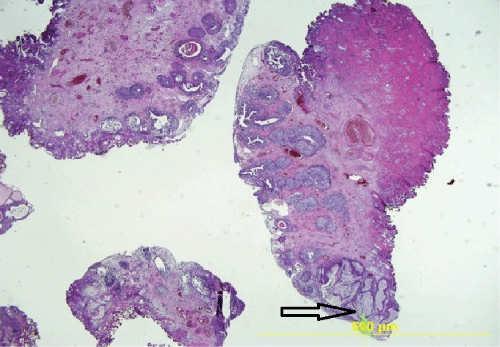
Figure 4: Histopathological examination with areas of intestinal type CCEG, (H-E, original magnification X20).
View Figure 4
.
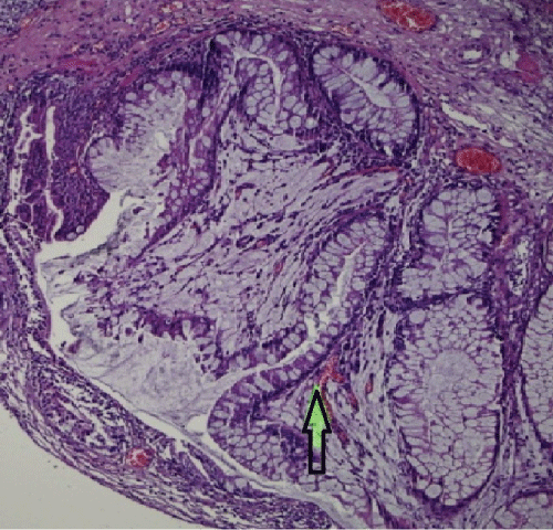
Figure 5: CCEG, intestinal type. The lesions consisted of a proliferation of glands in the lamina propria. The glands were lined by columnar epithelium, including goblet cells. Mucin production and focal mucin extravasation into the stroma was identified. There is no cytological atypia within the glands (H-E, original magnification X 100).
View Figure 5
Discussion
CCEG is a benign proliferative lesion of the bladder mucosa [1,2] which is defined as a metaplastic transformation of mucosal cells and was first reported by Yon Limberk in 1887 [6]. These 2 terms are often used interchangeably. Cystitis Cystica (CC) occurs with cystic dilatation of von Brunn's nests, where the nests acquire a luminal space and may become markedly dilated [1,2]. Cysistitis Glandularis (CG) is a common benign proliferative lesion of the bladder which is characterized with mucus producing glands within the mucosa and submucosa of urinary bladder epithelium [1-3].
The prevalence of cystitis cystica has been reported at 60% in normal bladders on autopsy [7]. Though it occurs in male and female adults, it is more common in women. The etiology remains unknown. It is believed that CCEG is due to chronic irritation to the bladder epithelium [2]. Many factors have been associated with CCEG; persistent infection, outlet obstruction, bladder exstrophy, bladder stones, indwelling catheterization, neurogenic bladder, or even tumors [1,4]. CCEG has been shown to be more frequent in patients with pelvic lipomatosis [5].
Morphologically, CG is subdivided into two subtypes: typical CG and intestinal CG [1,2]. Typical CG is characterized by the entity of structures coated by cubic or columnar epithelium and unaltered urothelial cells. These lesions are usually situated more superficially. The typical type of CG and CC rarely constitutes an actual diagnostic problem [2]. Intestinal type CG occurs when the epithelium contains intestinal type goblet cells interspersed among the columnar cells and morphologically look like colonic mucosa and not covered by urothelium [2]. The differential diagnoses of CG of intestinal type include adenocarcinoma, nephrogenic adenoma and endocervicosis [2,3].
Although this remains controversial, CCEG has been hypothesized as a potential precursor of adenocarcinoma. CCEG is often shown in relationship with carcinoma but no causal connection with carcinoma has been demonstrated [2]. There are a few reports of transformation of CG of the intestinal type into adenocarcinoma which may suggest that the intestinal CG has a higher risk of malignant transformation [1].
In most cases the course of CCEG is asymptomatic. CCEG is generally a microscopic detection and very seldomly present large appearing macroscopic lesions. However, some patients complain of hematuria, symptoms connected with the obstruction of the upper urinary tract, or LUTS [5,8]. The main symptoms of cystitis glandularis accompanied with upper urinary tract obstruction are renal colic and abdominal pain, and the symptom in most cystitis glandularis without upper urinary tract obstruction are frequency, urgency, dysuria, hematuria and fever. CCEG may cause obstructive urinary symptoms if it results in a mass that obstructs the bladder neck or ureter by infiltrating the periureteral submucosa [3]. In 2012 Zhu et al. [8] reported a rare case of recurrent florid cystitis cystica et glandularis (CCEG), common type, causing obstruction of the left ureterovesicle junction leading to renal colic and hydronephrosis [8]. In our case, the presenting symptom was recurrent macroscopic haematuria and bilaterale loin pain, due to bilaterale hydroureteronephrosis.
Most of the CCEG lesions are identified during urinary tract ultrasound imaging [1,3]. The condition is often diagnosed after cystoscopy and biopsy but the final diagnosis needs pathological examination [1-3].
The treatment of CCEG is usually performed by treating the underlying cause. Treatment of focal lesions in symptomatic patients generally involves removal by TUR, like we performed in our case [3,5,8]. Patients usually do not need supplementary treatment. The florid form is reported much rarely and usually necessitates extensive resection of the lesions. In patients with severe common form of CCEG the treatment is more difficult. In case of repetitive CCEG, which leads to obstruction of the upper urinary tract, the only way out may be to apply cystectomy with orthotopic neobladder [9]. There are no suggestions related to follow-up of patients with CCEG in the literature. Cystoscopy is indicated only in case of symptoms relapse.
Conclusion
Cystitis glandularis is a benign metaplasia of the bladder urothelium with a potential for malignancy due to the chronic inflammation. Although it is generally asymptomatic, the urinary tract obstruction is the main clinical symptom in patients with CCEG. In the medical protocol to CCEG presenting with upper urinary tract obstruction, the most important thing is to detect and remove the cause of the obstruction. According to previous published case reports CCEG can be considered as a premalignant condition of the bladder and these patients will require long-term surveillance in the form of cystoscopic evaluation if symptoms relapse.
References
-
Smith AK, Hansel DE, Jones JS (2008) Role of cystitis cystica et glandularis and intestinal metaplasia in development of bladder carcinoma. Urology 71: 915-918.
-
Clouston D, Lawrentschuk N (2013) Metaplastic conditions of the bladder. BJU Int 2: 27-31.
-
Kaya C, Akpinar IN, Aker F, Turkeri LN (2007) Large Cystitis glandularis: a very rare cause of severe obstructive urinary symptoms in an adult. Int Urol Nephrol 39: 441-444.
-
Lu Q, Jiang F, Xu R, Zhao XK, Zhong ZH, et al. (2013) A pilot study on intravesical administration of curcumin for cystitis glandularis. Evid Based Complement Alternat Med 2013: 269745.
-
Michajlowski J, Matuszewski M, Klacz J, Gibas A, Biernat W, et al. (2011) Acute urinary retention in a patient with extended cystitis glandularis. Cent European J Urol 64: 94-96.
-
Jost SP, Dixon JS, Gosling JA (1993) Ultrastructural observations on cystitis cystica in human bladder urothelium. Br J Urol 71: 28-33.
-
Wiener DP, Koss LG, Sablay B, Freed SZ (1979) The prevalence and significance of Brunn's nests, cystitis cystica and squamous metaplasia in normal bladders. J Urol 122: 317-321.
-
Zhu JX, Gabril MY, Sener A (2012) A rare case of recurrent urinary obstruction and acute renal failure from cystitis cystica et glandularis. Can Urol Assoc J 6: 72-74.
-
Black PC, Lange PH (2005) Cystoprostatectomy and neobladder construction for florid cystitis glandularis. Urology 65: 174.
