Clinical Medical
Reviews and Case Reports
Breast Cancer Adjacent to Nodular Fasciitis: A Case Report
Ryuji Takahashi1,2*, Ayaka Ishii1,2, Roka Namoto Matsubayashi1,2, Shino Nakagawa1,2, Momoko Akashi1,2, Kana Tsutsui1,2, Shino Harada1,2, Seiya Momosaki2,3 and Yoshito Akagi4
1Department of Breast Care Center, National Hospital Organization Kyushu Medical Center, Fukuoka, Japan
2Clinical Research Institute, National Hospital Organization Kyushu Medical Center, Fukuoka, Japan
3Department of Pathology, National Hospital Organization Kyushu Medical Center, Fukuoka, Japan
4Department of Surgery, Kurume University School of Medicine, Kurume, Japan
*Corresponding author:
Ryuji Takahashi, Kurume University School of Medicine, 67 Asahi-machi, Kurume, 830-0011, Japan, E-mail: ryuji@kyumed.jp
Clin Med Rev Case Rep, CMRCR-2-057, (Volume 2, Issue 9), Review Article; ISSN: 2378-3656
Received: July 20, 2015 | Accepted: September 10, 2015 | Published: September 12, 2015
Citation: Takahashi R, Ishii A, Matsubayashi RN, Nakagawa S, Akashi M, et al. (2015) Breast Cancer Adjacent to Nodular Fasciitis: A Case Report. Clin Med Rev Case Rep 2:057. 10.23937/2378-3656/1410057
Copyright: © 2015 Takahashi R, et al. This is an open-access article distributed under the terms of the Creative Commons Attribution License, which permits unrestricted use, distribution, and reproduction in any medium, provided the original author and source are credited.
Abstract
Nodular fasciitis is a benign proliferative lesion that is generally found in the soft tissue of the upper extremity and trunk. It has rarely been reported in the breast and that of the breast is indistinguishable from breast cancer.A 77-year-old woman visited our department to biopsy the swelled axillary lymph node. Contemporary, a breast tumor was found in her left breast.Mammography showed a high density mass with microlobulated margin. On ultrasonography, there was an irregular hypoechoic mass which was adjacent to a heterogeneous hyperechoic lesion. Needle biopsiedtissuesshowed a proliferation of spindle-shaped cells and suggested nodular fasciitis. On Magnetic Resonance Imaging (MRI), a lobulated mass was detected in the left breast. The mass showed iso Signal Intensity (SI) to mammary gland on T1-weighted images, while it showed intermingled high and low SI on T2-weighted images. Dynamic contrast-enhanced MRI showed rapid early enhancement followed by wash-out, but persistent enhancement was partly shown. Surgical excision of the breast tumor was performed in the present case. Pathologically, there was an invasive ductalcarcinoma adjacent to nodular fasciitis.By immunohistochemistry, these tumor cells showed diffusely positive for estrogen and progesterone receptors and negative for human epidermal growth factor receptor 2 (HER2). We propose that estrogen-related changes regarding mammary gland proliferation might play an important role in the etio pathogenesis of nodular fasciitis.
Keywords
Nodular fasciitis, Breast MRI, Breast cancer
List of Abbreviations
MRI: magnetic resonance imaging, SI: Signal intensity, HER2: human epidermal growth factor receptor 2, H.E: Eosin & Hematoxylin.
Introduction
Nodular fasciitis is a benign proliferation of myo fibroblasts usually found in the soft tissue of the upper extremities or the lower extremities, the head and thorax [1,2]. It has rarely been reported in the breast and has been advocated that indistinguishable from breast cancer.The history of trauma is elicited in approximately 5-15 percent of patients [2]. Herein we report a patient with breast cancer adjacent to nodular fasciitis in the breast.
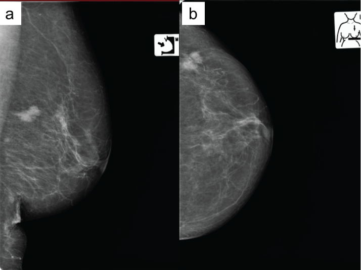
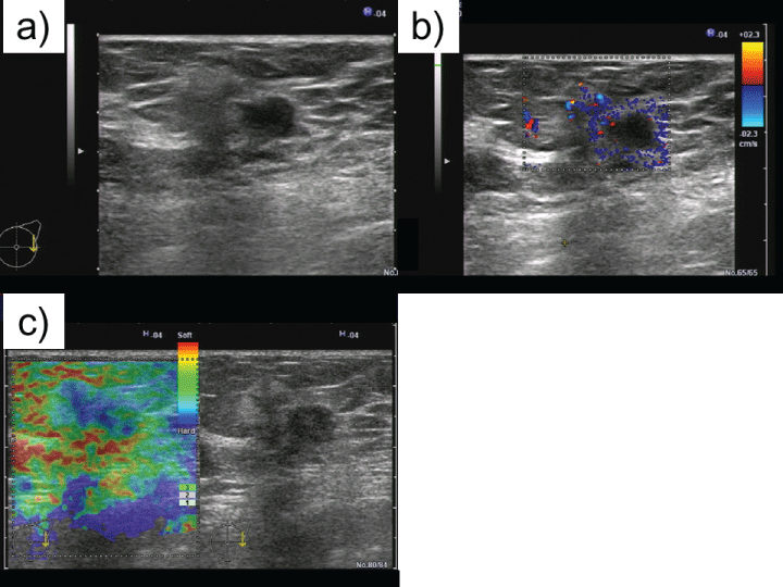
A 77-year-old woman visited our department to biopsy the axillary lymph node, because malignant lymphoma was clinically suggested. Her left breast had been bruised and a breast tumor was palpable in the left lateral lesion. Mammography showed a high density mass with microlobulated margin in the retromammary space (Figure 1a and Figure 1b). On ultrasonography, there was an irregular hypoechoic mass (15×9 mm in size) with an adjacent heterogeneous hyperechoic lesion (8×8 mm in size) (Figure 2a). Increased peri-tumoral vascular flow and hardness of the tumor measured by elastography suggested an invasive breast cancer which invaded the adipose tissues (Figure 2b and Figure 2c).
.
Figure 1: Mammography
Mammography showed a high density mass with microlobulated margin in the retromammary space (a: Mediolateral oblique view, b: Craniocaudal view).
View Figure 1
.
Figures 2: Ultrasonography
a: On ultrasonography, there was an irregular hypoechoic mass (15×9 mm in size) with an adjacent heterogeneous hyperechoic lesion (8×8 mm in size). b, c: Increased peri-tumoral vascular flow and hardness of the tumor measured by elastography suggested an invasive breast cancer which invaded the adipose tissue (b: Color Doppler mode, c: Elastography).
View Figures 2
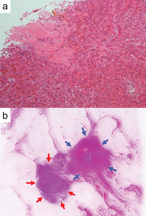
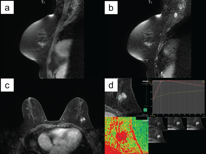
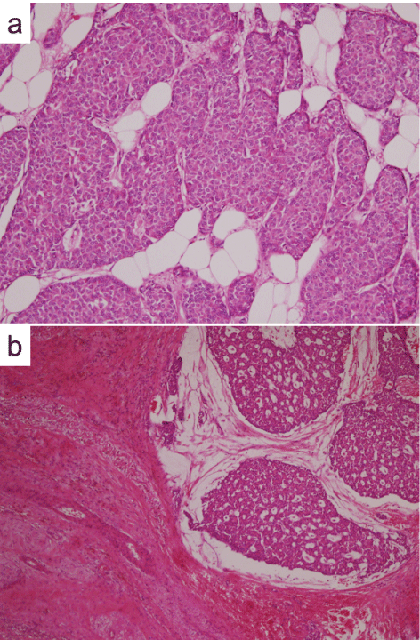
Needle biopsied tissues showed a proliferation of spindle-shaped cells without cellular atypia and suggested nodular fasciitis (Figure 3a). On magnetic resonance imaging (MRI), a lobulated mass was detected (15×15×14mm in size) in the left breast. The mass showe diso signal intensity (SI) to mammary gland on T1-weighted images, while it showed intermingled high and low SI on T2-weighted images (Figure 4a and Figure 4b). Dynamic contrast-enhanced MRI showed rapid early enhancement followed by wash-out, but steady persistent enhancement was partly shown (Figure 4c and Figure 4d). Although no malignant finding was shown in the tissue samples obtained by needle biopsy, malignant breast tumor including breast cancer was suggested by these imaging findings. Therefore, surgical excision of the breast tumor and axillary lymph node biopsy were performed. Pathologically, there was an invasive ductal carcinoma adjacent to nodular fasciitis (Figure 3b). Malignant epithelial cells proliferated mainly in the ducts arranged in solid-sheets, and focally invaded the adipose tissue (Figure 5a). The tumor cells also proliferated in the central part of the nodular fasciitis (Figure 5b). By immunohistochemistry, these tumor cells showed diffusely positive for estrogen and progesterone receptors and negative for human epidermal growth factor receptor 2 (HER2). No additional treatment but palliative care for advanced malignant lymphoma was administrated in the present case. She died after six months following surgery due to disease progression of malignant lymphoma.
.
Figure 3: Needle biopsied tissues and resected tumor
a: Needle biopsied tissues showed proliferation of spindle-shaped cells without cellular atypia and suggested nodular fasciitis [Eosin &Hematoxylin (H.E.) stain ×100]. b: Pathologically, there was an invasive ductal carcinoma adjacent to nodular fasciitis [H.E. stain ×5; red arrows show carcinoma, blue arrows show nodular fasciitis].
View Figure 3
.
Figure 4: Breast magnetic resonance imaging
The mass showe diso signal intensity (SI) to mammary gland on T1-weighted images, while it showed intermingled high and low SI on T2-weighted images (a: T1-weighted images, b: T2-weighted images). Dynamic contrast-enhanced MRI showed rapid early enhancement followed by wash-out, but steady persistent enhancement was partly shown [c: T1-weighted dynamic images (early phase), d: diffusion weighted images with dynamic curves).
View Figure 4
Discussion
Nodular fasciitis in the breast is very rare and only 25 cases have been reported worldwide to date (Table 1) [3-25]. The median age of the patients was 41 years (range 15-84 years) and the majorities of them were female. The median tumor size was 1.5 cm (range 0.5-5.6 cm), it seems not so large. Mammography and ultrasonography tend to reveal malignant tumor, thus immediate excision was performed in the most cases. Unexpectedly, invasive ductal carcinoma was complicated with nodular fasciitis in the present case. Regrettably, we had taken tissue samples only from the hypoechoic mass, so we should also have taken them from the hyperechoic lesion. Based on the correlation of pathological and ultra sonographic findings, the hyperechoic lesion reflected the invasive cancer while the hypoechoic lesion reflected the nodular fasciitis. Eventually, surgical excision was performed and then we diagnosed invasive ductal carcinoma adjacent to nodular fasciitis in the breast. To our knowledge, this is the first report of nodular fasciitis in the breast adjacent to breast cancer.
.
Figure 5: Histological findings of carcinoma and nodular fasciitis
a: Malignant epithelial cells proliferated mainly in the ducts arranged in solid-sheets, and focally invaded the adipose tissue (H.E. stain ×100) The tumor cells also proliferated in the central part of the nodular fasciitis (H.E. stain ×40).
View Figure 5
![]()
Table 1: Reported cases of nodular fasciitis in the breast
View Table 1
Histologically, nodular fasciitis is characterized by plump, immature spindle fibroblasts and myofibroblasts arranged in short irregular bundles [26]. Its benign nature has been well described in the literature, and it has been recently categorized as one of the benign mesenchymal tumors of the breast by the World Health Organization [27]. The histological differential diagnosis includes spindle-shaped cell tumors such as fibromatosis, myofibroblastoma, solitary fibrous tumor, phyllodes tumor, spindle cell carcinoma, fibrous histiocytoma, and various types of soft-tissue sarcomas. Nodular fasciitis can be discriminate from them based on cellularity, nuclear features, collagen content, and growth pattern [18]. Excisional, core needle, or vacuum-assisted biopsy is useful for histological diagnosis, and spontaneous regression and disappearance of the tumor was observed in the 5 cases (Case #4, #13, #23, #24, and #25 on Table 1) [6,15,24,25]. In addition, local recurrence following surgical excision rarely occurred (Case #13, #17 on Table 1) [15,18]. These characteristics of nodular fasciitis in the breast suggest that accurate histological diagnosis could avoid overtreatment. Although contrast-enhanced MRI could be helpful to discriminate nodular fasciitis from malignancy, only a few reports described MRI imaging in the breast [20,25]. Rapid early and persistent, or high contrast enhancement was shown on dynamic contrast-enhanced MRI. The present case showed rapid early enhancement followed by wash-out, concomitant partly persistent enhancement. We composed diffusion-weighted image with dynamic contrast-enhanced MRI, and it might be helpful to distinguish nodular fasciitis from breast cancer. By immunohistochemistry, these tumor cells showed diffusely positive for estrogen and progesterone receptors and negative for human epidermal growth factor receptor 2 (HER2). We propose that estrogen-related changesregarding mammary gland proliferation mightplay an important role in the etio pathogenesis of nodular fasciitis.
Conclusion
In conclusion, we presented a case of breast carcinoma adjacent to nodular fasciitis. We propose that estrogen-related changes regarding mammary gland proliferation might play an important role in the etio pathogenesis of nodular fasciitis.
Consent
Written informed consent was obtained from the nearest relative of the patient for publication of this Case report and any accompanying images. The patient had agreed to publish this case report as well, but we could not obtain her signature due to disease progression. A copy of the written consent is available for review by the Editor-in-Chief of this journal.
Competing Interests
The authors declare that they have no competing interests.
Authors' Contributions
RT and AI acquired the data and drafted the manuscript. MA, KT and SH participated in the design of the work. RNM evaluated the radiological images and revised the manuscript critically. SM evaluated the pathological findings. SN and YA supervised the design of the work. All authors read and approved the final manuscript.
Acknowledgements
The authors thank to Dr. TeruhikoFujii (Department of Breast Surgery, Asakura Medical Association Hospital, Asakura, Japan) for revising the manuscript critically for important intellectual content. The authors declare that they had no funding sources for this case study.
References
-
Dinauer PA, Brixey CJ, Moncur JT, Fanburg-Smith JC, Murphey MD (2007) Pathologic and MR imaging features of benign fibrous soft-tissue tumors in adults. Radiographics 27: 173-187.
-
Gelfand JM, Mirza N, Kantor J, Yu G, Reale D, et al. (2001) Nodular fasciitis. Arch Dermatol 137: 719-721.
-
Baba N, Izuo M, Ishida T, Okano A, Kawai T (1978) Pseudosarcomatous fasciitis of the breast simulating a malignant neoplasm: a case report and ultrastructural study. JpnJ ClinOncol 8: 169-180.
-
Fritsches HG, Muller EA (1983) Pseudosarcomatous fasciitis of the breast. Cytologic and histologic features. Acta Cytol 27: 73-75.
-
Törngren S, Frisell J, Nilsson R, Wiege M (1991) Nodular fasciitis and fibromatosis of the female breast simulating breast cancer. Case reports. Eur J Surg 157: 155-158.
-
Stanley MW, Skoog L, Tani EM, Horwitz CA (1993) Nodular fasciitis: spontaneous resolution following diagnosis by fine-needle aspiration. Diagn Cytopathol 9: 322-324.
-
Benson JR, Mokbel K, Baum M (1994) Management of desmoid tumours including a case report of toremifene. Ann Oncol 5: 173-177.
-
Black AJM, Kissin MW, Jackson P, Birdsall S, Shipley J, et al. (1994) Nodular fasciitis-Part I. Simulating breast carcinoma in an 84-year-old woman. Breast 3: 48-49.
-
Green JS, Crozier AE, Walker RA (1997) Case report: nodular fasciitis of the breast. Clin Radiol 52: 961-962.
-
Kontogeorgos G, Papamichalis G, Bouropoulou V (1988) Fibroblastic lesion of the breast exhibiting features of nodular fasciitis. Arch Anat Cytol Pathol 36: 113-115.
-
Maly B, Maly A (2001) Nodular fasciitis of the breast: report of a case initially diagnosed by fine needle aspiration cytology. Acta Cytol 45: 794-796.
-
Dahlstrom J, Buckingham J, Bell S, Jain S (2001) Nodular fasciitis of the breast simulating breast cancer on imaging. Australas Radiol 45: 67-70.
-
Polat P, Kantarci M, Alper F, Gursan N, Suma S, et al. (2002) Nodular fasciitis of the breast and knee in the same patient. AJR Am J Roentgenol 178: 1426-1428.
-
Tulbah A, Baslaim M, Sorbris R, Al-Malik O, Al-Dayel F (2003) Nodular fasciitis of the breast: a case report. Breast J 9: 223-225.
-
Brown V, Carty NJ (2005) A case of nodular fascitis of the breast and review of the literature. Breast 14: 384-387.
-
Porter GJ, Evans AJ, Lee AH, Hamilton LJ, James JJ (2006) Unusual benign breast lesions. Clin Radiol 61: 562-569.
-
Hayashi H, Nishikawa M, Watanabe R, Sawaki M, Kobayashi H, et al. (2007) Nodular fasciitis of the breast. Breast Cancer 14: 337-339.
-
Squillaci S, Tallarigo F, Patarino R, Bisceglia M (2007) Nodular fasciitis of the male breast: a case report. Int J Surg Pathol 15: 69-72.
-
Ozben V, Aydogan F, Karaca FC, Ilvan S, Uras C (2009) Nodular Fasciitis of the Breast Previously Misdiagnosed as Breast Carcinoma. Breast Care (Basel) 4: 401-402.
-
Iwatani T, Kawabata H, Miura D, Inoshita N, Ohta Y (2012) Nodular fasciitis of the breast. Breast Cancer 19: 180-182.
-
Paker I, Kokenek TD, Kacar A, Ceyhan K, Alper M (2013) Fine needle aspiration cytology of nodular fasciitis presenting as a mass in the male breast: report of an unusual case. Cytopathology 24: 201-203.
-
Son YM, Nahm JH, Moon HJ, Kim MJ, Kim EK (2013) Imaging findings for malignancy-mimicking nodular fasciitis of the breast and a review of previous imaging studies. Acta Radiol Short Rep 2: 2047981613512830.
-
Yamamoto S, Chishima T, Adachi S (2014) Nodular fasciitis of the breast mimicking breast cancer. Case Rep Surg 2014: 747951.
-
Samardzic D, Chetlen A, Malysz J (2014) Nodular fasciitis in the axillary tail of the breast. J Radiol Case Rep 8: 16-26.
-
Rhee SJ, Ryu JK, Kim JH, Lim SJ (2014) Nodular fasciitis of the breast: two cases with a review of imaging findings. Clin Imaging 38: 730-733.
-
Duncan SF, Athanasian EA, Antonescu CR, Roberts CC (2006) Resolution of nodular fasciitis in the upper arm. Radiol Case Rep 1: 17-20.
-
Tan PH, Ellis IO (2013) Myoepithelial and epithelial-myoepithelial, mesenchymal and fibroepithelial breast lesions: updates from the WHO Classification of Tumours of the Breast 2012. J Clin Pathol 66: 465-470.


