

Clinical Medical
Reviews and Case Reports
Some Aspect of Clinical Assessment of Carpal Unstability and Colapsus in Untreated Rheumatoid Arthritis
Dejan Spasovski*
Department of Rheumatology, University Clinical Centre, Skopje, Republic of Macedonia
*Corresponding author:
Dejan Spasovski, Department of Rheumatology, University Clinical Centre, Skopje, Republic of Macedonia, E-mail: drspasovski@yahoo.co.uk
Clin Med Rev Case Rep, CMRCR-2-055, (Volume 2, Issue 9), Research Article; ISSN: 2378-3656
Received: July 10, 2015 | Accepted: September 05, 2015 | Published: September 08, 2015
Citation: Spasovski D (2015) Some Aspect of Clinical Assessment of Carpal Unstability and Colapsus in Untreated Rheumatoid Arthritis. Clin Med Rev Case Rep 2:055. 10.23937/2378-3656/1410055
Copyright: © 2015 Spasovski D. This is an open-access article distributed under the terms of the Creative Commons Attribution License, which permits unrestricted use, distribution, and reproduction in any medium, provided the original author and source are credited.
Abstract
Introduction: The carpal instability is a biomechanical alteration with multiple pathogenesis. It ensues due to disturbance of the normal ligament and bone connections that control the wrist. There are four major types of carpal instability: 1 Dorsal Intercalated Segment Instability (DISI) 2.Volar Intercalated Segment Instability (VISI) 3.Ulnar translocation, and 4. Medial carpal instability. Instability in the wrist in Rheumatoid Arthritis (RA) appears as a result of incongruity between joint and bone surfaces, capsular and ligament laxity and muscle-tendon imbalance. Aim of this study is to evaluate carpal instability and collapse in patients with untreated RA and to determine the type of carpal instability using radiography.
Material and methods: In 30 patients with diagnosed RA we made anteroposterior and lateral radiographies in neutral position of both hands. On anteroposterior radiographies we measured carpal-ulnar distance. For evaluation of the bone set ups on lateral radiographies we used the axial Linscheid method for drawing carpal axes that enable measurements of angles which can define position of the carpal bones: scaphoid-lunate, radio-lunate and capitate-lunate angles.
Results: Of 60 rheumatoid hands in 50 we found carpal instability: ulnar translocation (32 hands), VISI (14 hands), DISI (4 hands). We detected isolated form of carpal instability in 38 hands, while in part we found complex carpal instability i.e. combination of two types of instability (12 hands).
Conclusion: This radiographic method enables evaluation of carpal instability and quantitative analysis for determination of the type of the carpal instability in RA.
Keywords
Wrist, Joint instability, Carpal bones, Carpal joint, Radiography, Arthritis, Rheumatoid
Introduction
Carpal instability is a biomechanical alteration with multiple pathogenesis. It occurs when there is disruption of the normal ligament and bone relations that control the wrist. The term carpal instability is introduced in 1967 by Dobyns [1] and is defined as follows: wrist not capable to endure physiological burden or has disturbed kinetics or both [2].
By radiography, the wrist is considered instable when there is deterioration in sagittal and/or anteroposterior placement of carpal bones out of the normal standards. In recent decades, together with the enhancement of the knowledge for biochemical mechanisms of the wrist, classifications were refined [3-5]. But, in everyday praxis according to the clinical and radiographic presentation there are four types of carpal instability: 1. DISI (dorsal intercalated segment instability) when lunate rotates in dorsiflexion and capitate is dislocated dorsally forming zig-zag radio-lunate-capitate line; 2. VISI (volar intercalated segment instability): opposite mechanism from DISI, when lunate rotates in palmar flexion; 3. Ulnar translocation -wrist is displaced towards ulna; 4. Medial carpal instability - displacement of normal palmar tilt of the wrist towards dorsally (most often after fracture of the distal radius). Instability usually occurs in RA, arthrosis or Morbus Kienböck, as well in some metabolic diseases [6-9]. Besides, about 10% of all carpal traumas end with instability [3,4]. Carpal instability in RA ensues as a result of incongruity between joint and bone surfaces, capsular and ligament laxity and muscle-tendon imbalance [7].
First diagnostic method for evaluation of the carpal instability in RA is radiography [3]. It is necessary to make objective measurements of the position of the carpal bones on anteroposterior and lateral radiography in neutral position, tending to get the right image of the carpal functional status. For that purpose there are several methods, but the axial method by Linscheid [10] has proven to be most accurate and most useful [5,11].
The aim of this study, using radiographic evaluation of the wrist, is to answer the questions whether there is carpal instability in RA and what type of instability is in the matter with radiographic evaluation.
Material and Methods
Standard routine Radiographs of the hands, wrists and feet were made in postero-anterior view and they were separately, in random order, scored in pairs by the two independent readers: rheumatologist and experienced radiologist. The radiologist was unaware of the patient's identity and the clinical activity of the disease, in prospective, singles centar study. All participants voluntarily participated in the study, so the ethic criteria for the preparation of the study were fulfilled. All study subjects participated voluntary after being informed of the risks, which were deemed minimal. The study was approved by the medical ethics committee for medicines and medical product, Medical Faculty, Ss. Cyril and Methodius University, Skopje, Republic of Macedonia.
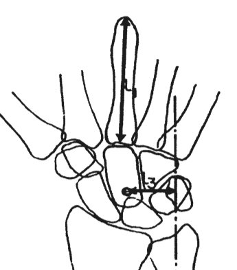
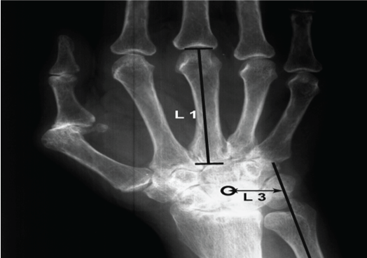
We made anteroposterior and lateral radiographies of both hands in 30 patients with RA. Patients with previous operations, fractures, congenital anomalies and symptoms and signs of other disease of the upper extremity different than RA, were excluded. Radiographies were made in carpal neutral position, elbow in flexion 90° and shoulder in abduction 90°. Positioning of the wrist in the right position (anteroposterior or lateral) was very important for further measurements. Radiographies were digitalized and enlarged in order to enable precise identification of the key anatomic indicators. On anteroposterior radiographies we measured carpal-ulnar distance. It is defined as perpendicular distance from the center of rotation for radio-ulnar deviation of the wrist (in the caput of the capitate) to the longitudinal axis of the ulna projected towards distally [12]. When this is divided with the length of the third metacarpal bone one gets the carpal-ulnar rate (Figure 1). Carpal-ulnar rate less than 0.25 denotes ulnar translocation. For determination of the bone placement we used the axial method of Linscheid [10] for drawing carpal axes on lateral radiographies.
.
Figure 1: Measurements of carpal-ulnar ratio L1 / L3
L1: Lenght of third metacarpal bone
L3: Carpal-ulnar distance
View Figure 1
The placement of the scaphoid bone in regard of lunate and proximal carpal line compared to radius and the distal carpal line could be defined by drawing axes (Figure 2):
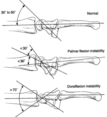
1. Longitudinal axes of the third metacarpal bone, capitate, lunate and radius (in normal wrist in neutral position these bones are in the same line) are drawn through the center of the caput of the third metacarpal bone, center of the caput of capitate, medial points of the proximal convex and distal concave bone surfaces of lunate and medial point of the distal radial joint surface (Figure 3).
2. Longitudinal axis of scaphoid is drawn through the middle of the proximal and distal pole of the bone (Figure 4).
With these axes one can measure angles to define position of the carpal bones. Scapho-lunate angle (SL angle) formed by the longitudinal axes of scaphoid and lunate is in average 46° (range 30-60°) in normal wrist. Angle > 70° or < 30° denotes carpal instability. Capitate-lunate angle (CL angle) formed by the longitudinal axes of capitate and lunate is normally 0°, but in carpal instability could be dorsal or palmar. Radio-lunate angle (RL angle) formed by axes of lunate and radius is a measurement for carpal dorsal or palmar flexion. When lunate related to radius or capitate related to lunate is in dorsiflexion, RL or SL angles are denoted as positive, but if they are in palmar flexion, angles are denoted negative. It is indicative for DISI if SL > 70°, dorsilexion of lunate (positive RL angle) and palmar flexion of capitate (negative CL angle). In VISI there is an opposite mechanism i.e. palmar flexion of lunate (negative RL angle), reduced SL angle and dorsiflexion of capitate in relation to lunate (positive CL angle).
We tested all these methods in our previous study in a group of normal individuals and concluded that our results for the normal population were in agreement with data from the literature. So, we standardized the radiography for carpal stability [13].
Results
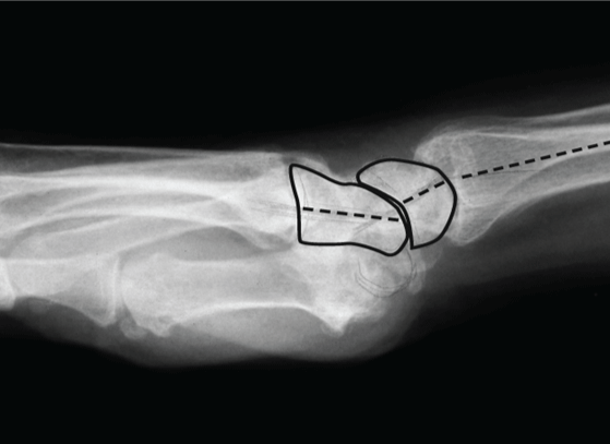
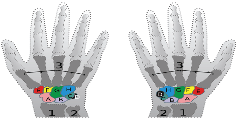
We made radiographic evaluation of the both wrists in 30 patients (21 female and 9 male). Mean age in patients with RA was 47.7 years (range 37-58 years) and mean disease duration was 11.7 years (5-19years). In table 1 are shown types of carpal instability determined by radiography evaluation in 60 normal wrists. Complex instability, i.e. combination of two types of instability was found in 12 wrists: ulnar translocation + VISI in 10 wrists and ulnar translocation + DISI in 2 wrists (Figure 5). In all patients where there was carpal instability and it was manifested bilaterally. Figures 3 and Figure 4 are shown radiographies of patients with carpal instability.
.
Figure 5: Proximal: A= Scaphoid, B= Lunate, C=Triquetrum, D= Pisiform
Distal: E= Trapezium, F= Trapezoid, G= Capitate, H= Hamate
1= Radius, 2= Ulna, 3= Metacarpals
View Figure 5
![]()
Table 1: Types of carpal instability determined by radiography evaluation in 60 normal wrists.
View Table 1
Discussion
The aim of this study was radiographic evaluation of carpal instability in rheumatoid patients and types of carpal instability. Of 60 patients with rheumatoid wrists in 50 we found carpal instability. The most often form of carpal instability was ulnar translocation (32 carpus), followed by VISI (14 carpus) and DISI (4 carpus). Isolated form of carpal instability was noted in 38 carpus, while the others had complex carpal instability i.e. combination of two types of carpal instability (12 carpus).
The pathogenesis of carpal instability in RA and the order of changes are yet unknown. Ulnar translocation is found very often in RA and rarely in other diseases of the wrist [4,14]. Pathognomonic for this type of instability is translocation of lunate and capitate towards ulna, followed by scaphoid (4). This leads to broadening of the space between styloid processus of the radius and scaphoid [4]. Pathogenetically, it seems that there is a direct relation between ulnar translocation of the metacarpophalangeal joints and ulnar translocation of the wrist [14].
For the other types, in the beginning, most probably due to the laxity of the intrinsic carpal ligaments increases the SL angle and ensues palmar flexion of lunate. Lately, gradually increases also the CL angle and capitate rotates towards dorsally [6]. In fact, the carpal instability in RA is complex and comprises laxity of the extrinsic and intrinsic carpal ligament apparatus. It was also shown in our study that great part of the patients showed combined carpal instability. Besides, in all patients with carpal instability both wrists were involved, which goes in favor of symmetric involvement of the wrist in RA [15].
Conclusion
Evaluation of the carpal instability with radiographic measurements is a routine protocol in orthopedic praxis all over the world [3-8]. This method is not yet in praxis in our country, but we think that evaluation and quantitative measurement of carpal instability in RA is of great importance. Although diagnosis for rheumatoid involvement of the wrist is easily established by clinical examination [15], it is very important this proces to be followed and re-evaluated for progression in order to choose among different treatment modalities or to make the right decision which surgical procedure would the be most apropriate in certain period of time. Our conclusion is that this method for evaluation of carpal instability enables evaluation and quantitative analysis of carpal instability in RA.
References
-
Dobyns JH, JC Perkins (1967) Instability of the carpal navicular. In Proceedings of the American Academy of Orthopaedic Surgeons. J Bone Joint Surg Am 49: 1014.
-
Definition of carpal instability. The Anatomy and Biomechanics Committee of the International Federation of Societies for Surgery of the Hand. J Hand Surg Am 24: 866-867.
-
De Filippo M, Sudberry JJ, Lombardo E, Corradi M, Pogliacom F, et al. (2006) Pathogenesis and evolution of carpal instability: imaging and topography. Acta Biomed 77: 168-180.
-
Taleisnik J (1988) Current concepts review. Carpal instability. J Bone Joint Surg Am 70: 1262-1268.
-
Bednar JM, Osterman AL (1993) Carpal Instability: Evaluation and Treatment. J Am Acad Orthop Surg 1: 10-17.
-
Muramatsu K, Ihara K, Tanaka H, Kawai S (2004) Carpal instability in rheumatoid wrists. Rheumatol Int 24: 34-36.
-
Resnick D and Niwayama G (1977) Carpal instability in rheumatoid arthritis and calcium pyrophosphate deposition disease. Pathogenesis and roentgen appearance. Annals of the Rheumatic Diseases 36: 311-318.
-
Hindley CJ, Stanley JK (1991) The rheumatoid wrist: patterns of disease progression. A review of 50 wrists. J Hand Surg Br 16: 275-279.
-
Hodgson SP, Stanley JK, Muirhead A (1989) The Wrightington classification of rheumatoid wrist X-rays: a guide to surgical management. J Hand Surg Br 14: 451-455.
-
Linscheid RL, Dobyns JH, Beabout JW, Bryan RS (1972) Traumatic instability of the wrist: Diagnosis, Classification and Pathomechanics. J Bone Joint Surg Am 54: 1612-1632.
-
Garcia-Elias M, An KN, Amadio PC, Cooney WP, Linscheid RL (1989) Reliability of carpal angle determinations. J Hand Surg Am 14: 1017-1021.
-
Youm Y, McMurthy RY, Flatt AE, Gillespie TE (1978) Kinematics of the wrist. I. An experimental study of radial-ulnar deviation and flexion-extension. J Bone Joint Surg Am 60: 423-431.
-
M Foteva Lj Karevski, A Poposka, A Adamov (2010) Determination of normal carpal angle measurements in the R. of Macedonia. Acta Morphologica.
-
McMurtry RY, Youm Y, Flatt AE, Gillespie TE (1978) Kinematics of the wrist. II. Clinical applications. J Bone Joint Surg Am 60: 955-961.
-
Daniel Aletaha, Tuhina Neogi, Alan J. Silman (2010) Rheumatoid Arthritis Classification Criteria. An American College of Rheumatology/European League against Rheumatism Collaborative Initiative. Arthritis & rheumatism 2569-2581.
