Trauma Cases and Reviews
I Can't Find the Left Iliac Artery! Abdominal Stab Wound Leading to Uncontrollable Hemorrhage
TL Freeborn, R Shinar, B Siegrist, N Keric, NY Patel and AJ Feinstein*
Trauma, General Surgery and Critical Care, Banner University Medical Center, Phoenix, USA
*Corresponding author: AJ Feinstein, Assistant Professor and Associate Surgical Clerkship Director, Trauma, General Surgery and Critical Care, Banner University Medical Center, Phoenix Integrated surgical residency, 925 East McDowell road, 2nd floor, AZ 85006, USA, Tel: 602-839-3339, Fax: 602-839-3339, E-mail: ara.feinstein@bannerhealth.com
Trauma Cases Rev, TCR-3-051, (Volume 3, Issue 1), Case Report; ISSN: 2469-5777
Received: May 27, 2016 | Accepted: February 25, 2017 | Published: February 28, 2017
Citation: Freeborn TL, Shinar R, Siegrist B, Keric N, Patel NY, et al. (2017) I Can't Find the Left Iliac Artery! Abdominal Stab Wound Leading to Uncontrollable Hemorrhage. Trauma Cases Rev 3:051. 10.23937/2469-5777/1510051
Copyright: © 2017 Freeborn TL, et al. This is an open-access article distributed under the terms of the Creative Commons Attribution License, which permits unrestricted use, distribution, and reproduction in any medium, provided the original author and source are credited.
Abstract
Introduction: A 32-year-old man presented with a single left lateral lower quadrant stab wound and pulsatile hemorrhage.
Methods: The patient was taken immediately to the operating room for laparotomy and was found to have an expanding retropertioneal hematoma. Aortic occlusion did not slow progression. The left iliac artery could not be located. A femoral cut down and proximal control of the femoral artery was also not helpful. The hematoma was entered directly and the hemorrhaging area in the lateral psoas muscle was ligated.
Results: The patient maintained palpable dorsalis pedis pulses throughout the procedure. Postoperatively, a CT (computed tomography) showed congenital absence of the left iliac artery with large retroperitoneal collateral vessels in the retroperitoneum that reconstituted in a normal femoral artery. The scan demonstrated the injury was to one of these large vessels. The patient was discharged home after and uneventful course.
Discussion: Congenital malformations of the iliac artery are rare, and most are discovered incidentally on imaging. We report a case of injury to a collateral vessel resulting from a left iliac atresia. This had implications for controlling hemorrhage in this case. Knowledge of vascular anomalies is important for management of traumatic injury.
Keywords
Vascular trauma, Congenital iliac hypoplasia, Iliac injury, Ileofemoral malformation
Introduction
A 32-year-old man presented to our Level I trauma center after being stabbed in the abdomen. Paramedics reported finding a significant amount of blood at the scene and he was transiently hypotensive during transport. On arrival, he was complaining of lightheadedness and he was diaphoretic and tachycardic. Examination revealed a 3 cm stab wound just medial to the left anterior superior iliac spine. When direct pressure was released from the wound, copious pulsatile hemorrhage was apparent. No other traumatic injuries were noted.
Materials and Methods
Large bore intravenous lines were placed, blood transfusion was initiated and the patient was taken emergently to the operating room. In the operating room, the patient was placed in a supine position, intubated and widely prepped from the neck to the knees. A midline laparotomy was performed while maintaining direct pressure on the wound. There was no free fluid in the abdomen. A large hematoma was noted in zone 2 on the left and zone 3 of the retroperitoneum. In order to achieve proximal control, a left medial visceral rotation was performed. The distal aorta and right iliac artery were localized, isolated and looped. The left ureter was visualized and preserved. The left iliac artery could not be located. Several attempts were made to follow the iliac artery distally from the aorta without success. The hematoma continued to expand laterally in zone 2 and into the pelvis. When the distal aorta was occluded, the hematoma appeared to expand more rapidly and did not diminish the pulsatile hemorrhage from the wound when pressure was released. During this time, another incision was made inferior to the inguinal ligament. The left proximal femoral artery was dissected and vessel loops were placed around it. Occlusion of the femoral vessel also did not diminish the hemorrhage from the wound when pressure was released.
The patient was becoming more hemodynamically unstable despite resuscitation and the decision was made to open the hematoma in the absence of proximal control. The hematoma was entered adjacent to the stab wound in the lateral inferior portion of zone 2. On exposure of the psoas muscle, a large amount of arterial hemorrhage was encountered from a defect in the muscle. Several large sutures were placed using absorbable suture around the defect until the arterial hemorrhage stopped. Pressure was released from the wound and there was minimal bleeding. There was a diminished but palpable pulse in the left foot. Vessel loops were removed and the retroperitoneum and groin were packed with laparotomy pads. A negative pressure dressing was placed in the abdomen and the patient was taken directly to the CT scanner.
Results
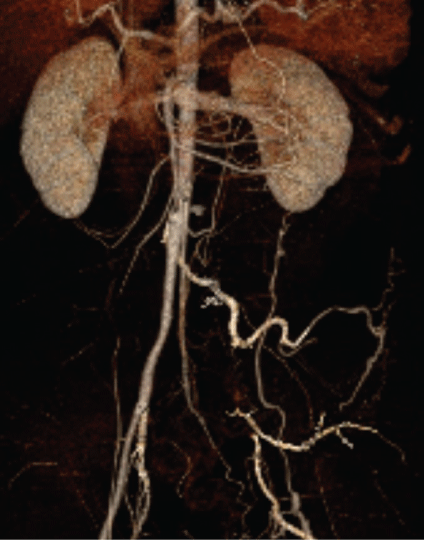
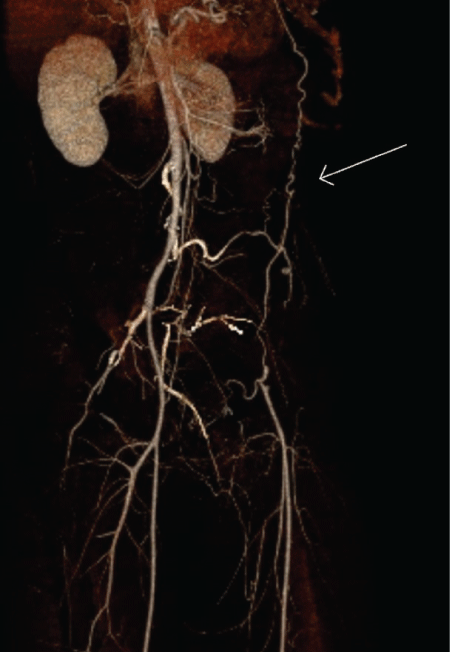
A contrast CT of the chest, abdomen and pelvis revealed congenital absence of the left iliac artery (Figure 1). Large collaterals, some arising from the thoracic aorta, reconstituted in the left femoral artery, which had good filling and runoff (Figure 2). The area of abdominal packing indicated the disruption of one or more of these collateral arterial vessels. The patient was taken back to the operating room the following day for removal of the abdominal packing and closure. His pulses remained intact and his hospital course was uneventful. He was discharged home on post-operative day seven.

.
Figure 1: Three-dimensional reconstruction of the postoperative CT demonstrated an absent left iliac artery.
View Figure 1

.
Figure 2: Collateral vessels, the largest branching from the thoracic aorta, reconstituted in a normal common femoral artery.
View Figure 2
Discussion
Congenital malformations of the iliofemoral arteries are rare [1]. The exact incidence is unknown. A series of 8,000 angiograms performed in symptomatic patients identified six patients with iliofemoral malformations [2]. Tamisier, et al classified these malformations into three groups [3]. Group 1 involves anomalies in the origin or course of the artery. These patients are unlikely to be symptomatic and often the anomaly is discovered at autopsy. Group 2 is hypoplasia or atresia of the artery compensated by a persistent sciatic artery. The sciatic artery is the main artery of the lower limb bud in the early embryo. It arises dorsally from the umbilical artery. Typically, it regresses as the ventral branch of the umbilical artery forms the external iliac artery. In a patient with a persistent sciatic artery, it will be visualized on angiogram as an enlarged internal iliac artery, which continues into the thigh posteriorly. Collaterals may develop to provide blood flow to the femoral artery. Group 3 is isolated hypoplasia or atresia of the artery with a residual cord. These groups of patients are most susceptible to chronic ischemia of the lower extremity, even in childhood and early adulthood. Patients develop collaterals to restore blood flow to the lower extremity. Examples of vessels that provide collateral flow include: anomalous vessels from the contralateral hypogastric artery or common femoral artery, prominent lumbar arteries, retroperitoneal vessels, inferior epigastric artery, deep circumflex iliac artery, middle sacral artery, or anomalous branches of the renal artery. This patient did not have a persistent sciatic artery, and thus would be categorized in Group 3. Despite this, he reported being asymptomatic and had been a competitive athlete. His collaterals were largely retroperitoneal, many with origins from the thoracic aorta.
Most ileofemoral malformations discussed in previous case reports were detected incidentally with imaging [4,5]. In one case, the anomaly was discovered after a surgical complication [6]. Osuro and colleagues reported a case where a patient developed limb ischemia after a left nephrectomy. The patient had congenital absence of the left iliac artery with collateral vessels branching from the left renal artery, which when ligated, limited perfusion to the extremity.
We present the first case, to our knowledge, of a penetrating injury to a collateral vessel resulting from congenital atresia of the iliac artery. Given the retroperitoneal course of the injured vessels, proximal control in the abdomen was virtually impossible. In fact, distal clamping of the aorta increased proximal pressure in these collateral arteries. Awareness of vascular anomalies is important in dealing with traumatic injuries, and while relatively rare, this anomaly presents a situation where proximal control of a bleeding artery is not feasible and thus requires ligation with concurrent hemorrhage.
References
-
Dabydeen DA, Shabashov A, Shaffer K (2015) Congenital Absence of the Right Common Iliac Artery. Radiol Case Rep 3: 47.
-
Greebe J (1977) Congenital anomalies of iliofemoral artery. J Cardiovasc Surg 18: 317-323.
-
Tamisier D, Melki JP, Cormier JM (1990) Congenital anomalies of the external iliac artery: Case report and review of the literature. Ann Vasc Surg 4: 510-514.
-
Palkhi E, Pathak S, Hostert L, Morris-Stiff G, Patel JV, et al. (2015) Complete absence of iliac arteries in the left hemipelvis in a case of deceased donor renal transplantation: case report. Case Reports in Transplantation 2015.
-
Koyama T, Kawada T, Kitanaka Y, Katagiri K, Ohno K, et al. (2003) Congenital anomaly of the external iliac artery: A case report. Journal of Vascular Surgery 37: 683-685.
-
GD Oduro, LH Cope, IM Rogers (1992) Case report: lower limb arterial blood supply arising from the renal artery with congenital absence of the ipsilateral iliac arteries. Clin Radiol 45: 215-217.


