Trauma Cases and Reviews
Bilateral Patella Sleeve Avulsions in an Otherwise Healthy Nine-Year-Old Girl: A Case Report and Review of the Literature
Shazaan F Hushmendy*, Timothy T Roberts, Elaine Tran and Garrett R Leonard
Department of Orthopaedic Surgery, Albany Medical Center, USA
*Corresponding author: Shazaan F Hushmendy, Department of Orthopaedic Surgery, Albany Medical Center, 47 New Scotland Ave, Albany, NY 12208, USA, Tel: 518-262-3125, E-mail: hushmendys@gmail.com
Trauma Cases Rev, TCR-3-049, (Volume 3, Issue 1), Case Report; ISSN: 2469-5777
Received: July 12, 2016 | Accepted: February 22, 2017 | Published: February 25, 2017
Citation: Hushmendy SF, Roberts TT, Tran E, Leonard GR (2017) Bilateral Patella Sleeve Avulsions in an Otherwise Healthy Nine-Year-Old Girl: A Case Report and Review of the Literature. Trauma Cases Rev 3:049. 10.23937/2469-5777/1510049
Copyright: © 2017 Hushmendy SF, et al. This is an open-access article distributed under the terms of the Creative Commons Attribution License, which permits unrestricted use, distribution, and reproduction in any medium, provided the original author and source are credited.
Abstract
A patella sleeve avulsion is a rare but devastating injury seen in children aged 8-to-12 years. This soft-tissue disruption is frequently missed upon initial presentation. The radiographic findings may be subtle and advanced imaging is often necessary to elucidate the pathology. Delayed diagnosis is linked to significant morbidity.
This report describes a nine-year-old healthy female with bilateral inferior pole patella sleeve avulsions. Only two other cases of bilateral patella sleeve avulsions are reported in the literature.
This patient was treated with a tension band construct using #2 Fiber wire (Arthrex; Naples, FL). She regained full and painless active range of motion of bilateral knees within one year of her surgery.
Case Report
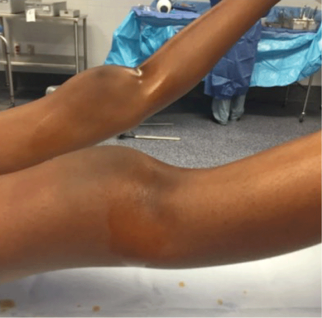
A 9-year-old African American female presented to the emergency department with bilateral knee pain after falling while running onto hyper flexed knees. She was otherwise healthy and denied antecedent knee pain. Her joints were not hyper mobile (Breighton score 0). On physical exam she had knee effusions and disruption of her extensor mechanisms bilaterally. Her patellae were high riding and defects could be palpated at the inferior poles (Figure 1).
.
Figure 1: Preoperative photograph of the patient’s knees. Notable palpable defect of the distal to the inferior pole of the left knee.
View Figure 1
Diagnosis
Bilateral Patella Sleeve Avulsions.
Introduction
The patella is the largest sesamoid bone in the human skeleton. It has 7 facets: 3 medial, 3 lateral, and one odd facet. Ossification occurs between 3 and 6 years of age, during which it is surrounded by a sleeve of cartilage. The quadriceps and patellar tendons are confluent with this cartilaginous sleeve.
The patella functions as a fulcrum for the powerful quadriceps. This places it at risk to injury from tensile forces. Incidence of patellar fractures in the pediatric population is low. It comprises less than 6.5% of all patellar fractures [1]. A sleeve avulsion fracture is a unique injury in children where a tendon is avulsed from the bone with an intact sleeve of cartilage or periosteum, with or without osseous fragments [2]. A patella sleeve avulsion is an osteochondral injury through the relatively weak cartilaginous sleeve. It most commonly occurs in children ages 8-to-12 years following an eccentric contraction of the extensor mechanism [1]. Patellar sleeve avulsion fractures account for almost half of all patellar fractures in this age group [3].
Patella sleeve avulsions of the inferior pole are more commonly described. However a few case reports have been published describing sleeve fractures of the superior pole [4-6]. The blood supply of the patella arises from the anterior surface of the distal pole. Thus injury to the anterior and distal pole may result in avascular necrosis of the proximal pole [7].
Physical Exam
Pediatric patients presenting with knee pain after a hyperflexion mechanism should raise concern for a patella sleeve avulsion. Clinical exam findings may be variable depending on severity of injury. Those with severe injuries may present with an acute hemarthrosis and inability to straight-leg-raise [8] as well as a high riding patella with a palpable inferior pole defect [9]. However, non-displaced or minimally displaced injuries with minimal exam findings can be difficult to diagnose and can be mistaken for a benign knee sprain, strain or contusion. Our patient demonstrated knee effusions and a disruption of her bilateral extensor mechanisms. Her patellae were high riding and defects could be palpated at the inferior poles (Figure 1).
The painful nature of the injury can preclude a complete and accurate physical exam. Physicians must have a high index of suspicion for patellar sleeve fractures to pursue the correct diagnostic imaging in lieu of a compromised physical exam.
Imaging
AP and lateral plain radiographs are a standard part of the workup for a pediatric patient with a history of knee trauma and pain. Sunrise and merchant views should be avoided since patient positioning may cause both displacement of the patella sleeve avulsion and significant discomfort.
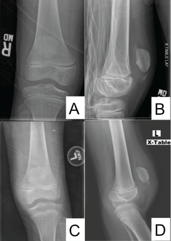
In many cases, radiographs may not be sufficient enough to diagnose patella sleeve avulsion fractures. The extensive cartilaginous anatomy of the patella in children prevents visualization of these injuries on plain radiographs. However, in our reported case, radiographs demonstrated a disruption of the inferior patellar pole (Figure 2).
.
Figure 2: A small osseous sleeve avulsion in noted on both lateral images. Bilateral knee effusions and soft tissue swelling is also visible.
A) AP of right knee; B) lateral of the right knee; C) AP of left knee; D) lateral of the left knee radiographs.
View Figure 2
Patella baja and alta are findings associated with superior and inferior patella sleeve avulsions respectively. The Insall-Salvati ratio is calculated using measurements from a lateral radiograph with the knee in 30 degrees of flexion. The ratio between (1) the distance from the inferior pole of the patella to the tibial tubercle and (2) the length of the patella is calculated. A ratio greater than 1.2 indicates patella alta while a ratio less than 0.8 indicates patella baja. Patella alta may be the only finding on plain radiographs in inferior patella sleeve avulsions [10].
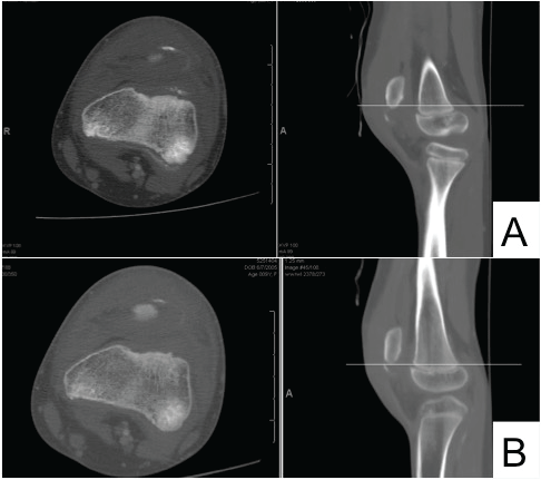
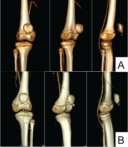
If there is clinical suspicion for patella sleeve fracture and no radiographic findings, advanced imaging like magnetic resonance imaging (MRI) may be used [11]. Ultrasound and computed tomography (CT) may also be used to aid with diagnosis. In this case, CT imaging confirmed bilateral inferolateral periosteal patella sleeve avulsions with approximately 1 cm of displacement (Figure 3). A three-dimensional CT rendering illustrates the displacement of the injury (Figure 4).
.
Figure 3: Pre-operative axial and sagittal CT of A) right; B) left knees. The small osseous sleeve avulsions are clearly demonstrated on both knees. There is no an additional fracture line or comminution visible.
View Figure 3
.
Figure 4: 3-dimensional CT reconstruction of the A) right; B) left knees.
This further reveals the fracture pattern of the sleeve fracture, confirming its diagnosis.
View Figure 4
Treatment
Indication for non-operative treatment is limited to minimally displaced (1-2 mm) fractures [3] with an intact knee extensor mechanism. The patient is immobilized in a long leg cast flexed to 0 to 10 degrees for 4 to 6 weeks. Indications for operative management include any patella sleeve avulsion with more than 2-3 mm of displacement or a disrupted extensor mechanism.
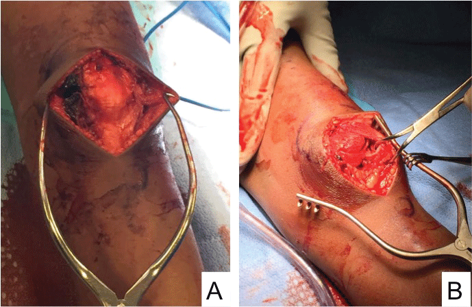
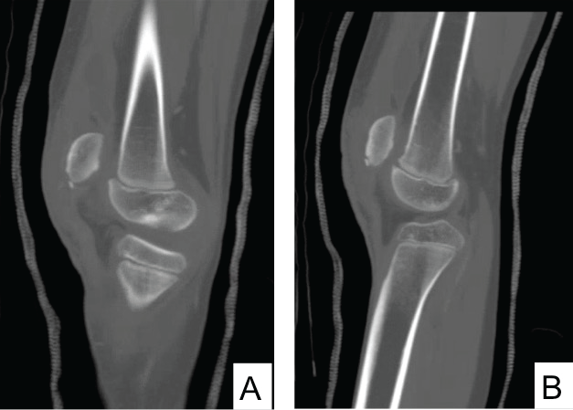
Several operative techniques have been described, including repair with trans-osseous non absorbable sutures, absorbable anchor sutures, and a tension band construction with Kirschner wires [2,12,13]. If patellotibial cerclage wiring is performed for protection of the primary repair, care should be taken not to disturb the proximal tibial apophysis as this may result in premature anterior physical arrest [8]. Concomitant injury to the surrounding soft tissue structures such as the medial and lateral retinaculae as well as the medial patellofemoral ligament (MPFL) have been described [10,14]. Thus when performing primary soft tissue repair of the extensor mechanism, it is important to address and reapproximate these structures in order to achieve a robust extensor repair. In this case, #2 Fiber wire suture (Arthrex; Naples, FL) was used to construct a tension band that reduced the avulsed fragments and primarily repaired the avulsed extensor tendon (Figure 5 and Figure 6).
.
Figure 5: A) right; B) left intraoperative images identifying the distal osseous fragment with its patella tendon attachment.
View Figure 5
.
Figure 6: Post-operative CT scan showing near-anatomic reduction of the fracture fragment in the A) right; B) left knees.
View Figure 6
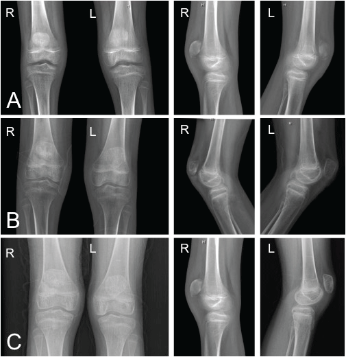
This patient had an uncomplicated perioperative course. She was placed in bilateral long leg casts for 2 weeks and transitioned to a brace for another 2 weeks. She was allowed to weight bear as tolerated in knee extension for the first 4 weeks. At 4 weeks follow-up, the brace was adjusted to permit movement between zero and 30 degrees with a gradual increase by 30 degrees every two weeks. The brace was removed at the three-month follow-up. At the 1-year post-operative period, the patient had full range of motion at bilateral knee joints. Figure 7 demonstrates radiographs obtained at 1-month, 6-months, and 1-year respectively. At the one year follow-up the patient was not required to return for a regular follow-up.
.
Figure 7: Follow-up AP and lateral radiographs of right and left knees at A) 1-month; B) 6-month; and at C) 1-year.
View Figure 7
Discussion
Patella sleeve avulsions are rare injuries in patients aged 8-to-12 years. These injuries are frequently misdiagnosed initially. This is likely due to a lack of awareness of this pathology in the health care community. Without a high index of suspicion, these injuries can be misconstrued as sprains, strains or contusions. Complications such as decreased knee flexion, ectopic bone formation, or avascular necrosis may occur as the result of delayed diagnosis [12,15].
Better education about the pathology, physical exam and imaging findings of patella sleeve avulsions will allow for more accurate and prompt diagnosis. Advanced imaging like ultrasound MRI and CT can help confirm a patella sleeve avulsion. Once diagnosed, there are several treatments, including our reported tension band construct, in a surgeon's armamentarium.
References
-
Ray JM, Hendrix T (1992) Incidence, mechanism of injury and treatment of fractures of the patella in children. J Trauma 32: 464-467.
-
Davidson D, Letts M (2002) Partial sleeve fractures of the tibia in children: an unusual fracture pattern. J Pediatr Orthop 22: 36-40.
-
Hunt DM, Somashekar N (2005) A review of sleeve fractures of the patella in children. Knee 12: 3-7.
-
Ro KH, Park JH, Kim MJ, Lee DH (2014) Rare sleeve fracture of the superior patella pole in an adult due to forceful passive physiotherapy following cast immobilization. Knee 21: 600-604.
-
Van Isacker T, De Boeck H (2007) Sleeve fracture of the upper pole of the patella: a case report. Acta Ortho Belg 73: 114-117.
-
Gettys FK, Morgan RJ, Fleischli JE (2010) Superior pole sleeve fracture of the patella: a case report and review of the literature. Am J Sports Med 38: 2331-2336.
-
Gao GX, Mahadev A, Lee EH (2008) Sleeve fracture of the patella in children. J Orthop Surg 16: 43-46.
-
Lin SY, Lin WC, Wang JW (2011) Inferior sleeve fracture of the patella. J Chin Med Assoc 74: 98-101.
-
Sullivan S, Maskell K, Knutson T (2014) Patellar sleeve fracture. West J Emerg Med 15: 883-884.
-
Majeed H, Datta P, dos Remedios I (2014) Sleeve fracture of the patella with lateral slip of the retinaculum: a case report in an 11-year-old child. J Pediatr Orthop B 23: 422-425.
-
Bates DG, Hresko MT, Jaramillo D (1994) Patellar sleeve fracture: demonstration with MR imaging. Radiology 193: 825-827.
-
Guy SP, Marciniak JL, Tulwa N, Cohen A (2011) Bilateral sleeve fracture of the inferior poles of the patella in a healthy child: case report and review of the literature. Adv Orthop 2011.
-
Kaar TK, Murray P, Cashman WF (1993) Transosseous suturing for sleeve fracture of the patella: a case report. Ir J Med Sci 162: 148-149.
-
Williams D, Kahane S, Chou D, Vemulapalli K (2015) Bilateral Proximal Tibial Sleeve Fractures in a Child: A Case Report. Arch Trauma Res 4: e27898.
-
Dai LY, Zhang WM (1999) Fractures of the patella in children. Knee Surg Sports Traumatol Arthros 7: 243-245.


