Trauma Cases and Reviews
Isolated Pancreas Injury by Blunt Trauma, a Case Report
Samet Sahin, Raziye Duruk, Cengiz Ceylan, Erdinc Cetinkaya* and Mesut Tez
Department of General Surgery, Ankara Numune Education and Research Hospital, Turkey
*Corresponding author: Erdinc Cetinkaya, MD, Department of Colorectal Surgery, General Surgery, Ankara Numune Education and Training Hospital, Sihhiye 06100, Ankara, Turkey, Tel: +905052918788, E-mail: drerdinccetinkaya@gmail.com
Trauma Cases Rev, TCR-2-046, (Volume 2, Issue 3), Case Report; ISSN: 2469-5777
Received: October 01, 2016 | Accepted: December 20, 2016 | Published: December 24, 2016
Citation: Sahin S, Duruk R, Ceylan C, Cetinkaya E, Tez M (2016) Isolated Pancreas Injury by Blunt Trauma, a Case Report. Trauma Cases Rev 2:046. 10.23937/2469-5777/1510046
Copyright: © 2016 Sahin S, et al. This is an open-access article distributed under the terms of the Creative Commons Attribution License, which permits unrestricted use, distribution, and reproduction in any medium, provided the original author and source are credited.
Abstract
We here in present our experience with the rare injury of main pancreatic duct because of the blunt trauma and managed by pancreatico-enterostomy. A 35-year-old male patient presented with a motor vehicle accident and was diagnosed isolated pancreas main duct ınjury by blunt trauma. Computerized tomography (CT) was used as a diagnostic method and were confirmed by Endoscopic retrograde cholangiopancreatography (ERCP). Intraoperative image was that necrosis developed in the anterior and lateral faces of pancreatic neck body tissues, and pancreatic duct was progressing towards necrosis in this area, but back wall was intact. Anterior Roux-en-Y pancreatico-jejunostomy was applied following the debridement of the wound area due to the intact posterior pancreatic parenchyma in the patient with proximal pancreatic duct injury in which pancreatic tissue was not transected. In a stable patients with isolated pancreatic duct injury, for not causing loss of organs such as the pancreas, spleen, duodenum, pancreatico-enterostomy is a safe option to reduce mortality and morbidity.
Keywords
Isolated pancreas main duct injury, Pancreatico-enterostomy, Pancreatic trauma
Background
Injury to the pancreas by a blunt trauma is very rare and accounts for less than 2% of all abdominal injuries [1,2]. It can present acutely or months later [2] As known CT is the major imaging method in the diagnosis of abdominal visceral injuries. We can confirm pancreatic main duct injuries with Magnetic Resonance Cholangiopancreatography (MRCP) or ERCP by contrast extravasation. ERCP has also been used therapeutically with transpapillary stenting across the pancreatic duct disruption or simply across the sphincter of Oddi aiming at a reduction of the intrapancreatic pressure gradient [2,3]. There are several surgery options for which can be employed to manage this injuries. According to the settelement and grade; distal pancreatectomy with or without splenectomy, pancreatico-jejunostomy, pancreatico-gastrostomy, whipple procedure are the options. In our case we preferred pancretico-jejunostomy [4].
Case Report
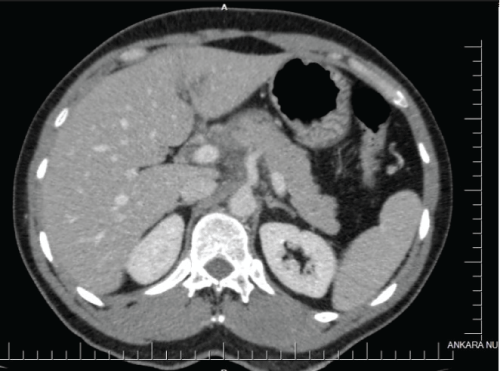
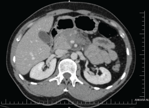
I.T., a 35-year-old male patient, was brought to the hospital via ambulance due to trauma as a result of accident at work. Patient had a history about striking his lower back by slide transport vehicle in his workplace, and remaining stuck between the wall and the vehicle. The patient had no known history of chronic disease, there was no routine drug usage and the patient had no surgery history except anterior mesh herniorrhaphy due to left inguinal hernia 20 years ago. When the patient arrived, he was evaluated as conscious, oriented cooperative and Glasgow coma scale was 15. Vital parametres were seen as; blood pressure: 125/85 mmHg, heart rate: 87/m, body temperature: 36.4 sO2: 98. laboratory evaluation of the patient that analyzed at emergency service were reported as, wbc: 12.700/ul, hb: 15.3 g/dl, plt: 228 thousand 1 u/l; biochemical parameters, ALT: 300 u/l, AST: 232 u/l amylase: 101 u/l, ldh: 543 u/l have been detected, with no electrolyte imbalance. Patient was admitted to the general surgery service for follow up and further investigations. IV contrast-enhanced thoracoabdominal CT was reported as grade 2 laceration in left lobe of liver, liquid collection in the vicinity of pancreas, 33 × 65 mm length were, surrounding SMA and SMV, was thought as hemorrhage and liquid reaching perihepatic 7 mm (Figure 1 and Figure 2).
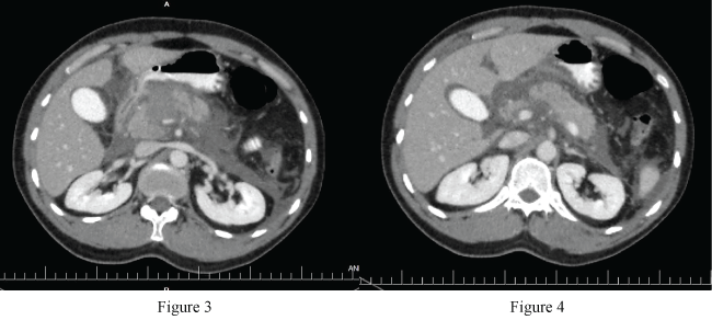
Oral intake of the patient was closed, and he was continued to be followed with IV hydration at the 12 hour of patient's follow up, body temperature was increased to 38.2, leucocytosis and hyperamilasemia were occur. (wbc: 14.200/ul, hb: 11.9 g/dl amylase: 578 u/l), therefore control IV contrast-enhanced abdominal CT has been performed to the patient. In left lobe of liver, a view compatible with grade 2 laceration was observed, and hypodense area in the neck of the pancreas was observed compared to surrounding pancreatic tissue. It was interpreted aspancreaticlaceration. Then, informed consent was obtained from the patient, and explorative laparotomy was planned with "acute abdomen" and "pancreatic laceration"? preliminary diagnosis (Figure 3 and Figure 4).
.
Figure 3-4: Hypodense area in the neck of the pancreas was observed compared to surrounding pancreatic tissue.
View Figure 3-4
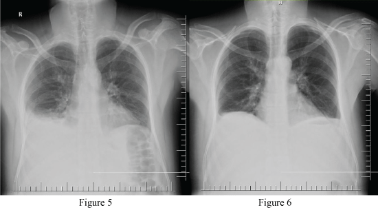
Patıent was operated after 2 days (nearly 50 hours) after he had been accepted to the hospital. During the exploration, 250 cc hemorrhagic collection was obresved in the abdomen, and it was aspirated. No signs of active bleeding were observed in laceration area in liver. Posterior of stomach was explored by openning gastrocolic ligament. Peripancreatic collection, ischemic foci and peripancreatic calcified fat necrosis were present in whole pancreas. However, major laceration area and necrotic regions were not detected in pancreas. Approximately 200 cc organized hematoma which was significant in paracaval and para-aortic region in retroperitonel field was observed. Hemostasis control was performed by draining hematoma. Upon absence of active bleeding, abdominal drain was placed to the patient, and the operation was ended. The patient was consulted on this stage about infectious diseases, and meropenem 3 × 1 g antibiotics was started. In post-operative follow-up, approximately 500 cc fluid per day came from right and left drains of the patienton post-operative day 1 (Figure 5). Upon presence of pleural effusion on the right in control PAAG of the patient with ongoing subfebrile fever post-operatively, the patient was consulted to the interventional radiology, thoracentesis was performed, and 300 cc serous transudate-qualified liquid was drained. After the thoracentesis it was seen in control PAAG that costophrenic recess blunt was lost (Figure 6).
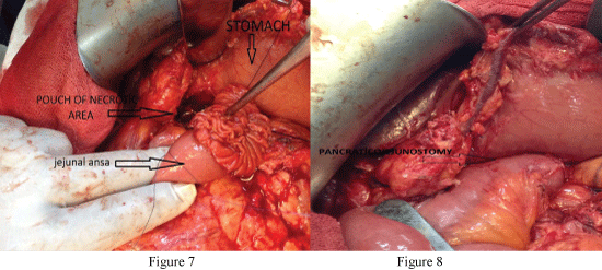
In biochemical evaluation performed with pleural effusion of the patient, it was evaluated as amylase: 1030 u/l, ldh: 3452 u/l. Upon no decrease in flow from the abdominal drains of the patient up to post-operative 5th day and post-operative 46020 u/l drain amylase level, IV contrast-enhanced CT was taken again. There was no continuity of neck-body junction of the pancreas in CT, and grade 4 pancreatic laceration was described, then the patient was consulted to gastroenterology, and ERCP was performed at post operative day 12. In ERCP, it was observed that there was staining in the head part of the pancreatic duct with the contrast agent, but there was no staining in body and tail parts. Contrast agent extravasation was detected from the head to the inside of abdomen and surgical drain. Pancreatic test was performed, and stent was tried to be placed, but it was not successful. As a result of ERCP performed in patient, full incision of duct not showing continuity in the pancreas body and neck junction was considered. Then, the consent of patient was taken, and the patient was operated post-operative day 16 again . In exploration, significant collection in the abdomen was not detected, but it was observed that necrosis developed in the anterior and lateral faces of pancreatic neck body tissues, and pancreatic duct was progressing towards necrosis in this area, but posterior wall of pancreas body was intact. In the operation proximal wirsung could not be revealed since the patient was re-operated, and cohesions in current region could not allow enough to expose. Since the patient was young, and distal part of the pancreas and head part in the neighborhood of the duedonum were intact, debridement of necrotic area and roux-en-y jejunostomy to this region were planned in order to reduce patient morbidity by abondoning whipple procedure or total pancreatectomy. After the debridement of necrotic tissues of described area, and hemostasis control, jejunal ans from 30 cm of pouch treitz ligament formed in the neck-body junction of pancreas was transected, it was advanced to poach area in a retrocolic way, and it was sutured in dublicate one by one with pancreaticojejunostomy 3.0 round vcryl (Figure 7 and Figure 8). Enteroenterostomy was also performed collaterally. The operation time was 1 hour and 15 minutes.
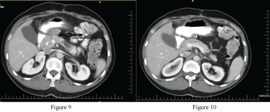
Patient was followed postoperatively for 1 day in intensive care unit, and patient was included in the service upon stable vital parameters and laboratory values. 150 cc, 100 cc and 70 cc came from abdominal drains on post-operative day 1, 2 and 3, respectively, it decreased to 20 cc on post-operative day 4, oral intake was opened on post-operative day 5, abdominal drains were taken after the tolerance of the patient, after the second operation there was no complication belongs to treatment and the patient was discharged with diet and medical treatment recommendations on post-operative day 8. The patient was admitted to hospital for control evalutions 6 weeks after discharge.There was no symptoms due to operation except anorexia which is seen sometimes andphysical examination wasnormal. Laboratory tests were also normal. IV contrast-enhanced abdominal CT was performed to patient and the CT was reported as "the head of pancreas is 32-33 mm and has homogenous appearance. From the corpus of pancreas, proximal wirsung is seen 4 mm and partially dilated. Also on the anterior face of pancreas there are sutur materials belongs to the anastomosis" (Figure 9 and Figure 10). After the evaluation, the patient discaharged with diet recomendations.
Discussion
In blunt abdominal traumas, pancreatic injuries are seen as less than 2%. Although there are accompanied solid organ injuries, it is quite rare as isolated [1]. Prognosis is influenced by the cause and complexity of the pancreatic injury, the amount of blood lost, duration of shock, speed of resuscitation and quality and nature of surgical intervention. Early mortality usually results from uncontrolled or massive bleeding due to associated vascular and adjacent organ injuries. Late mortality is a consequence of infection or multiple organ failure [5]. In blunt abdominal traumas, pancreatic duct injury must be assessed while deciding surgical or conservative treatments. While conservative treatment is considered in low grade (grade I-II) blunt abdominal traumas, surgical treatment will be required for high grade (grade III-IV and V) blunt abdominal traumas [6]. In the review of 94 studies involving 963 patients in which central pancreatectomy and distal pancreatectomy were compared, high postoperative morbidity (46% vs. 29%) and high incidence of pancreatic fistula (31% vs. 14%) but low endocrine deficiency (4% vs. 23%) in central pancreatectomy were found [7].
In our case report, anterior Roux-en-Y pancreaticojejunostomy was applied following the debridement of the wound area due to the intact posterior pancreatic parenchyma in the patient with proximal pancreatic duct injury in which pancreatic tissue was not transected (grade III). By means of this operation technique, the patient was protected from the complications of other operations described in previous paragraph. Also this technique was easier than Whipple procedure so operation time got reduced; by means of this, the patient can be protected from the complications of long term anesthesia. In this operation there was no necrotic tissue on posterior wall of pancreas so the anastomosis was smaller than dunking pancreatic anastomosis which is applied on Whipple procedure. Therefore the anastomosis leakage risk is less than the previous one. On the other hand, while applying this technique, it is important to be sure that there is no still necrotic tissue after the debritment because inadequate debritment increases the anastomosis leakage risk which is similar to the other operation techniques. However this operation technique may causes insufficent resection on patients with pancreas malignancies. To sum up, this procedure seems like to be preferable in young and non-malign patients.
References
-
Venkatesh SK, Wan JM (2008) CT of blunt pancreatic trauma: a pictorial essay. Eur J Radiol 67: 311-320.
-
Cogbill TH, Moore EE, Feliciano DV, Hoyt DB, Jurkovich GJ, et al. (1990) Conservative management of duodenal trauma: a multicenter perspective. J Trauma 30: 1469-1475.
-
Touloukian RJ (1983) Protocol for the nonoperative treatment of obstructing intramural duodenal hematoma during childhood. Am J Surg 145: 330-334.
-
Biffl WL, Moore EE, Croce M, Davis JW, Coimbra R, et al. (2013) Western Trauma Association critical decisions in trauma: management of pancreatic injuries. J Trauma Acute Care Surg 75: 941-946.
-
Krige JE, Beningfield SJ, Nicol AJ, Navsaria P (2005) The management of complex pancreatic injuries. S Afr J Surg 43: 92-102.
-
El-Boghdadly S, Al-Yousef Z, Al Bedah K (2000) Pancreatic injury: An audit and a practical approach. Ann R Coll Surg Engl 82: 258-262.
-
Iacono C, Verlato G, Ruzzenente A, Campagnaro T, Bacchelli C, et al. (2013) Systematic review of central pancreatectomy and meta-analysis of central versus distal pancreatectomy. Br J Surg 100: 873-885.


