

Trauma Cases and Reviews
Bilateral Simultaneous Rupture of the Quadriceps Tendons in Healthy Individuals
Takuro Moriya1,2* and Abe Yoshihiro1
1Department of Orthopaedic Surgery, Chiba Rosai Hospital, Japan
2Department of Orthopaedic Surgery, Chiba Kaihin Municipal Hospital, Japan
*Corresponding author: Takuro Moriya, Department of Orthopaedic Surgery, Chiba Rosai Hospital, 2-16 Tatsumidai-higashi, Ichihara 290-0003, Japan, Tel: +81-436-74-1111, Fax: +81-436-74-1151, E-mail: taku-m01@chibah.rofuku.go.jp
Trauma Cases Rev, TCR-2-043, (Volume 2, Issue 3), Case Report; ISSN: 2469-5777
Received: July 28, 2016 | Accepted: October 15, 2016 | Published: October 18, 2016
Citation: Moriya T, Yoshihiro A (2016) Bilateral Simultaneous Rupture of the Quadriceps Tendons in Healthy Individuals. Trauma Cases Rev 2:043. 10.23937/2469-5777/1510043
Copyright: © 2016 Moriya T, et al. This is an open-access article distributed under the terms of the Creative Commons Attribution License, which permits unrestricted use, distribution, and reproduction in any medium, provided the original author and source are credited.
Abstract
Quadriceps tendon rupture is an uncommon injury in healthy individuals. This paper presents two case reports of patients of bilateral quadriceps tendon rupture, who were misdiagnosed as muscle weakness of quadriceps with contusion of the knee joint. Subsequent physical examination showed a supra-patellar gap, moderate hemarthrosis of both knees, and failure of active knee extension. MRI showed bilateral rupture of the quadriceps tendons at the osteotendinous junction. Radiographs described the depression in the suprapatellar soft tissue, patella baja and an avulsion bony fragment on the patella. Surgery confirmed the MRI observation, so transosseous suturing and augmentation were undertaken. Both patients returned to a normal life with useful function. Adequate physical examination and correct understanding of both radiograph and MRI were required to prevent misdiagnosis.
Keywords
Bilateral quadriceps tendon ruptures, Healthy individuals
Introduction
Rupture of the quadriceps tendon is an uncommon injury, and bilateral rupture is very rare. It usually occurs in the elderly or in patients with chronic disease [1]. As these injuries are so rare, the diagnosis can be difficult and quite often is missed [2]. We report two cases of simultaneous bilateral quadriceps tendon ruptures in healthy individuals.
Case Report
Case 1
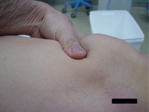
A 72-year-old man injured both knees after he slipped off a step. He was unable to stand up afterward and went to a clinic where his injury was diagnosed radiographically as contusions of both knees accompanied by muscle weakness. Five days later, clinical examination in our hospital showed a supra-patellar gap (Figure 1), moderate hemarthrosis of both knees, and failure of active knee extension. Based on this, the patient was diagnosed with bilateral rupture of the quadriceps tendons. Magnetic resonance imaging (MRI) confirmed the diagnosis, showing the quadriceps tendon rupture at the osteotendinous junction (Figure 2). Radiographs of his knees revealed the delle in the suprapatellar soft tissue, low-riding patella [3] and an avulsion fragment on the patella (Figure 3). The patellar heights of his right and left knees were 0.89 and 0.68, respectively, using the Insall-Saivati method. He did not have concomitant diseases.
.
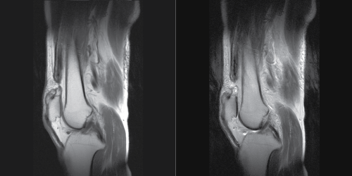
Figure 2: MRI on sagittal plane of Case 1. a) Proton density-weighted sequences; b) T2 weighted image.
View Figure 2
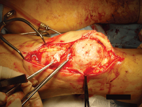
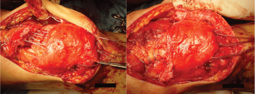
Five days after his injury, a correct diagnosis of bilateral rupture of the quadriceps tendons was made, and seventeen days after injury, the primary surgical repair was performed. At operation, both quadriceps tendons were found to be ruptured at the osteotendinous junction (Figure 4). A freshening of the edges, transosseous sutures were performed (Figure 5). A biopsy was taken from each injured side, and showed the rupture of the tendon, granulation formation, and patellar microfracture. Postoperatively both legs were placed in plaster casts in complete extension for 6 weeks with full weight bearing mobilization. At the end of 3 months, although the patient complained of weakness in both knees, he was walking independently with the aid of one crutch. He had bilateral flexion of 110 degrees and no extension lag on either knee. Radiographs showed patellar height of 1.00 on the right knee and 0.96 on the left knee using the Insall-Salvati method. At follow-up 26 months after repair, clinical examinations revealed a Lysholm score of 97 and a Tegnar activity score of 2, with full extension, no extension lag, flexion of 120 degrees on both knees, and no disturbance on walking without any aids.
.
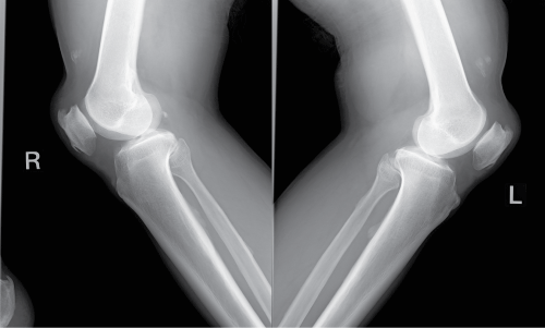
Figure 3: Radiographs on lateral view of Case 1. Patella baja and avulsion bony fragment.
View Figure 3
Case 2
A 69-year-old man fell down on a small hill when he was walking down a mountain. He complained of painful knees and was unable to extend his knees and stand up afterward. He went to a hospital and was diagnosed radiographically with muscle weakness and contusions on both knees. Two days following the fall, bilateral ruptures of the quadriceps tendons were diagnosed by clinical examination in our hospital. The diagnosis was based on the supra-patellar gap, moderate hemarthrosis of both knees, and failure of active knee extension. Magnetic resonance imaging (MRI) confirmed the diagnosis showing the quadriceps tendon rupture at the osteotendinous junction. Radiographs showed patellar baja and an avulsion of small bony fragments above the patella. The patellar height was 0.88 on the right knee and 0.87 on the left knee using the Insall-Salvati method. No systemic concomitant diseases were diagnosed.
A correct diagnosis of bilateral rupture of the quadriceps tendons was made, and thirteen days after injury the primary surgical repair was performed. At operation, complete ruptures of both quadriceps tendons were found at the osteotendinous junction. After freshening of the edges, both tendons were repaired with sutures passed through drill holes in the patella. After the operation, both legs were placed in plaster casts in complete extension for 6 weeks with full weight bearing mobilization. At the end of 4 months, the patient complained of weakness in both knees, with bilateral flexion of 110 degrees and no extension lag on either knee. He could walk independently without any aids. At follow-up 24 months after repair, he had flexion of 140 degrees and full extension without any extension lag on both knees. Radiographs showed patellar height of 1.10 on the right knee and 1.13 on the left knee. The Lysholm score was 99 and the Tegnar activity score was 3.
Discussion
Rupture of the quadriceps tendon is usually seen in patients older than 50 years who have concomitant diseases such as gout, hyperthyroidism, diabetes mellitus or chronic renal failure [1], or who have exposure to chronic corticosteroid use [4]. However, bilateral quadriceps tendon rupture is rare and occurs more frequently in healthy individuals [5].
The mechanism of rupture involves a sudden force, which combines contraction of the extensor mechanism and concurrent applied stretching of the quadriceps [6]. Landing with increased knee flexion angle leads to the increase of the knee extensor moment [7], and this produces sudden contraction force on quadriceps. Especially, during simultaneous landing with both legs, both legs are fixed on the ground at that moment, and ground reaction forces are conducted to both knees. Thus, these sudden contraction forces on the quadriceps at landing (i.e. large eccentric contractions) is thought to be the main cause of ruptures in these cases.
Diagnosis of quadriceps tendon rupture is based upon history and clinical examination [8]. History of the injury, such as a fall coupled with a violent contraction of the quadriceps, helps diagnosis. Additionally, patients describe sudden pain and a sensation of rupture on the injured site. A palpable suprapatellar gap, hemarthrosis of the knee and an inability to extend the knee actively are present on physical examination [9].
MRI is generally accepted as the most satisfactory way to diagnose tendon rupture. MRI can reveal not only the rupture but also the location of the injury site such as the osteotendinous junction or the musculotendinous area [10]. Therefore, MRI is used for diagnosis and also for preoperative planning [11]. Ultrasound is reported as a rapid way to investigate tendon rupture, which is a highly sensitive and specific means of assessment [12]. It is reported that Ultrasound can describe the location of the rupture and help differentiate complete from partial tears.
The patients reported here were misdiagnosed initially, in part because diagnosis was made using only radiographs. Radiographs can provide evidence of quadriceps tendon rupture by showing an obliteration of the quadriceps tendon shadow, a patella baja (low-riding patella), and a suprapatellar soft tissue mass representing retraction of torn tendons, and an avulsion of small bony fragments from the pole of the patella. However, radiographs are rarely sufficient to make the correct diagnosis [13]. It is reported that only 33% of quadriceps tendon ruptures were diagnosed correctly with radiography alone [13]. Bilateral quadriceps tendon rupture may be interpreted as a neurological deficit due to muscle weakness [14]. Both patients in this series were misdiagnosed as having muscle weakness and contusion with effusion and swelling. However, an adequate physical examination together with radiographic or MRI images can prevent misdiagnosis.
Surgical repair has been advocated as the treatment of choice. A variety of surgical procedures have been reported and the results were satisfactory [14]. Some investigators have reported that augmentation of quadriceps tendon ruptures should be performed [15], but this is controversial [16]. In our series, as both patients were elderly but had no concomitant chronic diseases which were reported as causes of weakening tendons, augmentation was not performed.
The duration from injury to surgical operation has been reported as a key factor in time to recovery after surgery. Neubauer, et al. reported that 50% of patients who had a repair within two weeks of injury showed complete recovery, whereas only 21.4% of patients whose repair was delayed more than two weeks achieved full recovery [17]. As any delay in diagnosis or repair is crucial for full recovery, early surgical repair of the tendon is recommended. As these two cases showed different clinical results with good functional recovery, case1 returned to daily life with slightly limited flexion range of both knee, case 2 showed full recovery to previous activity level without any deficit, this difference was thought to be the result of the difference of the duration to surgery.
Postoperatively, the standard of care has been to apply plaster casts for six weeks to immobilize the knee [17,18]. On the other hand, a few reports have described early mobilization with the use of a motion limited knee brace, to decrease the risk of thrombosis and flexion deficits [16]. However, no differences of function between early mobilization and immobilization have been reported [19]. In terms of weight bearing, full weight bearing gait with the knee locked in extension is considered as one of the most important program on rehabilitation, to prevent loss of muscle power of quadriceps [20] and to keep patient's activities in daily life higher even with any aids.
In conclusion, we report two cases of bilateral simultaneous rupture of the quadriceps tendons. To establish the correct diagnosis of quadriceps tendon rupture, it is important to identify the gap on the quadriceps tendons by physical examination and by imaging using radiography or MRI. Surgery within two weeks after the injury and early weight bearing with knee locked in extension, can provide excellent results and a return to daily normal activity in elderly people.
References
-
Goodfellow J, Hungerford DS, Zindel M (1976) Patello-femoral joint mechanics and pathology. 1. Functional anatomy of the patello-femoral joint. J Bone Joint Surg Br 58: 287-290.
-
Ramsey RH, Muller GE (1970) Quadriceps tendon rupture: a diagnostic trap. Clin Orthop Relat Res 70: 161-164.
-
Insall JN, Salvati E (1971) Patellar position in the normal knee joint. Radiology 101: 101-104.
-
Scuderi C (1958) Rupture of quadriceps tendon:study of 20 tendon ruptures. Am J Surg 95: 626-634.
-
Walker LG, Glick H (1989) Bilateral spontaneous quadriceps tendon ruptures. A case report and review of the literature. Otrhop Rev 18: 867-871.
-
Robert W Buchols, James D Heckman (1854) Fractures in adults (5th edn), Philadelphia, Lippincott 2002.
-
Podraza JT, White SC (2010) Effect of knee flexion angle on ground reaction forces, knee moments and muscle co-contraction during an impact-like deceleration landing: Implications for the non-contact mechanism of ACL injury. Knee 17: 291-295.
-
Sagiv P, Gepstein R, Amdur B, Hallel T (1989) Bilateral spontaneous rupture of the quadriceps tendon misdiagnoses as a "neurological condition". J Am Geriatr Soc 37: 750-752.
-
Mac Eachern AG, Plewes JL (1984) Bilateral simultaneous spontaneous rupture of the quadriceps tendons. J Bone Joint Surg (Br) 66: 81-83.
-
RS Bikkina, Chaljub G, Singh H, Allen SD (2002) Magnetic Resonance Imaging of Simultaneous Bilateral Quadriceps Tendon Rupture in a Weightlifter: Case report. J Trauma 52: 582-584.
-
Calvo E, Ferrer A, RObledo AG, Alvarez L, Castillo F, et al. (1997) Bilateral simultaneous spontaneous quadriceps tendons rupture: a case report studied by magnetic resonance imaging. Clin Imaging 21: 73-76.
-
Bianchi S, Zwass A, Abdelwahab IF, Banderali A (1994) Diagnosis of tears of the quadriceps tendon of the knee: value of sonography. AJR Am J Roetgenol 162: 1137-1140.
-
Kaneko K, DeMouy EH, Brunet ME, Benzian J (1994) Radiographic diagnosis of quadriceps tendon rupture analysis of diagnostic failure. J Emerg Med 12: 225-229.
-
Dhar S (1988) Birateral simultaneous spontaneous quadriceps tendon rupture. A report of 3 cases and review of the literature. Injury 19: 7-8.
-
Fujikawa K, Ohtani T, Matsumoto H, Seedhom BB (1994) Reconstruction of the extensor apparatus of the knee with the Leed-Keio ligament. J Bone Joint Surg Br 76: 200-203.
-
Konrath GA, Chen D, Lock T, Goitz HT, Watson JT, et al. (1998) Outcomes following repair of quadriceps tendon ruptures. J Orthop Trauma 12: 273-279.
-
Neubauer T, Wagner M, Potschka T, Riedl M (2007) Bilateral, simultaneous rupture of the quadriceps tendon: a diagnostic pitfall? Report of three cases and meta-analysis of the literature. Knee Surg Sports Traumatol Arthrosc 15: 43-53.
-
O'Shea K, Kenny P, Donovan J, Condon F, McElwain JP (2002) Outcomes following quadriceps tendon ruptures. Injury 33: 257-260.
-
Rougraff BT, Reeck CC, Essenmacher J (1996) Complete quadriceps tendon ruptures. Orthopedics 19: 509-514.
-
Berg HE, Dudley GA, Haggmark T, Ohlsen H, Tesch PA (1991) Effects of lower limb unloading on skeletal muscle mass and function in humans. J Appl Physiol 70: 1882-1885.
