Trauma Cases and Reviews
Subtrochanteric Fracture Non-Union with Implant Failure Managed with Exchange Intramedullary Nail with Autogenous Bone Graft and Supplement of Bone Morphogenic Protein
Jike Lu* and Masumi Maruo Holledge
Orthopaedics Section, Beijing United Family Hospital, Beijing, China
*Corresponding author: Jike Lu, MD, PhD, Orthopaedics Section, Beijing United Family Hospital, 2 Jiantailu, Chaoyan district, Beijing, 100016, China, Tel: +86-5927-7000, Fax: +86-5927-7407, E-mail: 13911095602@163.com
Trauma Cases Rev, TCR-2-041, (Volume 2, Issue 3), Case Report; ISSN: 2469-5777
Received: June 30, 2016 | Accepted: September 12, 2016 | Published: September 14, 2016
Citation: Lu J, Holledge MM (2016) Subtrochanteric Fracture Non-Union with Implant Failure Managed with Exchange Intramedullary Nail with Autogenous Bone Graft and Supplement of Bone Morphogenic Protein. Trauma Cases Rev 2:041. 10.23937/2469-5777/1510041
Copyright: © 2016 Lu J, et al. This is an open-access article distributed under the terms of the Creative Commons Attribution License, which permits unrestricted use, distribution, and reproduction in any medium, provided the original author and source are credited.
Abstract
We report unusual subtrochanteric non-union and metalwork failure after an index procedure of a trochanteric entry-point-locked cephalomedullary nailing. We treated this non-union case successfully with the exchange of a thicker and longer intramedullary (IM) nail, supplemented by autologous iliac crest bone graft and bone morphogenic protein 7 (BMP-7). We think the success is due to the optimisation of the mechanical environment (the revision of fixation) along with the enhancement of the biological pathways of bone healing. A single stage surgical revision can be used for recalcitrant atrophic non-union fracture with implant failure.
Keywords
Subtrochanteric femur fracture, Hip fractures surgery, Non-union, Bone morphogenic protein 7 (BMP-7)
Introduction
Subtrochanteric fractures account for 10-34% of all hip fractures, affecting young patients following high-energy trauma and older patients after low-velocity trauma, secondary to osteoporosis or metastatic pathological lesions [1,2]. The overall incidence of non-union, or delayed union, of subtrochanteric fractures and subsequent failure for any type of fixation, varies from 7% to 20% [3,4].
We report one case of unusual subtrochanteric non-union fracture and metalwork failure after an index procedure of a trochanteric entry-point-locked cephalomedullary nailing. We treated this non-union case successfully with a single stage surgical revision by optimising the mechanical and biological environment.
.
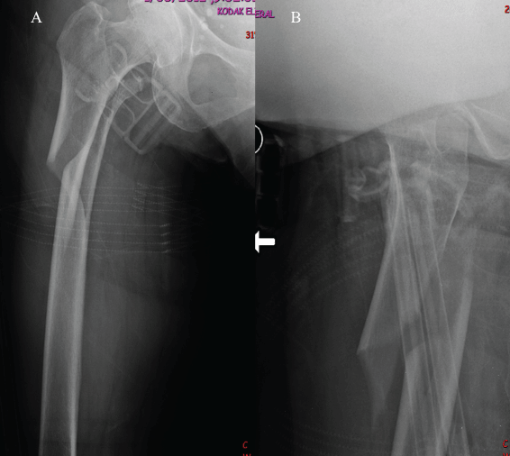
Figure 1: Initial injury radiographs showed subtrochanteric fracture of the proximal femur with reverse obliquity on AP and lateral views.
View Figure 1
.
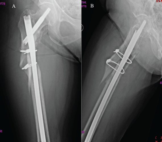
Figure 2: Immediate post-operative radiographs showed unreduced subtrochanteric fracture with two cerclage wires.
View Figure 2
Case Report
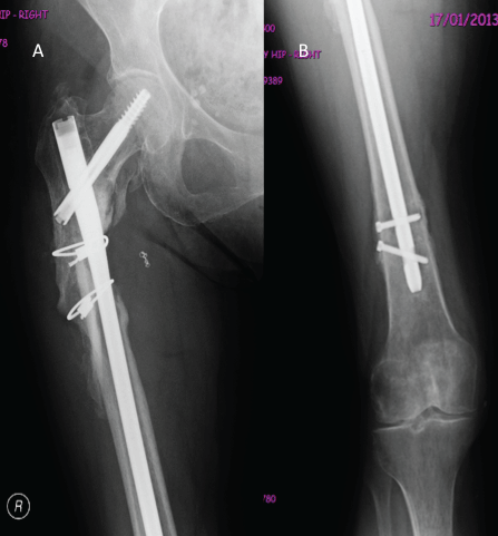
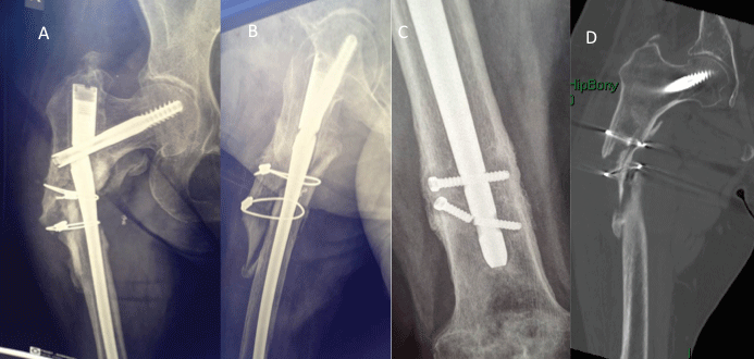
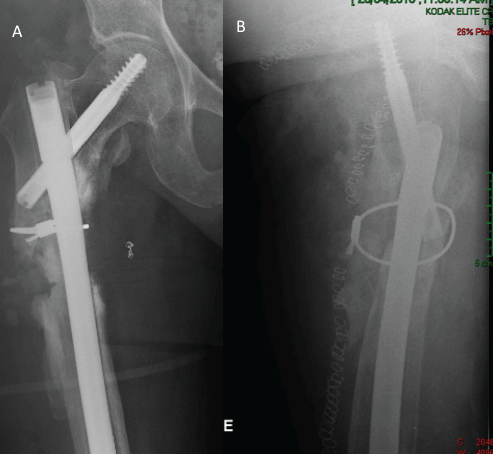
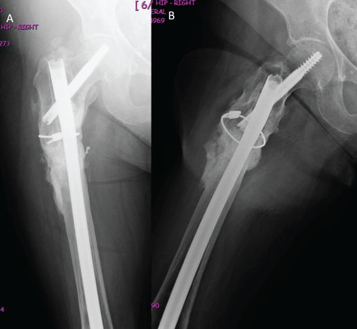
An 83-year-old female tripped and fell over while dancing with a group of elderly friends. Her initial injury radiographs showed subtrochanteric proximal femur spiral fracture (Figure 1). Immediately internal fixation was performed with the insertion of a long gamma nail. After insertion of the nail, open reduction was performed for the displaced fragment and an attempt was made to put in two cerclage wires (Figure 2). The cerclage wire caused active bleeding at the medial side of the proximal femur during surgery. The patient was immediately sent to a tertiary hospital with embolisation of active bleeding arteries, around the subtrochanteric region. Subsequently, the patient was kept in a non-weight bearing condition for six weeks after surgery. After six weeks post surgery, she had pain on mobilisation with assistance of a walker. The radiographs showed no sign of fracture healing. After nine months post-surgery, she still had pain on mobilisation. The radiographs showed atrophic non-union with the breakage of a distal locking screw (Figure 3). After 12 months post-surgery, the radiographs showed that the intramedullary nail was broken. There were no fracture healing signs either on radiographs or on CT scans (Figure 4). After 18 months the patient was introduced to our clinic, revision surgery was performed with the removal of broken implants, re-insertion of a thicker long gamma nail plus autologous iliac crest bone graft and supplemented by BMP-7 (Figure 5). After six weeks, the fracture started show signs of healing. After six months of revision surgery, complete healing was achieved (Figure 6).
.
Figure 3: After nine months post-operative radiographs showed no sign of fracture healing (atrophic non-union) with distal locking screws loosening.
View Figure 3
.
Figure 4: (a-c) After 12 months postoperative plain radiographs showed atrophic non-union; (d) CT scans did not reveal any sign of the fracture healing.
View Figure 4
Discussion
The management of subtrochanteric fractures is a challenging and technically demanding endeavour for surgeons. The fracture can be highly displaced and comminuted, because of the high concentration of stress in this area. The deforming forces for subtrochanteric fractures include flexion and external rotation from iliopsoas, abduction from the gluteal medius, adduction and shortening of the shaft from the hamstrings and the adductor muscle group. The degree of comminution of the medial cortical buttress, at the level of the fracture, will also be a surgical challenge.
.
Figure 5: (a) After 18 months from initial index surgery, we performed removed of broken implants, proximal femur vulgarised osteotomy, and re-insertion of longer and thicker long Gamma nail with autologous iliac crest bone graft and supplemented with the OP-1. There was no varus deformity on AP radiograph; (a,b) with anatomic reduction of the fracture after vulgarised osteotomy.
View Figure 5
.
Figure 6: After six months post-revision-surgery, radiographs showed complete healing of the fracture. The patient has no pain on her daily activities and clinical tests.
View Figure 6
The poor bone quality and the slow pace of bone healing in elderly patients result in high numbers of non-union and implant failures [5]. Regarding our case, there was a mal-reduction before the cephalomedullary nailing insertion, causing the fixation in mild varus and displaced (Figure 2), that is one of key factors in the later breakage of the nail. We think it was impossible to reduce the displaced subtrochanteric fracture after nail insertion. Bending stresses developing at the medial cortex along with the deficiency/comminution of the medial buttress could explain the failure of bone union, especially in the presence of varus mal-reduction [6,7]. In addition, the initial surgeon damaged the blood circulation at the fracture sites, by adding open reduction and cerclage wire fixation.
The 'self dynamization' of the initial reconstruction nail, as defined by the breakage of the distal locking screws, indicates some instability of the overall mechanical construction. The metalwork failure should be considered the consequence rather than the cause of the non-union. Breakage of the distal locking screws in a patient who is still symptomatic (feeling pain) at the fracture level should be considered a predictor of a pending failure. This should initiate action by the treating surgeon.
The diamond concept [8-10], include optimisation of the mechanical environment along with the enhancement of the multidimensional biological pathways of bone healing, has been proposed as the framework of a single-stage surgical revision for the recalcitrant problem of atrophic non-unions with implant failure. In the diamond concept, there is more emphasis on bone biology and the utilizing of growth factors scaffolding mesenchymal stem cells [9]. Still, the gold standard augmentation in the treatment of fracture non-unions is using autologous bone graft harvested from the iliac crest, highlighting the important role of structural stability in fracture healing.
The utilisation of composite bone grafts in non-unions, in combination with potent osteoinductive proteins (BMP-7), provides the rationale for using the full spectrum of fracture healing optimisation options for this complex case. Additionally, the treatment is cost effective by further strengthening the argument for single stage surgery, optimising the rates of eventual healing, avoiding prolonged follow-up, and most importantly, because there is no need for additional revisions. Exchanging a longer and thicker IM nail is one of the better options for the management of subtrochanteric non-union fractures, because the shorter the lever arm of the fixation, the better the load sharing, and the less bending movement across the fracture site and implant, the less soft tissue stripping, and the greater ease technically. However, as this case, anatomic reduction prior to nail insertion is critical to fracture healing and implant failure.
Simultaneous optimization of the mechanical and biological environment is a successful method for managing complex subtrochanteric atrophic non-unions with failed metalwork. However, multiple staged revision surgery is to be avoided with an elderly patient, therefore, careful planning optimizing fracture healing including autogenous bone graft supplement by BMP-7 is considered at index surgery. From one case alone, the usage of BMP-7 is not proven to be necessary. We will not be able to determine if re-fixation, exchange nailing and bone grafting alone without BMP-7 would have been sufficient to address the non-union. However, there is more emphasis in recent discussion on bone biology and the utilising of growth factors scaffolding mesenchymal stem cells [9]. Our management for subtrochanteric fracture non-union with implant failure with exchange intramedullary nail with autogenous bone graft in addition to supplement of bone morphogenic protein BMP-7 is an option when surgical decision-making process in the future with further study.
References
-
Yli-Kyyny TT, Sund R, Juntunen M, Salo JJ, Kroger HP (2012) Extra- and intramedullary implants for the treatment of pertrochanteric fractures - Results from a Finnish National Database Study of 14,915 patients. Injury 43: 2156-2160.
-
Loizou CL, McNamara I, Ahmed K, Pryor GA, Parker MJ (2010) Classification of subtrochanteric femoral fractures. Injury 41: 739-745.
-
Craig NJ, Sivaji C, Maffulli N (2001) Subtrochanteric fractures. A review of treatment options. Bull Hosp Jt Dis 60: 35-46.
-
de Vries JS, Kloen P, Borens O, Marti RK, Helfet DL (2006) Treatment of sub- trochanteric nonunions. Injury 37: 203-211.
-
Sims SH (2002) Subtrochanteric femur fractures. Orthop Clin North Am 33: 113-126.
-
Kuzyk PR, Bhandari M, McKee MD, Russell TA, Schemitsch EH (2009) Intramedullary versus extra-medullary fixation for subtrochanteric femur fractures. J Orthop Trauma 23: 465-470.
-
Park J, Yang KH (2010) Correction of mal-alignment in proximal femoral nailing - Reduction technique of displaced proximal fragment. Injury 41: 634-638.
-
Calori GM, Giannoudis PV (2011) Enhancement of fracture healing with the diamond concept: the role of the biological chamber. Injury 42: 1191-1193.
-
Giannoudis PV, Einhorn TA, Schmidmaier G, Marsh D (2008) The diamond concept - open questions. Injury 39: S5-8.
-
Giannoudis PV, Einhorn TA, Marsh D (2007) Fracture healing: the diamond concept. Injury 38: S3-6.


