
Trauma Cases and Reviews
Split Fracture and Displacement of Mandibular Lingual Cortical Plate of Mandibular Symphysis Requires Fixation
Masaki Fujioka1*, Kenji Hayashida2 and Hiroto Saijo2
1Clinical Professor, Department of Plastic and Reconstructive Surgery, Nagasaki University, Nagasaki, Japan
2Staff surgeon, Department of Plastic and Reconstructive Surgery, National Hospital Organization Nagasaki Medical Center, Japan
*Corresponding author:
Masaki Fujioka, Clinical Professor, Department of Plastic and Reconstructive Surgery, Nagasaki University, Nagasaki, and Director of the Department of Plastic and Reconstructive Surgery, Clinical Research Center, National Hospital Organization Nagasaki Medical Center, Nagasaki, Japan, E-mail: mfujioka@nagasaki-mc.com
Trauma Cases Rev, TCR-1-019, (Volume 1, Issue 4), Case Report; ISSN: 2469-5777
Received: November 17, 2015 | Accepted: December 01, 2015 | Published: December 03, 2015
Citation: Fujioka M, Hayashida K, Saijo H (2015) Split Fracture and Displacement of Mandibular Lingual Cortical Plate of Mandibular Symphysis Requires Fixation. Trauma Cases Rev 1:019. 10.23937/2469-5777/1510019
Copyright: © 2015 Fujioka M, et al. This is an open-access article distributed under the terms of the Creative Commons Attribution License, which permits unrestricted use, distribution, and reproduction in any medium, provided the original author and source are credited.
Abstract
The first priority in the management of comminuted mandibular fractures is the prevention of acute upper airway obstruction. Each division of mandibular fractures may cause respiratory obstruction. Among the variety of such fractures, split fracture of the symphyseal lingual cortical plate has a significant influence on the oropharyngeal and laryngopharyngeal airway spaces, and causes a markedly restricted ventilatory function. As the bony fragment is pedicled to the geniohyoid and genioglossus muscles, this type of fracture restricts hyoid movements, which may result in disturbance of deglutition.
We present two cases of split fracture and displacement of the mandibular lingual cortical plate of the mandibular symphysis, which were successfully treated with open reduction. We believe that precise reduction and fixation of the fragment of the symphyseal lingual cortical plate are indispensable for both relieving the airway obstruction and preventing disturbance of deglutition.
Keywords
Split fracture of mandibular, Displacement of fragment, Symphysis fracture, Open reduction
Introduction
The primary considerations in the treatment of comminuted mandibular fractures are open reduction and internal fixation to restore a satisfactory occlusion and shorten the period of oro-functional rehabilitation [1]. However, patterns of mandibular fracture vary; some dislocations involving fragmentation require special attention for the prevention of both acute upper airway obstruction and postoperative swallowing dysfunction [2]. We present two cases of split fracture and displacement of the mandibular lingual cortical plate of the mandibular symphysis, which caused obstruction of the upper airway and required open reduction.
Case Reports
Case 1
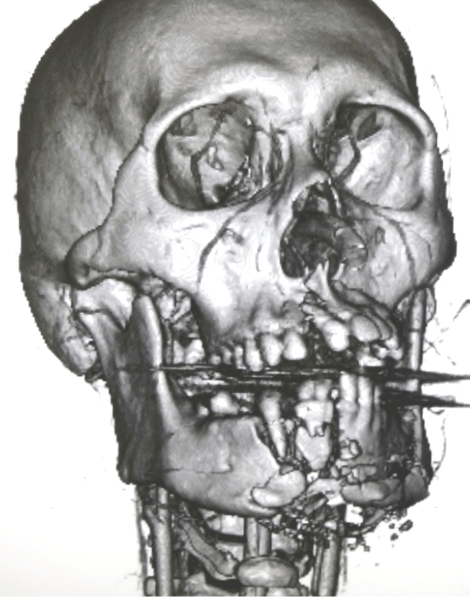
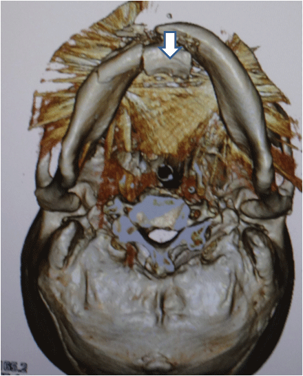
A 27 year old male was referred to our emergency unit complaining of marked neck swelling and airway obstruction resulting from a car accident. As the patient had extensive oral bleeding, he immediately underwent intra-tracheal intubation. An emergent cerebral angiogram revealed extravasation of the mental artery; thus, transcatheter arterial embolization was performed. Computed tomography (CT) showed both maxillary and mandibular open fractures (Figure 1a).
.
Figure 1a: Case 1. 3D-CT showed that both maxillary and mandibular open fractures.
View Figure 1a
.
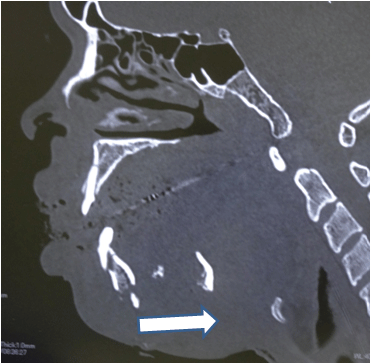
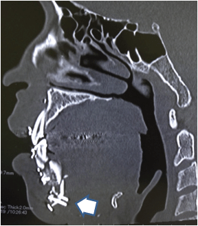
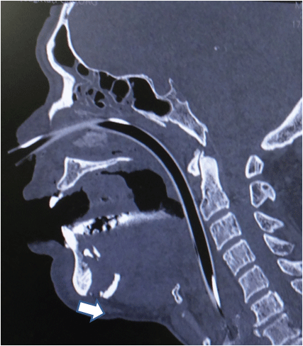
Figure 1b: Case 1. Sagittal slice CT showed that the fragment of the lingual cortical plate (arrow) was displaced 40 mm posteriorly.
View Figure 1b
The menton had fractured into pieces, and the largest fragment of the lingual cortical plate (45 × 20 mm) was displaced 40 mm posteriorly (Figure 1b and Figure 1c).
.
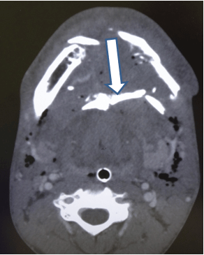
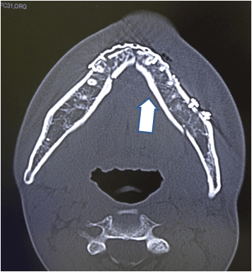
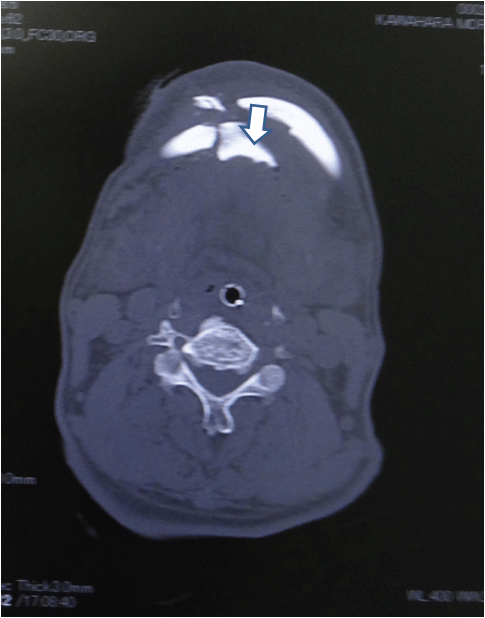
Figure 1c: Case 1. Axial slice CT showed that the fragment (arrow) was displaced posteriorly, and the upper airway was obstructed.
View Figure 1c
.
Figure 1d: Case 1. Axial slice CT showed that the fragment (arrow) was displaced posteriorly, and the upper airway was obstructed.
View Figure 1d
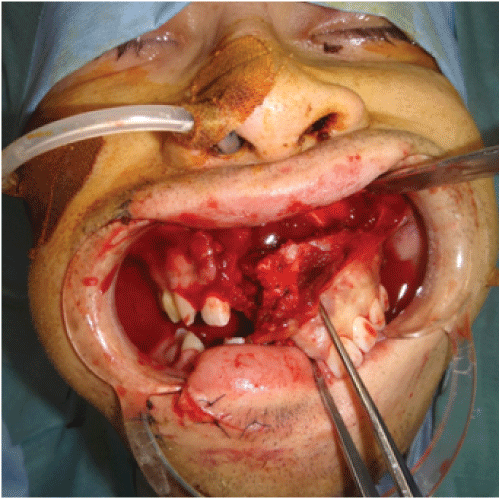
Six days later, open reduction was performed. AT first, osteosynthesis of the maxillary fracture and intermaxillary fixation using arch-bars were performed (Figure 1d). The comminuted mandibular fractures were exposed by both intra-oral buccal sulcus and mental skin incisions, and mandibular bone reduction and fixation with mini-plates (Figure 1e).
.
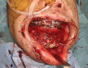
Figure 1e: Case 1: Intra-operative photography of repaired outer plates of mandibular fracture.
View Figure 1e
.
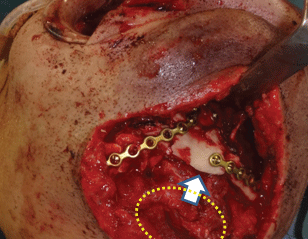
Figure 1f: Case 1: Intra-operative photography of the fragment of the lingual cortical plates, which was moved backward. The dotted line indicates the displaced fragment.
View Figure 1f
The fragment of the lingual cortical plates, which was moved backward by pulling the attached geniohyoid muscle, was securely fixed with lag screws (Figure 1f, Figure 1g and Figure 1h).
.
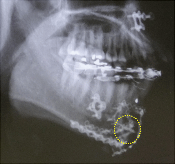
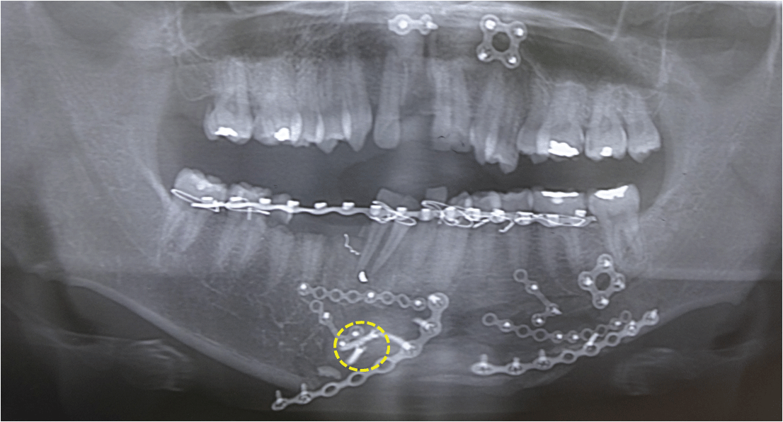
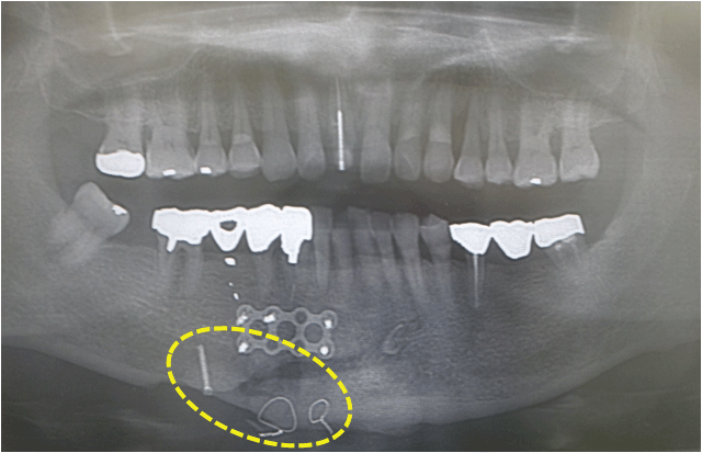
Figure 1g: X-ray photo 3 weeks after surgery showed 2 lag screws which fixed fragment (dotted circle).
View Figure 1g
.
Figure 1h: Panorama tomography 3 weeks after surgery showed 2 lag screws which fixed fragment (dotted circle).
View Figure 1h
Post-operative CT showed that the fragment of the lingual cortical plate was attached to the menton and formed favorable bone alignment. (Figure 1i and Figure 1j) Intermaxillary fixation was removed 2 weeks after surgery, and plates were removed after 6 months.
.
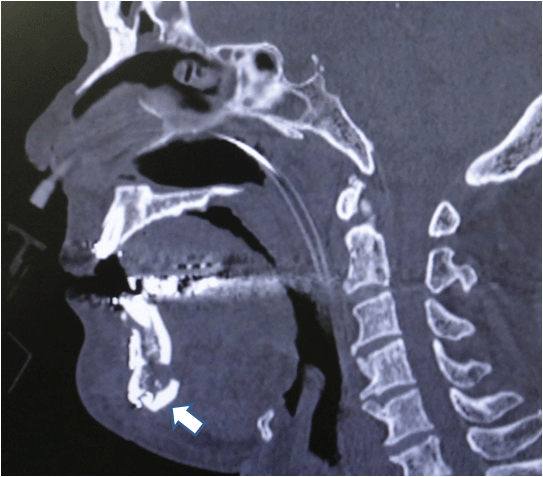
Figure 1i: Sagittal slice CT 3 weeks after surgery showed favorable reconstruction and rigid fixation with rag screws of the broken fragment (arrow).
View Figure 1i
.
Figure 1j: Axial slice CT 3 weeks after surgery showed favorable reconstruction of the broken fragment (arrow), which resulted in opening of the airway.
View Figure 1j
The postoperative diet and swallowing function were satisfactory.
Case 2
A 72-year-old male sustained a detrition injury to the maxilla and mandible due to being struck by the moving hook of a crane. He underwent immediate intra-tracheal intubation to relieve the airway obstruction. CT showed that the patient had a fracture of the mandible, and a fragment of lingual cortical plate (24 × 20 mm) was displaced 20 mm posteriorly (Figure 2a, Figure 2b and Figure 2c).
.
Figure 2a: Case 2: 3D-CT showed that mandibular open fractures. The fragment of the lingual cortical plate (arrow) was displaced 20 mm posteriorly.
View Figure 2a
.
Figure 2b: Case 2: Sagittal slice CT showed that the fragment (arrow) was displaced, and the upper airway was obstructed.
View Figure 2b
The patient underwent an immediate open reduction. The comminuted mandibular fractures were exposed by both intra-oral buccal sulcus incisions, and skin wound on the mental protuberance. Following intermaxillary fixation using arch-bars, osteosynthesis of the mandibular fracture and reduction of the fragment of the lingual cortical plate and fixation with long lag screw and wires (Figure 2d).
.
Figure 2c: Case 2: Axial slice CT showed that the fragment (arrow) was displaced, and the upper airway was obstructed.
View Figure 2c
.
Figure 2d: Panorama tomography 3 weeks after surgery showed a lag screw and 2 wires which fixed fragment (dotted circle).
View Figure 2d
Post-operative CT showed that the fragment of the lingual cortical plate was attached to the menton (Figure 2e). Intermaxillary fixation was removed 2 weeks after surgery. The postoperative diet and swallowing functions were satisfactory.
.
Figure 2e: Sagittal slice CT 2 weeks after surgery showed favorable reconstruction of the broken fragment (arrow), and opening of the airway.
View Figure 2e
Discussion and Conclusion
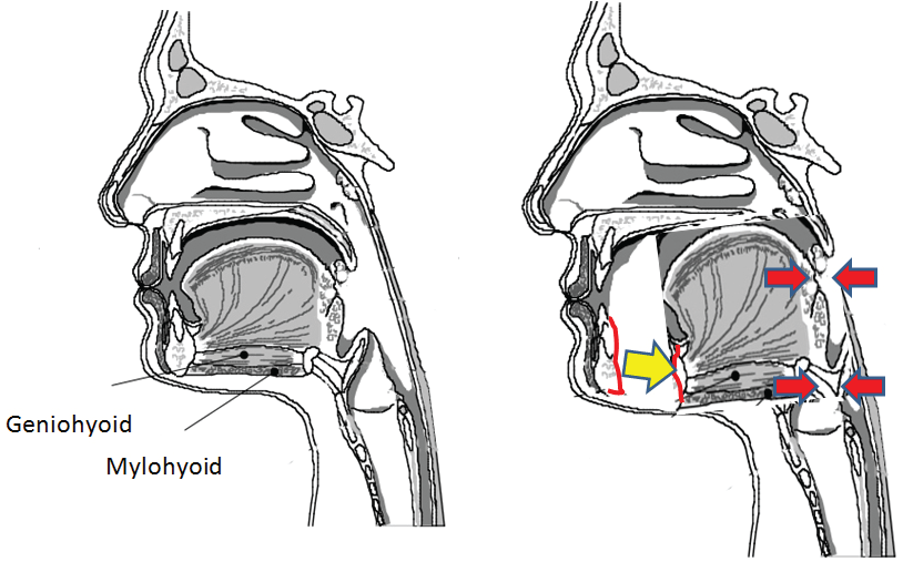
Mandibular fractures are common facial injuries [3-7]. They almost occur along with symphyseal, parasymphyseal, body, angle, vertical ramus, coronoid process, and condylar fractures [7]. In the oral cavity, displaced fragments, a sublingual hematoma, and swelling may press and obstruct the upper respiratory tract, and so, patients often require immediate intra-tracheal intubation to relieve the airway obstruction [2]. Among these mandibular fractures, bilateral mandibular fracture is known as a "flail mandible", which results in obstruction of the airway [8]. Bilateral parasymphyseal fracture has a significant influence on the oropharyngeal and laryngopharyngeal airway spaces, and causes a severely restricted the ventilator function [9]. The same phenomenon can occur with a split fracture of the symphyseal lingual cortical plate. This bony fragment is pedicled to the geniohyoid and genioglossus muscles, being the most likely to displace the hyoid in an anterior direction. Thus, displacement of symphyseal lingual cortical plate results in a serious airway obstruction (Figure 3) [10].
.
Figure 3: The figure shows that displacement of the symphyseal lingual cortical plate due to split fracture results in airway obstruction.
View Figure 3
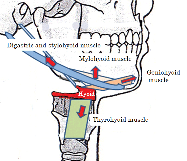
Furthermore, superior and anterior hyoid movements are important events in pharyngeal deglutition. The geniohyoid originates from the symphyseal lingual side of the mandible. The geniohyoid and thyrohyoid muscles have a greater structural potential to displace the hyoid anteriorly and superiorly, and are primarily responsible for opening the upper esophageal sphincter, which is essential for bolus entry into the esophagus (Figure 4) [10-13]. If a posteriorly displaced fragment of the lingual cortical plate is not fixed appropriately, anterior and superior movements of the hyoid may be insufficient and, consequently, impaired opening of the upper esophagus may cause suffocation.
.
Figure 4: The figure shows the muscles attached to the hyoid and their movements. The geniohyoid and thyrohyoid muscles have important roles in pharyngeal deglutition.
View Figure 4
In conclusion, we suggest that precise reduction and fixation of a fragment of the symphyseal lingual cortical plate are indispensable for both relieving airway obstruction in the acute phase, and preventing disturbance of deglutition later.
Disclosures
This manuscript has not benefited from any source of funding support or grants, and the author has no conflicting financial interest.
Ethical Considerations
This study was approved by the IRB of National Hospital organization Nagasaki Medical Center (27021, May 5th 2015), and all participants signed an informed consent agreement.
The content of the manuscript is original and it has not been published or accepted for publication, either in whole or in part, in any form. No part of the manuscript is currently under consideration for publication elsewhere.
References
-
Ellis E 3rd, Muniz O, Anand K (2003) Treatment considerations for comminuted mandibular fractures. J Oral Maxillofac Surg 61: 861-870.
-
Teichgraeber JF, Rappaport NH, Harris JH Jr (1991) The radiology of upper airway obstruction in maxillofacial trauma. Ann Plast Surg 27: 103-109.
-
Fujioka M, Nishida H, Daian T, Kondo M (2000) Analysis of 307 maxillofacial fractures: comparison with those of urban area. J Miyazaki medical society 24: 111-114.
-
Samieirad S, Tohidi E, Shahidi-Payam A, Hashemipour MA, Abedini A (2015) Retrospective study maxillofacial fractures epidemiology and treatment plans in Southeast of Iran. Med Oral Patol Oral Cir Bucal 20: e729-736.
-
Gaddipati R, Ramisetti S, Vura N, Reddy KR, Nalamolu B (2015) Analysis of 1,545 Fractures of Facial Region-A Retrospective Study. Craniomaxillofac Trauma Reconstr 8: 307-314.
-
Maliska MC, Lima Junior SM, Gil JN (2009) Analysis of 185 maxillofacial fractures in the state of Santa Catarina, Brazil. Braz Oral Res 23: 268-274.
-
Goldenberg DC, Alonso N, Ferreira MC (2009) Facial trauma. In: Guyuron B, Eriksson E, Persing J, Plastic surgery indications and practice. Saunders Elsevier: 619-644.
-
Bavitz JB, Collicott PE (1995) Bilateral mandibular subcondylar fractures contributing to airway obstruction. Int J Oral Maxillofac Surg 24: 273-275.
-
Chen LJ, Zhao MC, Pan XF, Wei YQ, Wang DY (2013) X-cephalometric study of different parts of the upper airway space and changes in hyoid position following mandibular fractures. West Indian Med J 62: 642-648.
-
Pearson WG Jr, Langmore SE, Zumwalt AC (2011) Evaluating the structural properties of suprahyoid muscles and their potential for moving the hyoid. Dysphagia 26: 345-351.
-
Matsuo K, Palmer JB (2008) Anatomy and physiology of feeding and swallowing: normal and abnormal. Phys Med Rehabil Clin N Am 19: 691-707, vii.
-
Cook IJ, Dodds WJ, Dantas RO, Massey B, Kern MK, et al. (1989) Opening mechanisms of the human upper esophageal sphincter. Am J Physiol 257: 748-759.
-
Perlman AL, Palmer PM, McCulloch TM, Vandaele DJ (1999) Electromyographic activity from human laryngeal, pharyngeal, and submental muscles during swallowing. J Appl Physiol (1985) 86: 1663-1669.
