

Obstetrics and Gynaecology Cases - Reviews
Ovarian Cancer in Early Pregnancy, an Unusual Presentation
Saniya M Eltayeb1*, Moza A AlKalbani2, Sarah O Abu Zaid2 and Saba Mubbashir2
1Department of Obstetrics and Gynaecology, College of Medicine, Sultan Qaboos University, Muscat, Oman
2Department of Obstetrics and Gynaecology, Sultan Qaboos University Hospital, Muscat, Oman
*Corresponding author: Saniya Eltayeb, MBBS, FRCOG, Department of Obstetrics and Gynaecology, College of Medicine and Health Sciences, Sultan Qaboos University, 123 Alkhod, Muscat, Oman, Tel: 96896118472; E-mail: Saniya.eltayeb@gmail.com
Obstet Gynecol Cases Rev, OGCR-1-006, (Volume 1, Issue 2), Case Report; ISSN: 2377-9004
Received: September 18, 2014 | Accepted: November 11, 2014 | Published: November, 13 2014
Citation: Eltayeb SM, AlKalbani MA, Zaid SOA, Mubbashir S (2014) Ovarian Cancer in Early Pregnancy, an Unusual Presentation. Gynecol Cases Rev 1:006. 10.23937/2377-9004/1410006
Copyright: © 2014 Eltayeb SM, et al. This is an open-access article distributed under the terms of the Creative Commons Attribution License, which permits unrestricted use, distribution, and reproduction in any medium, provided the original author and source are credited.
Abstract
The incidence of diagnosed ovarian carcinoma in pregnancy is rare, reported as 0.018-0.073/1000 pregnancies [1]. Very few cases are reported in the English literature regarding management of ruptured malignant ovarian cyst in pregnancy; almost all the cases reported described patients in the third trimester of pregnancy. We report a case of a young female who presented with acute abdomen at seven weeks gestation and was found to have an ovarian malignancy.
Keywords
Ovarian cancer, Pregnancy
Introduction
Ovarian malignancy is a rare entity during pregnancy, yet the rate is increasing as more women are deferring childbirth till later in life when malignancies are more frequent [2]. The routine use of ultrasound in early pregnancy has facilitated the early diagnosis and management of asymptomatic ovarian tumors. They may rarely present with complications such as torsion, rupture, hemorrhage, preterm labor and adverse prenatal outcome [3]. Such complications usually occur in the third trimester of pregnancy. Only two cases were reported in the literature of patients presenting with ruptured ovarian cancer in pregnancy. Malhotra et al. in 2010 reported a case of ruptured ovarian tumor presenting as an acute abdomen. The patient was at 30 weeks gestation and the tumor was a malignant mucinous cystadenocarcinoma [4]. The other case was reported by Segedi et al. in 2011, the patient was at eightweeks gestation and the tumor was a malignant serous cystadenocarcinoma at Stage 1 by the International Federation of Gynaecology and Obstetrics (FIGO) classification [5].
Case Report
A 27 ?year-old gravida two para one woman presented in clinical shock at seven weeks of gestation. She gave a history of vague lower abdominal pain for one week worsening over the last few hours and associated with fainting attacks. She had no significant past medical or surgical history. Apart from twins on her maternal side, there was no relevant family history. She was very pale, tachycardic with a thready pulse at 120 beats per minute, tachypnic with a respiratory rate of 44 per minute and blood pressure of 80/40 mmHg. There was tenderness and guarding on abdominal palpation. The initial hemoglobin was 4.1g/dl.
Vigorous resuscitation was started; a bedside ultrasound showed a viable intrauterine pregnancy equivalent to seven weeks gestation with a left-sided adnexal mass and significant amount of free fluid in the pelvis. The patient was taken immediately to the operating room for exploratory laparotomy which revealed a hemo-peritoneum of 2.2 liters of blood and blood clots, a left ovarian mass of 9x10 cm, twisted twice around the ovarian pedicle with a ruptured capsule and actively bleeding from the base (Figure 1). There was soft and velvety tissue inside the ruptured cyst (Figure 2). The left tube was twisted at the cornu and stretched over the mass. The left ovarian pedicle was clamped to control the bleeding and as there was no suspicion of malignancy macroscopically, left salpingo-oophorectomy was performed.
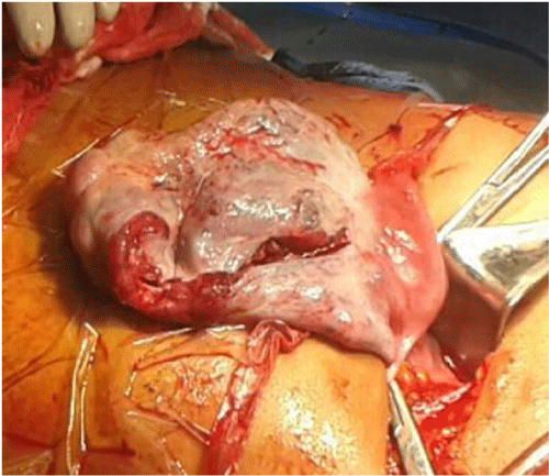 Figure 1: Photo taken at laparotomy. The left ovarian cyst after clamping the
pedicle, showing the site of capsule rupture.
View Figure 1
Figure 1: Photo taken at laparotomy. The left ovarian cyst after clamping the
pedicle, showing the site of capsule rupture.
View Figure 1
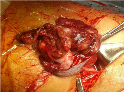 Figure 2: Showing the inside of the ruptured left-sided ovarian cyst after
clamping the ovarian pedicle.
View Figure 2
Figure 2: Showing the inside of the ruptured left-sided ovarian cyst after
clamping the ovarian pedicle.
View Figure 2
The patient received six units of packed red cells and eight units of fresh frozen plasma. The fetal viability was confirmed by an ultrasound scan on the first post- operative day. She had an uneventful post- operative course and discharged home on the fourth post- operative day. The post- operative hemoglobin was 9.2 gram/dl.
She was seen two weeks later in the outpatient department when she was found to have had a complete miscarriage confirmed by ultrasound scan. The histopathology revealed an invasive serous ovarian cystadenoma of high grade, with areas of benign serous cystadenoma with calcifications. Areas of lymphovascular invasion were present. Malignant tumor cells were present in the peritoneal wash. This put her as stage Ic by the FIGO classification.
She underwent a computerized tomography of the chest, abdomen, and pelvis for staging, which showed no obvious residual disease. The blood was tested for the level of the tumor marker cancer antigen 125 and the beta subunit of human chorionic gonadotropin, both were within normal limits.
She was referred to the multidisciplinary team for further management, yet she was lost to follow up for two months. When she returned three months after her initial operation, she opted for staging laparotomy and conserving the uterus and other ovary. Unfortunately she did not show up for the scheduled laparotomy and neither she nor her husband returned the hospital phone calls.
Discussion
Adnexal masses have been noted to occur in up to 1% of all gestations and ovarian cancers complicate one in 12- 20,000 pregnancies, yet they are often diagnosed incidentally during obstetrical ultrasound [4]. Most patients with ovarian cancer have no specific symptoms which makes early diagnosis difficult [6].
The widespread use of ultrasonography in early pregnancy has led to the early diagnosis of ovarian cancer during pregnancy hence improving the therapeutic approach and outcome [2]. Unfortunately our patient presented acutely with ruptured ovarian cancer early in pregnancy, prior to commencing her antenatal care, thus missing the chance for proper evaluation and planned management.
Ovarian epithelial tumors during pregnancy are usually reported as of low malignant potential, which is consistent with the age- matched non ? pregnant setting1. Primary carcinoma of the ovary occurs more commonly in women of low parity later in their reproductive years. Such carcinoma has been noted to occur in significantly older women than those with benign tumors or tumors of low malignant potential [1]. Contrary to this, our patient was in her twenties, yet had invasive ovarian carcinoma. She presented with an acute abdomen, necessitating urgent laparotomy before reaching a definitive diagnosis. The working diagnosis of heterotopic pregnancy with a ruptured ovarian component was made taking into account her age and family history of twin pregnancy. Ovarian cancer was at the bottom of our differential diagnosis list at the time of operation.
Torsion or rupture of ovarian masses is an important differential diagnosis of abdominal or pelvic pain during pregnancy, but it is often missed as the symptoms are mistaken as one of the symptoms of pregnancy. Moreover it is difficult to diagnose adnexal masses on ultrasound with increasing gestational age [7]. It is of high importance to evaluate the ovaries during the first trimester dating ultrasound scan as detection of ovarian tumors at such an early stage will facilitate correct and timely intervention which will improve both maternal and fetal outcome [4].
The optimal surgery for staging ovarian cancer includes bilateral salpingo-oophorectomy, total hysterectomy, omentectomy and pelvic and para-aortic lymphadenectomy. In addition to surgical treatment, the use of multi-agent chemotherapy during pregnancy has become widespread [2].
In our case the patient had a miscarriage which made immediate optimal treatment feasible as there was no fetus to consider, yet she opted for surgical staging and conserving the uterus and other ovary somehow compromising her chances of cure. Furthermore she did not even return to pursue her treatment. The pressure of her social and community setting influenced her decision to refuse treatment, as she desired to conserve her fertility whilst endangering her own life.
Such patients, by refusing the optimal management, pose a challenge for the treating doctor regarding ethics and medico-legal issues. They should be extensively counseled regarding the risks of suboptimal treatment of their ovarian cancer as they have poor prognosis due to the advanced stage of their disease [8].
Conclusion
Ovarian cancer should be suspected in any pregnant patient presenting with torsion or rupture of ovarian mass regardless of the maternal or gestational age
Conflict of Interest
We declare no conflict of interest. Consent for publication was obtained from the patient. This research received no specific grant from any funding agency in the public, commercial, or not-for-profit sectors.
Authorship Contribution
Saniya Eltayeb: Admitting and treating consultant, wrote the manuscript
MozaAlkalbani: Oncologist, took over the management after initial surgery
Sarah Abuzaid: Obtained the patient history and consent
Saba Mubbashir: Assisted in the initial surgery, done the literature review
References
-
Palmer J, Jivraj S, Galimberti A, Paterson M (2009) Serous ovarian carcinoma in pregnancy. BMJ Case Rep 2009.
-
Barut A, Arikan I, Barut F, Harma M, Harma MI, et al. (2011) Ovarian cancer during pregnancy. J Pak Med Assoc 61: 914-916.
-
Bakri YN, Ezzat A, Akhtar, Dohami, Zahrani (2000) Malignant germ cell tumors of the ovary. Pregnancy considerations. Eur J Obstet Gynecol Reprod Biol 90: 87-91.
-
Malhotra N, Sumana G, Singh A, Deka D, Mittal S (2010) Rupture of a malignant ovarian tumor in pregnancy presenting as acute abdomen. Arch Gynecol Obstet 281: 959-961.
-
Segedi LM, Mandic A, Segedi D, Kozarcic T (2011) Spontaneous rupture of malignant ovarian cyst in 8-gestation-week-pregnancy- a case report and literature review. Arch Oncol 19: 39-41.
-
Kwon YS, Mok JE, Lim KT, Lee IH, Kim TJ, et al. (2010) Ovarian cancer during pregnancy: clinical and pregnancy outcome. J Korean Med Sci 25: 230-234.
-
Sieroszewski P, Suzin J, Gottwald L, Karowicz-Bilinska A (2002) [The diagnostic value of ultrasound examination in detection of ovarian tumors during pregnancy]. Ginekol Pol 73: 376-378.
-
Behtash N, Karimi Zarchi M, Modares Gilani M, Ghaemmaghami F, Mousavi A, et al. (2008) Ovarian carcinoma associated with pregnancy: a clinicopathologic analysis of 23 cases and review of the literature. BMC Pregnancy Childbirth 8: 3.
