

Obstetrics and Gynaecology Cases - Reviews
Urinary Incontinence as a Presenting Symptom of Bladder Cancer
Elena Vilar González*, Marta Perez de la Fuente, Enrique González Díaz
Pelvic Floor Unit, Department of Obstetrics and Gynaecology, Complejo Asistencial Universitario de Leon (CAULE), Spain
*Corresponding author: Enrique González Díaz, Servicio de Obstetricia y Ginecología, Complejo Asistencial Universitario de León (CAULE), Calle Altos de Nava S/N, 24080, León, Spain, Email: enriquegonzalezdiaz@hotmail.com
Obstet Gynecol Cases Rev, OGCR-3-080, (Volume 3, Issue 2), Case Report; ISSN: 2377-9004
Received: July 13, 2015 | Accepted: March 22, 2016 | Published: March 25, 2016
Citation: González EV, de la Fuente MP, Díaz EG (2016) Urinary Incontinence as a Presenting Symptom of Bladder Cancer. Obstet Gynecol Cases Rev 3:080. 10.23937/2377-9004/1410080
Copyright: © 2016 González EV, et al. This is an open-access article distributed under the terms of the Creative Commons Attribution License, which permits unrestricted use, distribution, and reproduction in any medium, provided the original author and source are credited.
Abstract
Urinary incontinence is a very common complaint among women. We should think of potentially serious aetiologies when there is no response to empiric treatments, especially in postmenopausal women. We report a case of a 69 year old woman, referred to our pelvic floor unit with urinary incontinence and nocturia. After failing with anticholinergic treatment, a transvaginal ultrasound scanning shows an irregular solid lesion of 6 mm, which was diagnosed by further urological investigation as a transitional cell carcinoma.
Keywords
Urinary incontinence, Bladder cancer, Transvaginal ultrasound
Introduction
Urinary incontinence is very common in women and it often remains undetected and undertreated. Women use to think it is a normal related to age problem, so many times they don't tell about it to their doctors. Its prevalence increases with age, although estimates vary widely, becoming an important and very prevalent problem among postmenopausal women.
The initial evaluation of urinary incontinence includes the description and classification of the type of incontinence, which can be done by a direct questioning about symptoms, identifying potentially reversible causes of incontinence and identifying the underlying conditions, such as neurologic disorders or malignancy. Bladder carcinoma is a rare but possible findings in patients who have urinary incontinence.
Initial diagnosis does not require any invasive test [1]. We have to consider additional testing in those high risk patients [2], especially when there is no response to empirical treatments. In our gynaecologic check-up, after the initial pelvic examination and during the transvaginal ultrasound, one may insist on ultrasound scanning of the bladder. This bladder examination becomes especially important in those patients with potentially serious conditions, such as bladder cancer, or when no gynaecological pathology is found. Our ultrasound scanning should start before emptying the bladder or be repeated after its proper filling.
Case Report
The patient was a 69 year old white female, smoker, gravida 3 para 3, postmenopausal for 19 years, referred to our pelvic floor department due to urinary incontinence and nicturia. She had ischaemic heart disease as the result of an acute myocardial infarction treated by angioplasty in 2006. She also presented a depressive disorder and dyslipidemia.
Previous gynecological examinations presented no pathological signs. The patient reported urge incontinence and nicturia. Clinical examination was unremarkable: the evaluation of sensibility and reflexes was normal and pelvic organ prolapse or urinary incontinence were absent during Valsalva maneuvers. Urinalysis did not present any pathologic findings. According to these facts, anticholinergic therapy was prescribed (solifenacin, 5 mg/24 h).
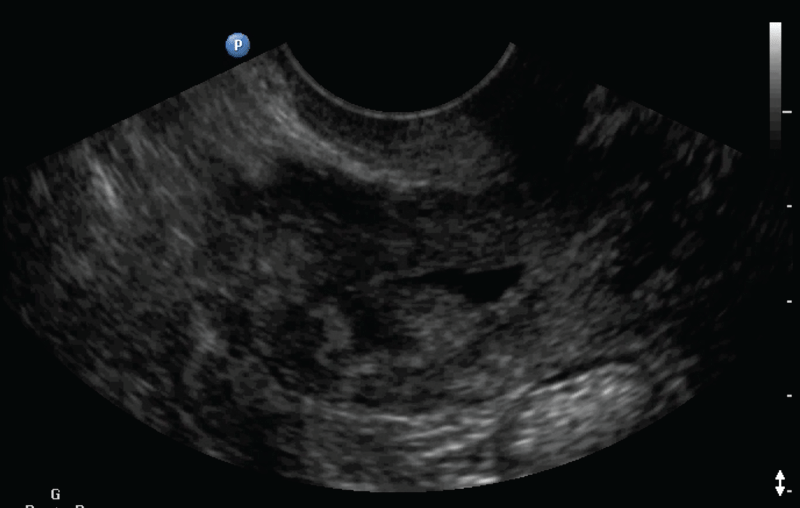
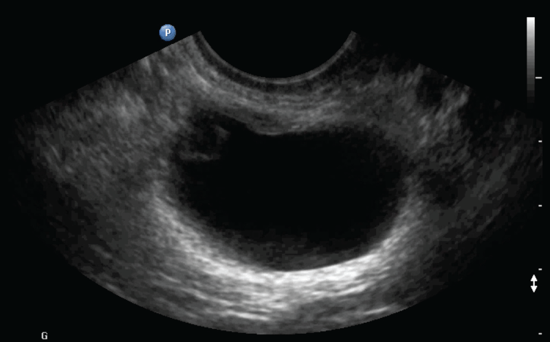
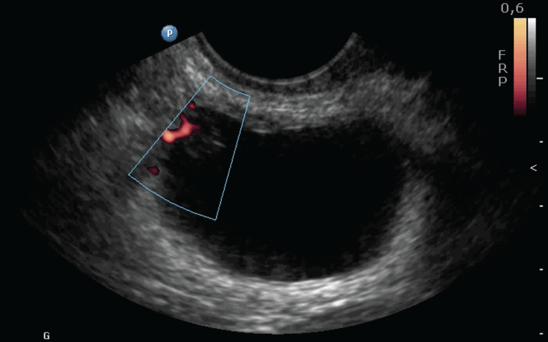
During the next consultation, the patient had withdrawn treatment due to the lack of improvement in symptoms, and she described one episode of hematuria. Transvaginal ultrasound was performed, as we always do in our pelvic floor unit patients in their second visit [2], showing a thickened endometrium (Figure 1) and irregularities in the cavity contour. The ovaries had both proper size and aspect for her age and no free fluid was identified in the pouch of Douglas. The bladder was full during the examination, which allowed the identification of a polypoid, irregular lesion (Figure 2) of 6 mm located in the top right angle. It appeared on color Doppler imaging as highly vascular (Figure 3).
.
Figure 2: Two dimensional imaging of the full bladder showing the irregular lesion.
View Figure 2
Subsequent to these findings, the urology service run a cystoscopy, describing a papillary tumor of 1 cm on the right wall of the bladder. A transurethral resection (TUR) was carried out to remove the lesion, being the histopathological diagnosis of low grade transitional cell carcinoma (pTa G1).
.
Figure 3: Two dimensional power Doppler of the bladder showing the highly vascularised lesion.
View Figure 3
In addition to that, the gynecology service run a histeroscopy and a endometrium curettage getting an histopathological report of well differentiated endometrial adenocarcinoma, succeeding in the performance of complete histerectomy and double anexectomy.
In the patient successive checks carried out by urologists, she presented signs of recurrence of the transitional cell carcinoma, leading to a new TUR and Bacillus Calmette-Guerin instillation. Due to the intolerance to the intravesical treatment, the patient needed a laparoscopic radical cystectomy with external urinary derivation by bilateral percutaneous nephrostomy.
Regarding her gyneacological pathology, the patient kept disease-free during the next controls.
Discussion
Urinary incontinence is defined as the complaint of involuntary leakage of urine. It is a very common problem among women, affecting approximately 36% of women over 60 years [3], altering significantly their quality of life. An increase in urinary incontinence prevalence with age is caused by multiple factors including an incremented incidence of comorbidities such as obesity and diabetes, polypharmacy, and an age-related cognitive and functional decline [4].
Urinary incontinence can be caused by many different disorders. First of all, due to its high prevalence, we should consider several external factors as possible causes [2]. The first one is urinary tract infection. Alcohol and pharmaceuticals can contribute to this condition.
The physiological changes of the genitourinary tract related to age [4] may be the origin of incontinence, as well as genital atrophy, pelvic organ prolapses or involuntary detrusor contractions.
Urinary incontinence can respond to systemic disorders, being a common complaint in obese patients [5]. Nycturia is a typical manifestation of congestive cardiac failure, neurologic diseases as multiple sclerosis or some other neuropathies as the diabetic neuropathy. Other severe pathologies like malignancies can induce the incontinence [1]. They should be suspected specially when no other explanation is found. UI is the most common initial symptom in urethral cancer [6], and can be the presenting symptom in bladder cancer too, as shown in our case report.
Bladder cancer is the most common malignancy of the urinary tract [7], and urothelial (transitional cell) carcinoma is the predominant histologic type. The classic presentation of bladder cancer in most patients is hematuria, grossy visible or microhematuria. It is present at initial diagnosis in the 85% of patients [8]. The remaining small subgroup of newly diagnosed bladder cancer patients will present the complex of symptoms of bladder irritability and urinary frequency, urgency, and dysuria [8]. Hematuria associated with irritative symptoms such as frequency, dysuria, and urgency, should be carefully examined after infection and neurogenic bladder have been dismissed because it may point to relatively advanced tumors or carcinoma in situ that extends to the bladder neck or the urethra.These voiding symptoms in bladder cancer are more common in patients with carcinoma in situ [9], and they are usually the result of a functional decrease of the storage capacity, detrusor overactivity, invasion of the trigone or obstruction of the bladder neck or urethra.
The diagnosis is often delayed because of the similarity of these symptoms to those presented in benign processes as common as urinary tract infections or interstitial cystitis [1]; delays can lead to a worse prognosis due to advanced stage at diagnosis.
Men are about 3 to 4 times more likely to get bladder cancer during their lifetime [10] than women, drawing the attention of this proportion in Spain, where it raises up to 7 [11]. The excess of bladder cancer in men compared to women is not completely clarified by discrepancies in smoking addiction and occupation [12] (the 2 most well-known risk factors for bladder cancer). Surveys about the incidence and mortality propose that multiparous women have a lower risk of getting bladder cancer than nulliparous women, probably due to the hormonal changes related to gestation, and that the risk may be reduced with increasing pregnancies [13]. It has been proved that female patients with bladder cancer have a worse prognosis than male patients [14].
There is evidence that suggests that poorer survival in women can be related to a later diagnosis, as women are more used to suffer urinary incontinence due to physiological changes related to age [2]. Women postpone consulting and clinicians often exclude benign pathologies before thinking about malignancies [14]. We have to consider the possibility of bladder cancer in those high risk patients, especially when there is no response to empirical treatments, as said before; as it was our patient case, voiding symptoms due to bladder cancer characteristically does not improve with anticholinergic treatment.
The initial diagnosis of an urinary incontinence does not require any invasive test. In our gynaecologic examination, the initial physical exam of the pelvis will be followed by a transvaginal ultrasound [2,15], that offers the opportunity of examining the whole pelvis. The ultrasound would be follow by an urodynamic study and a cystoscopy [2]. In high risk patients, or when no gynaecologic pathology is found, one may insist on ultrasound scanning of the bladder. In our case, the episode of hematuria that the patient referred in the second check was seen as a reason enough to suspect bladder pathology, but even without this episode, thickened endometrium does not explain voiding symptoms.
While the main goal of a gynaecological ultrasound is excluding gynaecologic pathology, examination of the bladder should become part of the standard examination procedure [15], especially in postmenopausal women, and women with unexplained symptoms as our patient. For the correct bladder examination, one may initiate the scanning prior to emptying the bladder, or repeat the examination after proper filling. The bladder should be filled with, at least, about 50 mL of urine, that will act as a negative contrast agent [16]. We may be able with proper contrast to examine the bladder wall, and visualize any irregularity or solid suspicious lesion. Among them, big size and vascularization (Doppler positive) are the worst prognosis signs. Suspicious bladder lesions, as in our case, are an absolute for urgent cystoscopy indication.
While cystoscopy is the gold standard for bladder cancer diagnosis, ultrasonography has been shown to have 63% specificity and 99% sensitivity for the detection of bladder tumours [17]. Underfilling and trabeculation due to bladder wall thickening are the main causes of false positive results.
Authors' Contribution
E Vilar Gonzalez: Data collection, and Manuscript writing; M Perez de la Fuente: Manuscript writing; E Gonzalez Diaz: Concepcion and Supervision.
References
-
Nitti V, Taneja S (2005) Overactive bladder: achieving a differential diagnosis from other lower urinary tract conditions. Int J Clin Pract 59: 825-830.
-
M Espuna, M. Fillol (2013) Documentos de consenso Sociedad Espanola de Ginecología y Obstetricia, 2013: Diagnóstico de las disfunciones de suelo pélvico. Exploración especializada de las disfunciones de suelo pélvico 42-43.
-
Anderson KM, Davis K, Flynn BJ (2015) Urinary incontinence and pelvic organ prolapse. Med Clin North Am 99: 405-416.
-
Smith PP (2010) Aging and the underactive detrusor: a failure of activity or activation? Neurourol Urodyn 29: 408-412.
-
Moreno-Vecino B, Arija-Blázqueza A, Pedrero-Chamizo R, Julián Alcázar, Alba Gómez-Cabello, et al. (2015) Associations between obesity, physical fitness, and urinary incontinence in non-institutionalized postmenopausal women: The elderly EXERNET multi-center study. Maturitas 82: 208-214.
-
Amin MB, Young RH (1997) Primary carcinomas of the urethra. Semin Diagn Pathol 14: 147-160.
-
Ploeg M, Aben KK, Kiemeney LA (2009) The present and future burden of urinary bladder cancer in the world. World J Urol 27: 289-293.
-
Marshall VF (1956) Current clinical problems regarding bladder tumors. Cancer 9: 543-550.
-
Farrow GM, Utz DC, Rife CC, Greene LF (1977) Clinical observations on sixty-nine cases of in situ carcinoma of the urinary bladder. Cancer Res 37: 2794-2798.
-
Torre LA, Bray F, Siegel RL, Ferlay J, Lortet-Tieulent J, et al. (2015) Global cancer statistics, 2012. CA Cancer J Clin 65: 87-108.
-
Asociación Espanola Contra el Cáncer, 2015. //www.aecc.es/ Incidencia del cancer de vejiga.
-
Freedman ND, Silverman DT, Hollenbeck AR, Schatzkin A, Abnet CC (2011) Association between smoking and risk of bladder cancer among men and women. JAMA 306: 737-745.
-
Davis-Dao CA, Henderson KD, Sullivan-Halley J, Ma H, West D, et al. (2011) Lower risk in parous women suggests that hormonal factors are important in bladder cancer etiology. Cancer Epidemiol Biomarkers Prev 20: 1156-1170.
-
Mitra AP, Skinner EC, Schuckman AK, Quinn DI, Dorff TB, et al. (2014) Effect of gender on outcomes following radical cystectomy for urothelial carcinoma of the bladder: a critical analysis of 1,994 patients. Urol Oncol 32: 52.
-
Tunn R, Petri E (2003) Introital and transvaginal ultrasound as the main tool in the assessment of urogenital and pelvic floor dysfunction: an imaging panel and practical approach. Ultrasound Obstet Gynecol 22: 205-213.
-
Betsas G, Van den Bosch T, Deprest J, Bourne T, Timmerman D (2008) The use of transvaginal ultrasonography to diagnose bladder carcinoma in women presenting with postmenopausal bleeding. Ultrasound Obstet Gynaecol 32: 959-960.
-
Datta SN, Allen GM, Evans R, Vaughton KC, Lucas MG (2002) Urinary tract ultrasonography in the evaluation of haematuria--a report of over 1,000 cases. Ann R Coll Surg Engl 84: 203-205.
