

Obstetrics and Gynaecology Cases - Reviews
Postmenopausal Calcified Pedunculated Large Subserous Leiomyoma: A Case Report
Ozhan Ozdemir*, Cemal Resat Atalay, Mustafa Erkan Sari, Ertugrul Sen and Mehriban Nebioglu
Department of Obstetrics and Gynecology, Ankara Numune Education and Research Hospital, Turkey
*Corresponding author: Ozdemir Ozhan, Department of Obstetrics and Gynecology, Ankara Numune Training and Education Hospital, 06010, Ankara, Turkey, Tel: +905052255078, E-mail: seyozi@hotmail.com
Obstet Gynecol Cases Rev, OGCR-2-054, (Volume 2, Issue 4), Case Report; ISSN: 2377-9004
Received: May 21, 2015 | Accepted: August 01, 2015 | Published: August 03, 2015
Citation: Ozdemyr O, Atalay CR, Sary ME, Sen E, Nebyoglu M (2015) Postmenopausal Calcified Pedunculated Large Subserous Leiomyoma: A Case Report. Obstet Gynecol Cases Rev 2:054. 10.23937/2377-9004/1410054
Copyright: © 2015 Ozdemyr O, et al. This is an open-access article distributed under the terms of the Creative Commons Attribution License, which permits unrestricted use, distribution, and reproduction in any medium, provided the original author and source are credited.
Abstract
A 72-year-old postmenopausal and nullipar woman presented with abdominopelvic pain and a palpable mass in the right lower quadrant. Ultrasonography showed a large, hyperechoic, solid mass. Contrast-enhanced computed tomography (CT) of the abdomen demonstrated a well-circumscribed mass. Rutine laboratuary tests and tumour markers revealed as normal. Total abdominal hysterectomy and bilateral salpingo-oophorectomy is the surgery of choice in this case. Histopathological examination confirmed a calcified leiomyoma. The majority of uterine leiomyomas are confidently diagnosed sonographically. However, large, degenerated tumourslike in our case may be a diagnostic challenge and postmenopausal uterine leiomyoma with degeneration mimicking ovarian malignancy. A calcified pedunculated subserous leiomyoma in a postmenopausal woman is rare and CT may help further characterize large pelvic masses and determine their organ of origin.
Keywords
Subserous, Calcified, Pedunculated, Leiomyoma
Introduction
Fibroids are classified by their location in the uterus and subserosal fibroids, which originate from the serosal surface of the uterus, can be pedunculated. A calcified pedunculated subserous leiomyoma in a postmenopausal woman is rare [1]. Although fibroids are mainly a problem in the reproductive years, there are reports of problems from fibroids in postmenopausal women. Torsion and degeneration of a pedicle of a pedunculated myoma usually leads to pelvic pain subsequent to ischemia and necrosis within the tumor [2]. The majority of uterine leiomyomas are confidently diagnosed sonographically. However, large, degenerated tumours like in our case may be a diagnostic challenge and postmenopausal uterine leiomyoma with degeneration mimicking urologic stone, ovarian malignancy and teratoma.
Case Report
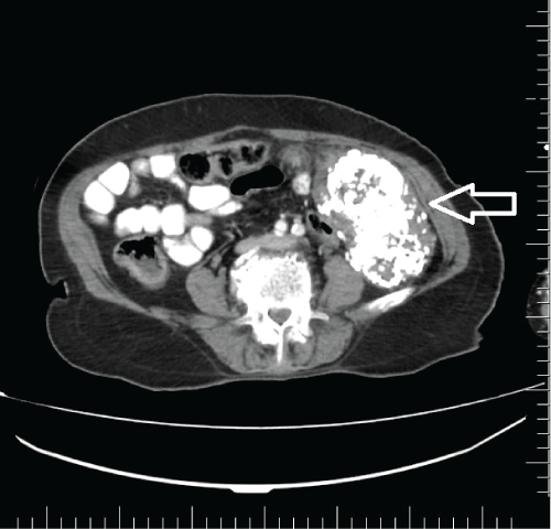
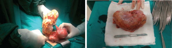
A 72-year-old postmenopausal and nullipar woman presented with abdominopelvic pain and a palpable mass in the right lower quadrant from 3 years. There was no history of surgery or medical illness. Per abdominal examination revealed a mobile hard pelvic mass in the right lower quadrant. Abdominal ultrasound examination showed a 15x10x8cm, hyperechoic, solid mass in the right lower quadrant. X ray of the abdomen showed a radioopaque mass in the same area. Contrast-enhanced computed tomography of the abdomen demonstrated a well-circumscribed mass (Figure 1). Laboratory tests including tumour markers and serum hormonal assays were normal in case. The patient was underwent total abdominal hysterectomy and bilateral salpingo-oophorectomy. On gross inspection, the removed omental parasite vascular supply with pedunculated subserous mass (Figure 2). The mass was described as benign by frozen analysis and histopathological examination confirmed a calcified leiomyoma (Figure 3). The patient was discharged four days after the surgery and on follow-up, there were no further problems noted.
.
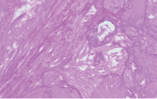
Figure 3: Microscopic evaluation; Calcification and smooth muscle cells (HE stain x 200)
View Figure 3
Discussion
Leiomyomas arise from overgrowth of the smooth muscle and connective tissue of the uterus. Around the menopause, leiomyomas decrease in size because their growth is thought to be estrogen dependent, but leiomyomas may still be newly diagnosed in postmenopausal women.
A calcified pedunculated leiomyoma in a postmenopausal woman is extremely rare; in such cases it is more difficult to predict the clinical symptoms and physical findings. Typical fibroids are easily recognized on imaging, but atypical presentation caused by degenerative changes can cause diagnostic confusion in postmenopausal women [2].
As leiomyomas enlarge, they may outgrow their blood supply, resulting in various types of degeneration: hyaline, myxomatous, calcific, cystic or red degeneration. In general, hyaline degeneration is the most common (63%) form of degeneration, while the others occur less frequently, such as myxomatous changes (13%), calcification (8%), mucoid changes (6%), cystic degeneration (4%), red degeneration (3%), and fatty changes (3%) [3]. The finding of a calcified leiomyoma is more common in postmenopausal woman. In our case, the pedunculated leiomyoma might be thought to be coiled on its uterine pedicle. Over time, the blood supply within the myoma might decrease, and the tissue becomes ischemic. Calcium is deposited in the peripheral portion of the leiomyoma. As the degenerative changes progress, the leiomyoma may become solidly calcified. The leiomyoma may separate completely from the uterus and develop an alternative blood supply from another source, such as omentum and adipose tissue [3]. The finding of a calcified leiomyoma is more common in postmenopausal women and only few cases of postmenopausal severely calcified leiomyomas have been reported in the literature [3-5].
Most uterine leiomyomas may be confidently diagnosed using sonography. However, degenerative changes may create heterogeneous or unusual appearances which contribute to diagnostic confusion. A pedunculated calcified leiomyoma in a postmenopausal woman is difficult to predict the clinical symptoms and physical findings. Pedunculated leiomyomas can have obscure origins and may be mistaken for a lesion of ovarian origin. A sonographic diagnosis of a pedunculated, subserosal leiomyoma can be made if a vascular pedicle is demonstrated. However, these features may not always be detected sonographically [6]. Computed tomography is not the primary modality for diagnosing leiomomas but may facilitate characterization of giant calcified pelvic masses and determine the organ of origin [7].
In the literature, case reports of calcified pedunculated subserous leiomyoma in a postmenopausal woman is rare. Pedunculated leiomyomas with calcified degeneration should be considered in the differential diagnosis of a solid and calcified adnexal mass. CT may help further characterize large pelvic masses and determine their organ of origin.
References
-
Owen C, Armstrong AY (2015) Clinical management of leiomyoma. Obstet Gynecol Clin North Am 42: 67-85.
-
Ciarmela P, Ciavattini A, Giannubilo SR, Lamanna P, Fiorini R, et al. (2014) Management of leiomyomas in perimenopausal women. Maturitas 78: 168-173.
-
Hwang JH, Modi GV, Jeong Oh M, Lee NW, Hur JY, et al. (2010) An unusual presentation of a severely calcified parasitic leiomyoma in a postmenopausal woman. JSLS 14: 299-302.
-
Samal SK, Rathod S, Rani R, Anandraj R (2014) An unusual presentation of a severely calcified subserous leiomyoma in a postmenopausal woman: a case report. Int J Reprod Contracept Obstet Gynecol 3: 463-465.
-
Singh K, Prasad D, Pankaj S, Suman S, Kumar A, et al. (2014) Postmenopausal massive subserous calcified fibroid: a case report. J of Evolution of Med and Dent Sci 3: 2255-2257.
-
Caoili EM, Hertzberg BS, Kliewer MA, DeLong D, Bowie JD (2000) Refractory shadowing from pelvic masses on sonography: a useful diagnostic sign for uterine leiomyomas. AJR Am J Roentgenol 174: 97-101.
-
Rajanna DK, Pandey V, Janardhan S, Datti SN (2013) Broad ligament fibroid mimicking as ovarian tumor on ultrasonography and computed tomography scan. J Clin Imaging Sci 3: 8.
