

Journal of Musculoskeletal Disorders and Treatment
Appropriateness and Suggested Use of MRI in Management of Shoulder Pain
Alan W Reynolds, AB and April D Armstrong, MD, FRCSC*
Department of Orthopaedics and Rehabilitation, Penn State College of Medicine, USA
*Corresponding author:
April D Armstrong, MD, FRCSC, Department of Orthopaedics and Rehabilitation, Penn State College of Medicine, Milton S. Hershey Medical Center, 30 Hope Drive, PO Box 859, Hershey, PA, 17033, USA, Tel: 717-531-5638, E-mail: aarmstrong@pennstatehealth.psu.edu
J Musculoskelet Disord Treat, JMDT-3-028, (Volume 3, Issue 1), Case Report
Received: July 10, 2016 | Accepted: January 20, 2017 | Published: January 24, 2017
Citation: Reynolds AW, Armstrong AD (2017) Appropriateness and Suggested Use of MRI in Management of Shoulder Pain. J Musculoskelet Disord Treat 3:028.
Copyright: © 2017 Reynolds AW, et al. This is an open-access article distributed under the terms of the Creative Commons Attribution License, which permits unrestricted use, distribution, and reproduction in any medium, provided the original author and source are credited.
Abstract
Magnetic resonance imaging (MRI) accounts for a significant proportion of the cost to manage patients with shoulder pain. Improved decision making for MRI use could result in a meaningful reduction in the total number of studies ordered and thereby costs of treating shoulder pain. This study aimed to document MRI ordering patterns for patients with shoulder pain and to propose a protocol to guide efficient management of these patients.
A retrospective study was conducted of all new patient visits for shoulder pain to an academic medical center shoulder and elbow surgery practice between July 2012 and June 2013. A total of 491 patients were included in the study, of which 196 (40%) had an MRI study prior to presenting to the shoulder and elbow clinic. An additional 79 MRI studies were ordered by the clinic as part of patient evaluation and treatment. For all 275 patients who were evaluated with MRI, 98 (36%) did not receive any treatment with injections or physical therapy prior to imaging, and 182 (66%) did not have any form of surgery.
Based on a retrospective application of our proposed management protocol, MRI studies could have been avoided for 76 patients without putting them at risk of additional harm. This would have prevented at least $26,381.88 of wasteful spending over 12 months, for the 491 new patients by following an appropriate progression of evaluation and treatment.
Keywords
Shoulder pain, MRI, Management, Cost, Protocol
Introduction
Shoulder pain is a highly prevalent ailment in the U.S., affecting a self reported 16-34% of the population [1,2], with rotator cuff pathologies alone accounting for more than 4.5 million physician visits per year [3]. The evaluation and treatment of these patients can be very costly, with imaging often accounting for a majority of these costs [4]. Magnetic resonance imaging (MRI), is a very useful diagnostic tool for evaluating shoulder pain and identifying pathology, however, discretion is highly encouraged when considering its use due to its high cost [5,6]. Many patients with shoulder pain have common pathology that can be evaluated and treated effectively based solely on a careful history and physical exam, and without the use of MRI [7]. In these cases, the use of advanced imaging represents an unneeded expense. With current and impending changes in healthcare reimbursement placing more and more emphasis on the delivery of cost-efficient care, a consistent and efficient process to evaluate shoulder pain will be of value to health systems.
The purpose of this study was to evaluate MRI ordering patterns for patients with shoulder pain. The second purpose was to propose a protocol for cost-efficient evaluation and treatment of patients presenting with shoulder pain. It was hypothesized that there is a high rate of unnecessary MRI studies ordered prior to the patient seeing the surgical specialist, representing a significant opportunity for cost savings.
Methods
Data source and inclusion criteria
A retrospective study was conducted of all new patient visits to an orthopedic surgery shoulder and elbow specialty practice for the 12-month period beginning in July 2012 and ending in June 2013. Patients were identified by searching the Cerner Powerchart scheduling system, and all patients presenting with a chief complaint of shoulder pain and 18 years of age or older were included in the study. Patient medical records were examined and the following information was recorded: sex, age at the time of the new patient visit; MRI studies and their findings done within 6 months prior or 12 months following the new patient visit; physical therapy or injections received for treatment of shoulder pain in the 12 months prior to the new patient visit; and any shoulders surgery within 12 months following the new patient visit. The study was granted approval by the Institutional Ethics Review Board.
Exclusion criteria
Aside from patients not meeting the inclusion criteria above, patients with less common diagnoses were also excluded from the study. The rationale for this is that the majority of patient complaints and therefore costs are due to common etiologies, including rotator cuff pathology, glenohumeral arthritis, adhesive capsulitis and non-specific shoulder pain. In addition to this, other, less common pathology is more complex to workup and diagnose and often done by the specialist. A list of diagnoses that were excluded are shown in table 1. In addition, patients presenting to the clinic without a chief complaint of shoulder pain (elbow pain or other upper extremity complaint) were also excluded.
![]()
Table 1: Uncommon diagnoses that were excluded from the study.
View Table 1
Results
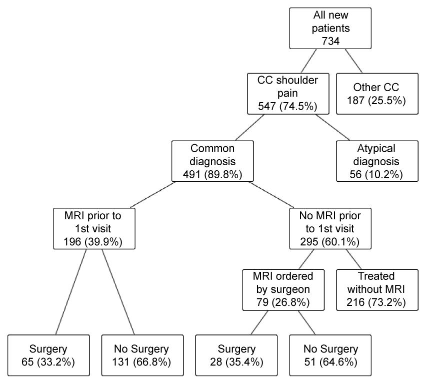
Over the 12-month period, 734 new patients were identified, of which 547 had a chief complaint of shoulder pain and 491 of those had a common shoulder diagnosis and were further evaluated by the study. Common diagnoses were considered to be all types of rotator cuff tear, rotator cuff tendinitis, glenohumeral arthritis, adhesive capsulitis and general shoulder pain. A total of 56 patients with other diagnoses were excluded from the study based on the exclusion criteria.
The use of MRI in evaluating these patients is represented in figure 1. Among all new patients presenting to the Shoulder and Elbow Specialty Program 196 (39.9%) had already been evaluated by MRI. Of the 295 new patients presenting without a prior MRI study, 79 (26.8%) were referred for a new MRI.
.
Figure 1: Distribution of patients by MRI use and surgical outcome.
CC: chief complaint.
View Figure 1
Treatment patterns for new patients who were evaluated with MRI are presented in table 2 by final diagnosis, based on MRI findings, clinical evaluation and, where applicable, surgery. Table 3 depicts specific subgroups among those who had an MRI. Prior treatment was considered to be at least 1 injection or a trial of physical therapy within the 6 months prior to the MRI study. Surgery was documented when performed within the practice and in the 12 months following the initial visit.
![]()
Table 2: Treatment patterns for all patients with MRI by final diagnosis, as determined by MRI or by surgical finding when applicable.
View Table 2
![]()
Table 3: Notable trends in MRI ordering and treatment patterns.
View Table 3
Based on a generalized application of our suggested protocol, 96 out of the 275 MRIs that were ordered and reviewed in this study fell outside of the suggested indications. Out of these 96 patients, 20 eventually underwent surgery. Among the 20 patients who underwent surgery that would not have required an immediate MRI according to our protocol, 6 had a diagnosis of arthritis and 14 had partial thickness rotator cuff tears. The rates for each group within the protocol to undergo surgery are shown in table 4.
![]()
Table 4: Percentage of patients undergoing surgery based on MRI protocol group.
View Table 4
Discussion
The results of this study highlight the opportunity for increased discretion to be used when ordering MRI studies for patients presenting with shoulder pain, as was hypothesized. Although each case requires unique clinical judgment, general trends can help identify patient groups where such discretion could be particularly warranted. One in four patients who had an MRI ordered to evaluate their shoulder pain did not have any formal treatment beforehand and also did not receive surgical treatment (Table 3). This category of patients was mainly composed of those who had a diagnosis of partial thickness rotator cuff tear, rotator cuff tendinitis, benign shoulder pain or arthritis. It is possible that for this subgroup of patients, a well-focused physical exam, steroid injection or trial of physical therapy could greatly reduce the number of patients in this group who require an MRI to be successfully treated.
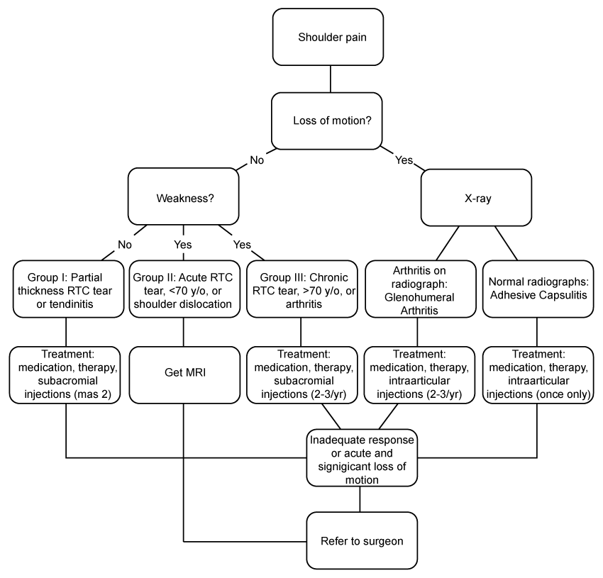
A suggested protocol to evaluate patients presenting with common shoulder pain and when an MRI may be necessary is presented in figure 2. In general, all patients should attempt non-operative treatment prior to having an MRI, with the exception of young patients presenting acutely with rotator cuff weakness, particularly those who have had a shoulder dislocation. This protocol is obviously not exhaustive for all shoulder pathology, such as trauma, but it can be used as an initial starting point to help create more discretion with ordering MRI's for both primary care physicians and the specialist. The authors recognize that in order for this protocol to be further implemented into clinical practice, it would require a collaborative effort between the specialist and the primary care referral physicians, especially to review pertinent clinical exam findings and to make it more exhaustive for the more uncommon shoulder pathologies.
.
Figure 2: Suggested protocol for evaluation and treatment of shoulder pain. Loss of motion refers to passive range of motion.
RTC: rotator cuff.
View Figure 2
Considering the 96 patients who were imaged prematurely as per the protocol, 68 of the studies were ordered by referring physicians (35.0% of the 196 ordered), and 28 were ordered by a shoulder and elbow surgeon (35.5% of the 79 ordered). This illustrates the potential for increased discretion in MRI ordering for surgeons in addition to referring physicians.
Out of these 96 patients who could have been treated without MRI or benefited from a trial of therapy prior to MRI imaging, 20 did eventually undergo surgery, indicating that at least 76 patients may have never required an MRI. These studies can be considered unnecessary since they did not change the management of these patient's conditions, either directly or by influencing their plan of care. All of these patients were diagnosed with pathology that does not require an MRI for diagnosis and treatment, and none of them required advanced imaging for surgical planning. Considering that Medicare reimbursement for an upper extremity joint MRI without contrast is $347.53, at least $26,381.88 can be considered a wasteful expense for new patients presenting to this practice with shoulder pain over one year, as no value was provided by these unnecessary studies [8].
There are several other indirect mechanisms by which unnecessary MRI studies increase the costs of treating patients with shoulder pain, namely that abnormalities are often found in asymptomatic individuals [9,10], and that early advanced imaging can lead to more unnecessary referrals to specialists [11]. In fact, an argument could be made that advanced imaging and possible referral to a surgeon would increase patient anxiety and costs while lowering satisfaction, in the majority of cases where non-operative treatment is indicated [3].
Although this study presents quantitative data on MRI ordering patterns it is important to keep in perspective that it is generalizing some broad guidelines to a subset of patients who visit a shoulder and elbow surgery specialty practice. There are undoubtedly exceptions to our categorizations and certainly some patients who's need for an MRI may not have been accurately captured by chart review. Other reasons for MRI ordering not accounted for in our study include cases where a patient may be adamant that they have the study and where a placebo affect from an MRI study plays a role in treatment. Finally, it is worth noting that many patients presenting with shoulder pain are managed non-operatively without any advanced imaging, and also that many patients who have a clean MRI may never see a surgeon. This represents an abundance of cases that would exemplify well-managed shoulder pain as well as cases of inappropriately ordered MRI studies that were not included in this study. Additionally, patients who elected to have surgery outside of this health system may have been missed by this study. In terms of future study, it would be of value to repeat a similar study following the implementation of our suggested protocol, in order to quantify its effectiveness.
Conclusion
It is clear that there is room for improvement in MRI ordering habits for patients presenting with shoulder pain. There are a number of conditions, including glenohumeral arthritis, where imaging is almost never required, and many other cases where non-operative treatment may resolve shoulder pain without the need for MRI studies. Given the high cost of these studies, this represents an area where costs savings are attainable in conjunction with improving care for patients with shoulder pain. Use of a proposed management protocol could aid in decision making for MRI use and likely reduce the costs of treating shoulder pain.
References
-
Silverstein BA, Viikari-Juntura E, Fan ZJ, Bonauto DK, Bao S, et al. (2006) Natural course of nontraumatic rotator cuff tendinitis and shoulder symptoms in a working population. Scand J Work Environ Health 32: 99-108.
-
Urwin M, Symmons D, Allison T, Brammah T, Busby H, et al. (1998) Estimating the burden of musculoskeletal disorders in the community: the comparative prevalence of symptoms at different anatomical sites, and the relation to social deprivation. Ann Rheum Dis 57: 649-655.
-
Oh LS, Wolf BR, Hall MP, Levy BA, Marx RG (2007) Indications for rotator cuff repair: a systematic review. Clin Ortho Relat Res 455: 52-63.
-
Yeranosian MG, Terrell RD, Wang JC, McAllister DR, Petrigliano FA (2013) The costs associated with the evaluation of rotator cuff tears before surgical repair. J Shoulder Elbow Surg 22: 1662-1666.
-
Lenza M, Buchbinder R, Takwoingi Y, Johnston RV, Hanchard NC, et al. (2013) Magnetic resonance imaging, magnetic resonance arthrography and ultrasonography for assessing rotator cuff tears in people with shoulder pain for whom surgery is being considered. Cochrane Database Syst Rev CD009020.
-
Nazarian LN, Jacobson JA, Benson CB, Bancroft LW, Bedi A, et al. (2013) Imaging algorithms for evaluating suspected rotator cuff disease: Society of Radiologists in Ultrasound consensus conference statement. Radiology 267: 589-595.
-
Mitchell C, Adebajo A, Hay E, Carr A (2005) Shoulder pain: diagnosis and management in primary care. BMJ 331: 1124-1128.
-
Virta L, Joranger P, Brox JI, Eriksson R (2012) Costs of shoulder pain and resource use in primary health care: a cost-of-illness study in Sweden. BMC Musculoskelet Disord 13: 17.
-
Gill TK, Shanahan EM, Allison D, Alcorn D, Hill CL (2014) Prevalence of abnormalities on shoulder MRI in symptomatic and asymptomatic older adults. Int J Rheum Dis 17: 863-871.
-
Sher JS, Uribe JW, Posada A, Murphy BJ, Zlatkin MB (1995) Abnormal findings on magnetic resonance images of asymptomatic shoulders. J Bone Joint Surg Am 77: 10-15.
-
Ostör AJ, Richards CA, Prevost AT, Speed CA, Hazleman BL (2005) Diagnosis and relation to general health of shoulder disorders presenting to primary care. Rheumatology (Oxford) 44: 800-805.
