

International Journal of Surgery Research and Practice
Fibrous Dysplasia of the Inferior Turbinate
Huseyin Baki Yilmaz1, Sevtap Akbulut1*, Mustafa Paksoy1, Arif Sanli1 and Kayhan Basak2
1Department of Otolaryngology Head and Neck Surgery, Dr. Lutfi Kirdar Kartal Education and Research Hospital, Turkey
2Department of Pathology, Dr. Lutfi Kirdar Kartal Education and Research Hospital, Turkey
*Corresponding author: Sevtap Akbulut, Department of Otolaryngology, Dr.Lutfi Kirdar Training and Research Hospital, Semsi Denizer Cad. E-5 Karayolu Cevizli Mevkii, Kartal, 34890 Istanbul, Turkey, Tel: +90216 458 30 00, E-mail: sevtap.akbulut@gmail.com
Int J Surg Res Pract, IJSRP-1-016, (Volume 1, Issue 1), Case Report; ISSN: 2378-3397
Received: October 14, 2014 | Accepted: November 25, 2014 | Published: November 30, 2014
Citation: Yilmaz HB, Akbulut S, Paksoy M, Sanli A, Basak K (2014) Fibrous Dysplasia of the Inferior Turbinate. Int J Surg Res Pract 1:016. 10.23937/2378-3397/1410016
Copyright: © 2014 Yilmaz HB, et al. This is an open-access article distributed under the terms of the Creative Commons Attribution License, which permits unrestricted use, distribution, and reproduction in any medium, provided the original author and source are credited.
Abstract
Fibrous dysplasia is a developmental abnormality of the skeletal system. Craniofacial involvement has been reported in about 10% -27% of the cases with monostotic disease. Involvement of the sinonasal tract has very rarely been reported. In this report, a 14 year old boy that has been treated for 6 months for acute sinusitis and allergic rhinitis is reported. Careful nasal endoscopic examination and obtaining a CT scan led to a diagnosis of unilateral fibrous dysplasia of the inferior turbinate.
Keywords
Fibrous dysplasia, Inferior turbinate, Nasal obstruction, Rhinorrhea
Introduction
Fibrous dysplasia (FD) is a developmental abnormality of bone. It is a rare benign disease that is characterized by replacement of the healthy bony tissue by fibrotic tissue and trabecular bone like a network [1].
Involvement of the inferior turbinate as a single bone is extremely uncommon and only three similar cases have been reported in the literature [2]. Here we present a pediatric patient with monostotic FD of the inferior turbinate.
Case Report
A 14-year old boy was referred to our outpatient clinic with the complaint of unilateral rhinorrhea and nasal obstruction that started a year ago. Nasal endoscopic examination showed mucoid discharge in the left inferior meatus and a hypertrophic left inferior turbinate that was firm with palpation with a curette (Figure 1).
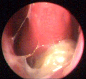 Figure 1: Preoperative endoscopic examination revealed inferior turbinate
hypertrophy firm with palpation.
View Figure 1
Figure 1: Preoperative endoscopic examination revealed inferior turbinate
hypertrophy firm with palpation.
View Figure 1
There was little amount of serous discharge and mild deviation in the right nasal cavity. On paranasal sinus tomography, left inferior turbinate and a small part of the medial wall of the left maxillary sinus had a ground glass appearance with normal mucosa. The diagnosis was FD of the inferior turbinate (Figure2a,2b).
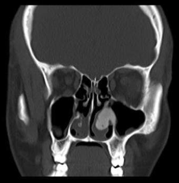
Figure 2a: Coronal CT scan of the patient. Left inferior turbinate and a
small part of the medial wall of the left maxillary sinus have a ground glass
appearance with normal mucosa.
View Figure 2a
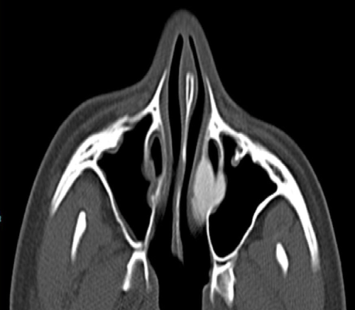
Figure 2b: Figure 2B: Axial CT scan of the patient.
View Figure 2b
Serum alkaline phosphatase levels and urine analysis were normal. The mass was removed en-bloc leaving the nasal mucosa overlying the inferior turbinate intact. Macroscopically, the mass was solid, whitish-grayish in color (Figure 3). On histopathological examination, the mass composed of woven bone formation in a fibrous stroma without osteoblastic cells and immature bony trabeculae (Figure 4).
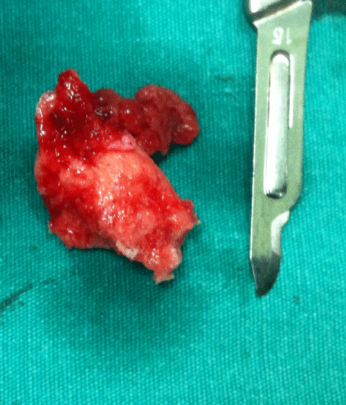 Figure 3: Figure 3: Macroscopically the mass is solid and white-grayish in color.
View Figure 3
Figure 3: Figure 3: Macroscopically the mass is solid and white-grayish in color.
View Figure 3
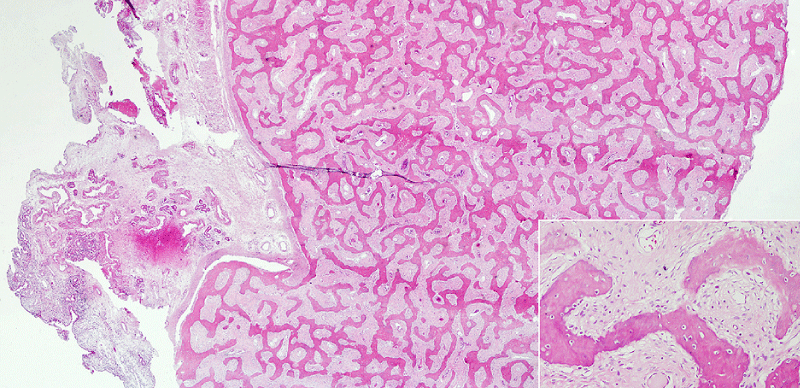 Figure 4: Histopathologic appearance of the lesion; woven bone formation
in a fibrous stroma without osteoblastic cells and immature bony trabeculae
(H&E, x40 ve x200).
View Figure 4
Figure 4: Histopathologic appearance of the lesion; woven bone formation
in a fibrous stroma without osteoblastic cells and immature bony trabeculae
(H&E, x40 ve x200).
View Figure 4
Patient was free of symptoms and no recurrence was observed at postoperative 6th month (Figure 5). Since malignant transformation has been reported for FD, the patient was instructed to be under follow-up regularly.
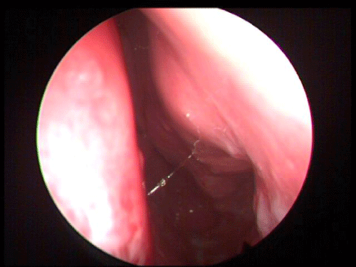
Figure 5: Endoscopic examination after endoscopic resection.
View Figure 5
Discussion
FD has been shown in the rib of a young Neandertal who lived about 120,000 years ago, in what is now present-day Krapina, Croatia [3]. FD evidently is a disease that has been present for thousands of years. The incidence is between 1/4000-1/10000. It accounts for approximately 2.5% of all bone tumors and 7% of benign bone tumors [4].
FD can be classified into 4 types; Monostotic type with a single bone involvement (70%), polyostotic type with multiple bones involvement (30%), McCune Albright syndrome (polyostotic FD, Caf� au lait pigmentations and endocrine pathologies) and Mazabraud syndrome (FD and soft tissue myxomas). Approximately 50% of patients with polyostotic disease and 10% -27% with monostotic disease have craniofacial involvement [5]. Craniofacial FD typically presents at around 10 years of age and then progresses throughout adolescence [3].
To our knowledge only three cases with isolated fibrous dysplasia of the inferior turbinate has previously been reported [2]. Of these cases, 2 were adults, and one was a child with acute leukemia. Our patient was an otherwise healthy 14-year old child with no symptoms other than one sided nasal obstruction and unilateral rhinorrhea for a year. He underwent many physical exams including �the gold standard� nasal endoscopy. He used various medications with different diagnostics for 6 months, but the definite pathology was not identified until he had a computerized tomography of his paranasal sinuses.
The CT appearance of fibrous dysplasia is pathognomonic. In the early stages of the disease the bony matrix is radiolucent, as the disease progresses it gets more opaque [5]. The bony cortex is thin with regular margins. Differential diagnosis includes simple bony cysts, multiple enchondromatosis, hemangiomas, vascular malformations, eosinophilic granulomas, Paget�s disease, osteoblastic metastasis and neurogenic tumors (schwannomas, neurofibromatosis and traumatic neuroma) [6].
Management of fibrous dysplasia depends on where the lesions are located. Asymptomatic lesions that do not cause functional problems can be observed [7]. In our patient, since the lesion was confined to the nasal cavity we chose to make an endoscopic resection preserving the overlying mucosa. Malignant transformation has been reported in about 1% of the cases with FD, most of which occurred after radiation therapy [8].
Conclusion
FD must be considered in the differential diagnosis of unilateral nasal obstruction in pediatric patients, especially those over 10 years of age. In a pediatric patient with unilateral nasal obstruction and rhinorrhea, the otolaryngologist should perform an endoscopic examination. If there is unilateral turbinate hypertrophy, it is mandatory to feel the texture of the turbinate with an instrument in order to make a distinction between bony and mucosal hypertrophy. If bony hypertrophy is strongly suspected, it is of real importance to order a CT (computerized tomography) of the paranasal sinuses in order to rule out FD. Early detection of this disease in the nasal cavity will enable the otolaryngologist to remove the pathology easily with an endoscopic approach. However, it is mandatory to keep in mind that since FD has been reported to have malignant transformation, pediatric patients with this diagnosis must have repeated nasal endoscopic exams and long term radiologic follow up.
References
-
Lietman SA, Levine MA (2013) Fibrous dysplasia. Pediatr Endocrinol Rev 10 Suppl 2: 389-396.
-
Park HJ, Cho MS, Lee SS (2013) Fibrous dysplasia of the inferior turbinate. Int J Clin Exp Pathol 6: 531-535.
-
Monge J, Kricun M, Radovcic J, Radovcic D, Mann A, et al. (2013) Fibrous dysplasia in a 120,000+ year old Neandertal from Krapina, Croatia. PLoS One 8: e64539.
-
Assaf AT, Benecke AW, Riecke B, Zustin J, Fuhrmann AW, et al. (2012) Craniofacial fibrous dysplasia (CFD) of the maxilla in an 11-year old boy: a case report. J Craniomaxillofac Surg 40: 788-792.
-
Menon S, Venkatswamy S, Ramu V, Banu K, Ehtaih S, et al. (2013) Craniofacial fibrous dysplasia: Surgery and literature review. Ann Maxillofac Surg 3: 66-71
-
Stathopoulos IP, Balanika AP, Baltas CS, Lampropoulou-Adamidou K, Koromila T, et al. (2013) Fibrous dysplasia; confirmation of clinical diagnosis by DNA tests instead of biopsy. J Musculoskelet Neuronal Interact 13: 120-123.
-
Kruse A, Pieles U, Riener MO, Zunker Ch, Bredell MG, et al. (2009) Craniomaxillofacial fibrous dysplasia: a 10-year database 1996-2006. Br J Oral Maxillofac Surg 47: 302-305.
-
Van Rossem C, Pauwels P, Somville J, Camerlinck M, Bogaerts P, et al. (2013) Sarcomatous degeneration in fibrous dysplasia of the rib cage. Ann Thorac Surg 96: e89-90.
