

International Journal of Ophthalmology and Clinical Research
Optic Disk Size Assessment Techniques: Photo Essay
Sourabh Arora1, Helen Chung2 and Karim Damji1*
1Department of Ophthalmology, University of Alberta, Canada
2Division of Ophthalmology, University of Calgary, Canada
*Corresponding author: Dr. Karim Damji, Royal Alexandra Hospital, 2317, 10240, Kingsway Avenue, Edmonton, Alberta, T5H 3V9, Canada, Tel: (780) 735-4200, Fax: (780) 735-5242, E-mail: kdamji@ualberta.ca
Int J Ophthalmol Clin Res, IJOCR-2-009, (Volume 2, Issue 1), Case Report; ISSN: 2378-346X
Received: October 14, 2014 | Accepted: January 20, 2015 | Published: January 25, 2015
Citation: Arora S, Chung H, Damji K (2015) Optic Disk Size Assessment Techniques: Photo Essay. Int J Ophthalmol Clin Res 2:009. 10.23937/2378-346X/1410009
Copyright: © 2015 Arora S, et al. This is an open-access article distributed under the terms of the Creative Commons Attribution License, which permits unrestricted use, distribution, and reproduction in any medium, provided the original author and source are credited.
Abstract
Measuring optic disc size is an essential part of the optic nerve head for evaluation for glaucoma. Large discs that appearing to have large cups may in fact be physiologic because of normal neuroretinal rim area. Small discs may appear to have a normal cup to disc ratio, but have decreased neuroretinal rim area in association with glaucoma.We illustrate relevance of these points with respect to glaucoma diagnosis in two case examples.We then present clinically relevant techniques to assess disc size including direct ophthalmoscopy, slit lamp biomicroscopy, disc macula: disc diameter ratio, planimetric assessment of fundus photographs, and imaging modalities such as OCT as well as HRT.
Keywords
Disk size, Glaucoma, Cup-disk ratio, Neuroretinal rim
Introduction
Measuring optic disk size is an essential part of the optic nerve head for evaluation for glaucoma. The average human optic disc dimensions are 1.88 mm vertically by 1.77 mm horizontally, and can be grouped by vertical diameter into small (1.2-1.7 mm), average (1.87-1.96 mm), and large (2.03-2.27 mm) disc sizes [1]. This classification is clinically significant because a large disc can have a physiologically large cup with a healthy neuroretinal rim, and needs to be distinguished from a glaucomatous nerve with a large cup and thinning of neuroretinal rim tissue. Likewise, pathology may be missed in small discs, where a seemingly 'normal' cup-disk ratio (CDR) can still indicate glaucomatous damage. We present clinically relevant techniques to assess disc size and illustrate relevance with respect to glaucoma in two cases examples (small and large discs).
Case 1
A 37 year old female with elevated IOP was referred by an optometrist for assessment of glaucoma. The patient had no visual complaints or past ocular history. She was otherwise healthy, and taking no medications. There was no family history of glaucoma. On examination, her visual acuity was 20/20 OU, and intra-ocular pressure was 22 OU measured by Goldman applanation. Angles were open bilaterally. Stereoscopic fundus imaging revealed that both optic nerves were smaller than average with small a cup (vertical CDR 0.35 OU) (Figure 1). This patient had abnormal visual fields bilaterally, with MD -9.96 OD and PSD 8.21 OD, while MD was -14.04 OS, and PSD 7.22 OS. There had been progression since the last HVF completed 16 months earlier (MD -4.72 OD, PSD 7.14 OD, MD -6.35 OS, PSD 5.74 OS).
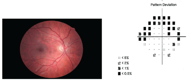
Figure 1: Disk photos for small disk with accompanying visual fields. (A) Right disk photo showing vertical cup to disk ratio 0.35 with health
appearing neuroretinal rim (B) Right visual field showing superior arcuatescotoma.
View Figure 1
Case 2
A second patient, a 25 year old female, was referred for assessment of glaucoma. She had no visual concerns and no prior ocular history. She was otherwise healthy, and taking no medications. There was no family history of glaucoma. On examination, her visual acuity was 20/20 OU, and intra-ocular pressure was 15 OU measured by Goldman applanation. As seen with a 60D lens, her optic nerves were larger than average (2.2 mm) (Figure 2) with a large cup (vertical CDR 0.75 OU) (Figure 3). According to HRT, the linear cup-disk ratio was elevated at 0.69 (0.27-0.55), and her cup/disk area ratio was also elevated 0.47 (0.07�0.3). Mean RNFL thickness was decreased to 0.15 mm OD while it was borderline normal at 0.20 mm OS (normal 0.20-0.32).
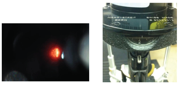 Figure 2: (A) Slit lamp photo of optic disk as seen through 60D lens, and (B) Slit lamp reticule used to measure disk size.
View Figure 2
Figure 2: (A) Slit lamp photo of optic disk as seen through 60D lens, and (B) Slit lamp reticule used to measure disk size.
View Figure 2
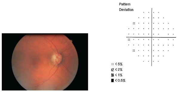 Figure 3: Disk photos for large disk with accompanying visual fields. (A) Right disk photo, vertical cup-disk ratio 0.75 with healthy
neuroretinal rim (B) Right visual field appears normal.
View Figure 3
Figure 3: Disk photos for large disk with accompanying visual fields. (A) Right disk photo, vertical cup-disk ratio 0.75 with healthy
neuroretinal rim (B) Right visual field appears normal.
View Figure 3
Discussion and Literature Review
Case 1 demonstrated an important principle about smaller disks requiring a more thorough assessment. The patient had a seemingly normal neuroretinal rim area and CDR, however was experiencing glaucomatous visual field loss. Case 2 on the other hand had a larger disk, with an elevated CDR. However this was diagnosed as physiological cupping. This was confirmed by the normal visual field results.
There are many techniques for clinically measuring disc size. Drance and Gross describe operating the 5 degree light spot of a direct ophthalmoscope which is 1.5 mm when projected over the disc. To determine which size of light spot is appropriate, one may stand 1m from the wall and the spot will have a diameter of 85 - 95 mm. When this spot size is projected over the optic nerve, if the disc fits on that target, the disc is normal. If the disc is smaller than that target, the disc is small. And if the disc is larger than the target, it is a large disc [2]. This technique works well for eyes that are within -5D to +4D of refractive error. A slit lamp can also be used with various high-powered lenses, where one adjusts a vertical beam of light onto the disc and records the size. Correction factors for a 60D, 78D, and 90D lens is 1�, 1.1� and 1.3�, respectively [3]. It appears that the distance from the center of the fovea to the center of the disc is a fixed value in most eyes (3840um). Disk macula to Disk diameter ratio (DM: DD) decreases with macrodiscs and increases in optic nerve head hypoplasia.
More advanced imaging technology such as optic disc by optical coherence tomography (OCT) and Heidelberg Retina Tomograph (HRT) are often used and yield more specific measurements. Correcting for disc size also improves the sensitivity of imaging techniques such as the (HRT) in identifying abnormal discs [4]. However, occasionally there are errors in the automated marking of disc margins and so clinical examination of the optic nerve as another check is warranted. Scleral ring identification is an essential component to assessing disk size for observers and imaging modalities. The observer should be aware of pigmentary and atrophic changes, as well as tilting of the disk that can make it difficult to delineate these margins, and hence further compound the difficulty in assessing for glaucoma. In the future, basement membrane opening may be another useful parameter.
Diagnostic scales such as the Disc Damage Likelihood Scale (DDLS) [5] and the Optic Disc Damage Staging System (ODDSS) [6] may be useful in glaucoma management and consider the disk size in evaluating for glaucoma risk and stage. When we apply the DDLS to case 1, the DDLS stage is 2 (small disc < 1.50mm and narrowest rim/disc ratio=0.25), indicating �possible damage�. For case 2, the DDLS stage is 0b (large disc >2.00mm and narrowest rim/disc ratio=0.2) indicating�probably no damage�.
Large disks appearing to have cupping may be considered physiologic because of normal neuroretinal rim area. Small disks may appear to have a normal CDR, but have decreased neuroretinal rim area. Common techniques for estimating disk size include direct ophthalmoscopy, slit lamp microscopy, consideration of disk macula:disk diameter ratio, planimetric technique with the fundus photograph, fundus imaging modalities such as OCT and HRT. Neuroretinal rim area, focal changes and adjacent RNFL should also be considered in assessing the optic nerve head. Accurate identification of the disk margin is essential for observers and imaging devices.
References
-
Quigley HA, Brown AE, Morrison JD, Drance SM (1990) The size and shape of the optic disc in normal human eyes. Arch Ophthalmol 108: 51-57.
-
Gross PG, Drance SM (1995) Comparison of a simple ophthalmoscopic and planimetric measurement of glaucomatous neuroretinal rim areas. J Glaucoma 4: 314-316.
-
Ansari-Shahrezaei S, Maar N, Biowski R, Stur M (2001) Biomicroscopic measurement of the optic disc with a high-power positive lens. Invest Ophthalmol Vis Sci 42: 153-157.
-
Mardin CY, Horn FK (1998) Influence of optic disc size on the sensitivity of the Heidelberg Retina Tomograph. Graefes Arch Clin Exp Ophthalmol 236: 641-645.
-
Spaeth GL, Henderer J, Liu C, Kesen M, Altangerel U, et al. (2002) The disc damage likelihood scale: reproducibility of a new method of estimating the amount of optic nerve damage caused by glaucoma. Trans Am Ophthalmol Soc 100: 181-185.
-
Brusini P, Zeppieri M, Tosoni C, Parisi L, Salvetat ML (2010) Optic disc damage staging system. J Glaucoma 19: 442-449.
