International Journal of Ophthalmology and Clinical Research
Macular Hole Formation Following Choroidal Neovascularization Treatment in A High Myopic Eye with Epiretinal Membrane
Yusaku Yoshida*,Manabu Yamamoto, Takeya Kohno and Kunihiko Shiraki
Department of Ophthalmology and Visual Science, Osaka City University Graduate School of Medicine, Japan
*Corresponding author: Yusaku Yoshida, MD, Department of Ophthalmology and Visual Science, Osaka City University Graduate School of Medicine, 1-4-3 Asahi-machi, Abeno-ku, Osaka 545-8585, Japan, Fax:+81-666-343-873, E-mail: m1158667@med.osaka-cu.ac.jp
Int J Ophthalmol Clin Res, IJOCR-2-008, (Volume 2, Issue 1), Case Report; ISSN: 2378-346X
Received: December 19, 2014 | Accepted: January 18, 2015 | Published: January 21, 2015
Citation: Yoshida Y, Yamamoto M, Kohno T, Shiraki K (2015) Macular Hole Formation Following Choroidal Neovascularization Treatment in A High Myopic Eye with Epiretinal Membrane. Int J Ophthalmol Clin Res 2:008. 10.23937/2378-346X/1410008
Copyright: ©2015 Yoshida Y, et al. This is an open-access article distributed under the terms of the Creative Commons Attribution License, which permits unrestricted use, distribution, and reproduction in any medium, provided the original author and source are credited.
Abstract
Purpose: To report on the development of macular hole after treatment of choroidal neovascularization (CNV) in a patient with high myopia and epiretinal membrane (ERM).
Methods: Retrospective single case report.
Patients: A 60-year-old man presented with reduced vision in his right eye. On the initial examination, the right eye was high myopic with decimal best-corrected visual acuity (BCVA) of 0.3.
Results: A subretinal grayish-white lesion was seen at the macula with hemorrhage, and optical coherence tomography showed a poorly demarcated, solid subretinal lesion and ERM in the central fovea. Fluorescein fundus angiography revealed classic CNV. The CNV was treated with intravitreal bevacizumab injection and photodynamic therapy (PDT). Three months after the treatment, the CNV had contracted but BCVA remained at 0.2. Since an exudative change persisted, additional treatment with intravitreal ranibizumab injection (IVR) and PDT was performed. Three months after the second treatment, BCVA was 0.4 and the exudative changes had disappeared. However, a macular hole was observed.
Conclusion: The contraction of the CNV appeared to have ripped apart areas with and without ERM, which led to the development of the macular hole. During the course of CNV regression, a cautious approach is necessary in patients with high myopic eyes with ERM, as macular holes may develop.
Keywords
Choroidal neovascularization; Epiretinal membrane; High myopia; Macular hole
Introduction
Macular Hole (MH) is one of several sight-threatening complications such as chorioretinal atrophy (CRA), subretinal hemorrhage (SRH), choroidal neovascularization (CNV), epiretinal membrane (ERM), and foveoschisis that can occur in high myopic eyes. However, it has been reported that macular hole could occur irrespective of high myopia in several other diseases with CNV such as age-related macular degeneration (AMD) [1-5]. We report a case of macular hole development where post-treatment regression of CNV seemed to have played a major role in a high myopic eye with ERM.
Case
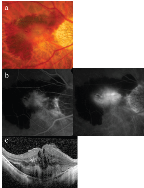
A 60-year-old man was examined in the Department of Ophthalmology of Osaka City University Hospital for reduced vision in his right eye in January 2009. On initial examination, decimal best-corrected visual acuity (BCVA) was 0.3 in the right eye and 1.5 in the left eye, while intraocular pressures were 13 and 14 mmHg in the right and left eye, respectively. His previous medical and family histories included nothing of note. Fundus examination showed a subretinal grayish-white lesion with surrounding SRH in the right macula, and optical coherence tomography (OCT) showed a poorly demarcated, subretinal lesion and cystic macular edema in addition to ERM at the temporal side of the fovea (Figure 1). Fluorescein fundus angiography (FA) revealed a classic CNV, with profound dye leakage from the early phase. After diagnosis of CNV in the high myopic eye, a combination therapy using intravitreal bevacizumab (IVB) injection and photodynamic therapy (PDT) was performed.

Figure 1: (a) A color fundus photograph showed a subretinal, grayishwhite
lesion and subretinal hemorrhage in the right macula. (b) The early
(45 seconds) and late (10 minutes) fluorescein angiograms showed classic
CNV, with profound dye leakage. (c) Optical coherence tomography (OCT)
showed a poorly demarcated, solid subretinal lesion. An epiretinal membrane
was seen just temporal to the foveal depression with cystic macular edema.
View Figure 1
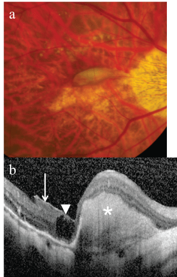
Three months after the treatment, the right BCVA remained at 0.2, the SRH had almost completely resolved. OCT showed a subfoveal, well-demarcated mound extending nasally, indicating a fibrous tissue after the CNV regression. In addition, the OCT showed an intraretinal cyst just temporal to the fibrous mound with concomitant ERM temporal to the cystic lesion (Figure 2). Since the cystic edema and slight subretinal hemorrhage persisted, an additional treatment with an intravitreal ranibizumab (IVR) injection and PDT was performed.
 Figure 2: (a) A color fundus photograph showed a localized, subfoveal,
fibrous mound extending nasally. Hard exudates and slight subretinal
hemorrhage were present. (b) OCT showed a sub- and juxtafoveal, welldemarcated
mound (*) extending nasally and an intraretinal cyst (arrowhead)
just temporal to the fibrous mound with concomitant ERM (arrow) temporal
to the cystic lesion.
View Figure 2
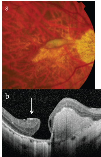
Three months after the additional treatment, exudative changes such as hard exudates and subretinal hemorrhage had disappeared and the right BCVA was 0.4. However, MH developed at the temporal side of the subretinal fibrous tissue. While ERM was still noted at the edge of the temporal side of the MH, it was not found at the nasal side (Figure 3).
 Figure 3: (a) A color fundus photograph showed a similar subretinal, fibrous
mound. (b) OCT showed a macular hole located at the right temporal side
of the fibrous mound. ERM (arrow) was seen at the edge of the temporal
side of the macular hole.
View Figure 3
Discussion
With respect to the underlying mechanism of MH, Gass et al. [6] reported that contraction of the vitreous imposed traction tangentially to the retina, while Gaudric et al. [7] stated diagonal traction due to a combination of the anterior-posterior and tangential traction was involved. Shimada et al. investigated macular hole and foveoschisis in 181 high myopic eyes [1]. They found that the retina around the CNV was extremely thin within a CRA lesion, particularly at the edge of the CNV that occurred within an area of more than 1 disc diameter of the CRA. Moreover, they found that there was strong adhesion between the CNV and retina as a result of the CNV fibrovascular membrane. They hypothesized that contraction of the CNV membrane during its regression might impose traction on the extremely thinned retina of the CRA and cause tearing of the retina to produce MH at the edge of the CNV area.
There have been other studies that have examined the development of MH after treatment for CNV with high myopia or AMD [2-5]. Mechanisms responsible for the development of MH include incarceration of the vitreous at the injection site of intravitreal anti-VEGF agents, which results in vitreoretinal traction. In addition, the effect of the anti-VEGF agents themselves can cause contraction of the CNV and intensify the tangential traction on the retina. Moreover, PDT can cause choroidal swelling leading to dehiscence of the fovea, the tangential traction, cystic macular edema with coalescence of cysts.
In the case reported here, ERM was present over the temporal side of the CNV, and cystic macular edema was also evident in the fovea. It was likely that the treatment such as intravitreal anti-VEGF agents and PDT caused the CNV contraction on the nasal side of the fovea, which led to the formation of MH at the temporal side of the CNV.
As to the underlying mechanism, it is possible that contraction of the CNV generated tangential traction on the retina. During the contraction, the surrounding retina adhering to the CNV was pulled and shifted along with the CNV. Thus, the temporal side of the fovea was pulled toward the nasal side. However, since the retina temporal to the fovea and under the ERM was of poor extensibility, it stayed in place, thereby tearing a hole at the fragile site of the cystic edema of the macula. Since the CRA was localized but not extensive around the area of the MH in this patient, this suggests that thinning of the retina caused by the CRA may not have had a major contribution to the formation of the MH. As in previous reports, while the traction due to the contraction of CNV contributed to MH development, local differences in the extensibility of the retina due to the presence of ERM and cystic edema of the macula may have been an additional causative factor.
This case suggests that MH formation should be considered as a potential complication when treating CNV in an eye with ERM.
References
-
Shimada N, Ohno-Matsui K, Yoshida T, Futagami S, Tokoro T, et al. (2008) Development of macular hole and macular retinoschisis in eyes with myopic choroidal neovascularization. Am J Ophthalmol 145: 155-161.
-
Moisseiev E, Goldstein M, Loewenstein A, Moisseiev J (2010) Macular hole following intravitreal bevacizumab injection in choroidal neovascularization caused by age-related macular degeneration. Case Rep Ophthalmol 1: 36-41.
-
Rishi P, Kasinathan N, Sahu C (2011) Foveal atrophy and macular hole formation following intravitrealranibizumab with/without photodynamic therapy for choroidal neovascularization secondary to age-related macular degeneration. Clin Ophthalmol 5: 167-170.
-
Querques G, Souied EH, Soubrane G (2009) Macular hole following intravitreal ranibizumab injection for choroidalneovascular membrane caused by age-related macular degeneration. Acta Ophthalmol 87: 235-237.
-
Cho JH, Park SE, Han JR, Kim HK, Nam WH (2011) Macular hole after intravitreal ranibizumab injection for polypoidal choroidal vasculopathy. Clin Exp Optom 94: 586-588.
-
Gass JD (1988) Idiopathic senile macular hole. Its early stages and pathogenesis. Arch Ophthalmol 106: 629-639.
-
Gaudric A, Haouchine B, Massin P, Paques M, Blain P, et al. (1999) Macular hole formation: new data provided by optical coherence tomography. Arch Ophthalmol 117: 744-751.


