The novel coronavirus (SARS-CoV-2) pandemic continues to surge across the globe with no signs of slowing down. SARS-CoV-2 infections (Covid-19) affect multiple organ systems with varied clinical presentation. Common clinical respiratory signs and symptoms associated with Covid-19 include fever, cough and shortness of breath. However, clinicians should be aware that cutaneous rashes could be the only clinical sign and symptom. Recognition of cutaneous rashes without other symptoms should be considered as part of the clinical presentation of Covid-19 infection and could easily be overlooked. Prompt recognition could lead to early clinical testing for the coronavirus and appropriate management to mitigate community spread of the virus.
The primary target organ of the severe acute respiratory syndrome coronavirus (SARS-CoV-2) and the most commonly reported symptoms reported by patients who are infected with SARS-CoV-2 involve the respiratory tract. Such clinical symptoms include coughing, sneezing, shortness of breath and congestion [1-3]. SARS-CoV-2 is a multisystem disorder that can affect the cardiovascular, gastrointestinal, neurologic and renal systems [1,4-6].
Reports of isolated cutaneous lesions have recently been reported, such as erythematous rash, confluent urticarial wheals, purpuric rash, chickenpox-like vesicles, petechiae rash, transient livedo reticularis and red papules on fingers [7-9]. Such dermatologic manifestations could be the only initial presenting sign and symptom and should be considered as part of the clinical presentation in the diagnosis of SARS-CoV-2 [8,10]. This case report describes a patient who presented with a cutaneous rash on the right lower extremity without other symptoms three weeks prior to hospitalization for management of severe Covid-19. The patient denied experiencing any fever, cough, shortness of breath or other clinical symptoms associated with Covid-19. The patient presumed that the rash was due to a fungal infection from cutting his toenails and then immediately scratching his right leg. At the time of initial presentation, the patient was not tested for the coronavirus, but tested positive at the time of hospital admission.
A 61-year-old Asian male presented with a rash on the right lower extremity (Figure 1A and Figure 1B). He states that the rash developed about five day ago and is intensely itchy. Based on the clinical presentation, urticarial vasculitis was suspected. Medical history reported by the patient included hypertension, and hypercholesterolemia. In 2014, the patient was treated for a bilateral pulmonary embolism. The patient reported that he was prescribed the following medications: Metoprolol 100 mg three times per day, atorvastatin 40 mg daily and Apixaban 5 mg daily.
 Figure 1: (A) Confluent macular rash of right lower extremity; (B) Note the edematous and erythematous wheals commonly present with intense pruritis. Rash was only symptom reported by the patient.
View Figure 1
Figure 1: (A) Confluent macular rash of right lower extremity; (B) Note the edematous and erythematous wheals commonly present with intense pruritis. Rash was only symptom reported by the patient.
View Figure 1
The patient believes that the leg rash may have developed from fingernail scratching as the rash was very itchy. Antifungal ointment was applied to the area of the rash twice per day for a period of 10 days. After two weeks, there was complete resolution of the rash. On September 15, the patient began to experience coughing and development of a fever two days later. On September 18, the patient complained of dyspnea and chest pain. He presented to the emergency room that afternoon and was admitted to the hospital for management of a suspected pulmonary embolus. The patient tested positive for the coronavirus during the hospital admission. On Computed Tomography (CT) pulmonary angiography of the chest after administration of intravenous contrast material, multifocal consolidative and ground glass opacities of the bilateral lung were present consistent with Covid-19 pneumonia.
Treatment for severe Covid-19 infection consisted of supportive care, intravenous remdesivir 200 mg on day one, followed by 100 mg for the next four days, intravenous dexamethasone 6 mg for five days, and convalescent plasma on two consecutive days. Anticoagulation management consisted of daily subcutaneous injection of enoxaparin 40 mg. On October 05, the patient was discharged from the hospital. Eight days after discharge from the hospital, he experienced intense pruritis in the area below the stomach (Figure 2). The skin became erythematous with development of a purpuric rash characterized by petechiae and erythematous purpuric papules. The rash was treated with prednisone 20 mg for 5 days. The skin rash resolved but continues to sporadically recur.
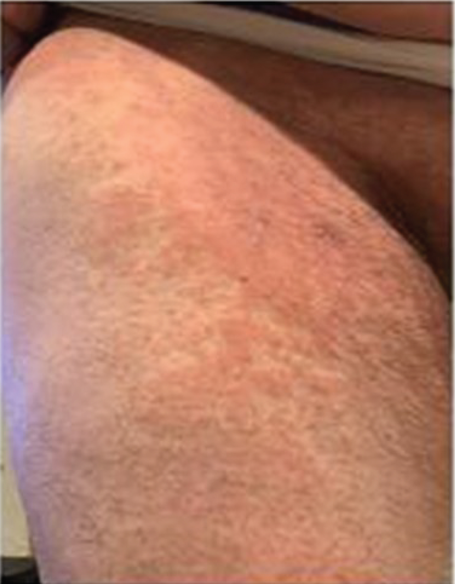 Figure 2: Erythematous-purpuric papular rash on right thigh of patient after patient diagnosed with Covid-19.
View Figure 2
Figure 2: Erythematous-purpuric papular rash on right thigh of patient after patient diagnosed with Covid-19.
View Figure 2
As Angiotensin-Converting Enzyme 2 (ACE2) is the primary functional host receptor for SARS-CoV-2, it plays a key role in the pathogenesis of Covid-19 infections as it is expressed in various tissues of the human body, including the heart, intestine, kidney, and pulmonary alveolar (type II) cells [10-12]. Entry of the coronavirus into human cells occurs through the interaction of a receptor-binding domain on the viral spike glycoprotein ectodomain with the ACE2 receptor [12,13].
ACE2 has also been found to be expressed in the basal epidermal layer of the skin and mucosa of the oral and nasal cavities [10]. Although the mechanism for cutaneous lesions is unknown, high levels of ACE2 receptors in the skin may explain the dermatologic manifestations of COVID-19 infection [14]. Cutaneous manifestations should be considered as part of the clinical presentation of Covid-19 infection. In a study by Guan, et al. [1] two out of 1099 patients reported a rash. Recalcati [7] reported that 18 out of 88 hospitalized patients (20.4%) reported a rash either at the onset of diagnosis or after hospitalization. Of the 18 patients, the rash was present from the onset of viral infection and was the initial presenting symptom. The most common location of rash is the trunk and extremity with pruritis.
Without histopathology, it remains unclear if such cutaneous abnormalities are due to the coronavirus or other pathogenic mechanism. Although the etiology remains unclear for development of rash in patients infected with the coronavirus, one hypothesis is a direct effect of the virus on the skin due to high concentrations of lymphocytes, lymphohistiocytic infiltrates and papillary dermal edema [15,16]. A second theory is activation of the complement system resulting in a diffuse microvascular vasculitis [17].
Such abnormalities of the skin should be considered as part of the spectrum of the coronavirus disease and could be the only presenting sign and symptom that the patient has been infected with the coronavirus before the presence of respiratory symptoms as in this case report. What remains unclear is if such cutaneous abnormalities are a definite pathognomonic feature of Covid-19 and the importance of this relationship relative to diagnosis and management of Covid-19.
As this case report demonstrates, Covid-19 infection should be suspected and included as part of the clinical diagnosis when a patient presents with a cutaneous rash as the only presenting symptom. Dermatologic consultation and skin biopsy should be considered for histopathologic diagnosis to further obtain an understanding of the relationship between Covid-19 infections and the reported cutaneous manifestations.
None.