Though long considered to be ectopic breast tissue representing the caudal remnants of the milk ridges, anogenital mammary-like glands are nowadays believed to represent a normal constituent of the anogenital area. Lesions involving these glands are histologically similar to their mammary counterparts and can develop the same benign and malignant tumors as the normally located breast. Because of their infrequent presentation, they represent a challenge for physicians.
We present the case of a 38-year-old woman who developed bilateral and painful labial masses a few days after giving birth to her fourth child. Histologic examination revealed ectopic breast tissue of the vulva and a benign tumor (fibroadenoma). We describe the differential diagnoses, treatment and follow-up.
Ectopic mammary tissue may occur anywhere along the milk line, which extends bilaterally from the axilla to the groin. The frequency of ectopic breast tissue in females is 1 to 6% and is relatively common in the axilla or the chest wall but is unusual in the vulva [1].
Ectopic breast lesions of the vulva are rare and difficult to distinguish from other labial masses upon physical examination, although they have been documented in the literature as far back as 1872. Back then, they were considered to derive from rudiments of the embryonic milk lines. Recently, there has been more focus on mammary-like anogenital glands as a possible source. Like the normal breast, this tissue is hormonally sensitive and can grow during pregnancy. Aside from the cosmetic inconvenience, the clinical significance of extramammary breast lesions in the vulva resides in their vulnerability to the same pathologic changes as the normal breast [2].
Vulvar fibroadenoma is an infrequent and extremely rare mammary like fibroepithelial lesion. Its etiology is uncertain. It affects women of all ages, usually after puberty and enlarges gradually. The clinical presentation, histopathological features and prognosis is similar to that of breast fibroadenoma [3].
A 38-year-old woman, gravida 4, para 4, presented for evaluation at the Department of Vulvar Diseases of the Hospital Italiano of Buenos Aires, six days after giving birth. She complained of vulvar pain and swelling that had started a few days after the cesarean section. This swelling was first noticed as small bilateral nodules which grew considerably in size, coincidentally with the beginning of the breastfeeding. Unlike with her previous children, she was currently breastfeeding, which indicated a possible hormonal effect.
Physical examination revealed multiple and bilateral labial masses of approximately 1 centimeter in the right labia majora and of 2 cm in the left labia majora. The tumors were compressible, soft and non-tender. No discharge elicited from the mass (Figure 1). Vulvar ultrasound informed a multiloculated collection, with thick walls and liquid content of 72 × 17 millimeters. (Diagnosis of particulated lymphedema was made).
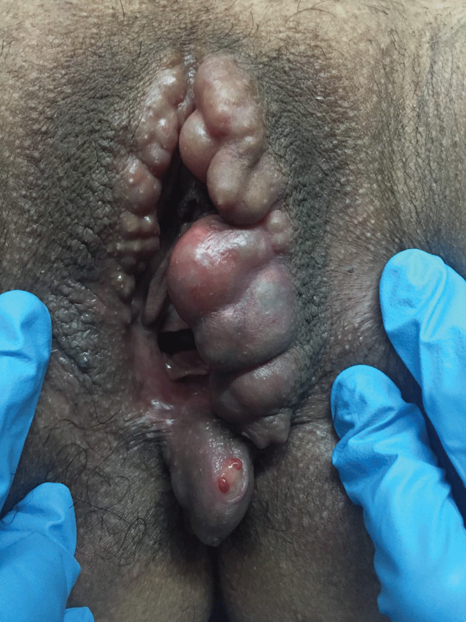 Figure 1: Bilateral multi-cystic masses of the labia majora that developed five days postpartum. View Figure 1
The patient complained of persistent vulvar pain and discomfort, which led to fine-needle aspiration of the masses on several occasions (Figure 2). However, the masses tended to redevelop, and the symptoms recurred immediately. To reach a definitive diagnosis, a biopsy of one of the labial masses was performed. Histopathologic findings were compatible with ectopic mammary tissue with extensive lactational changes (Figure 3).
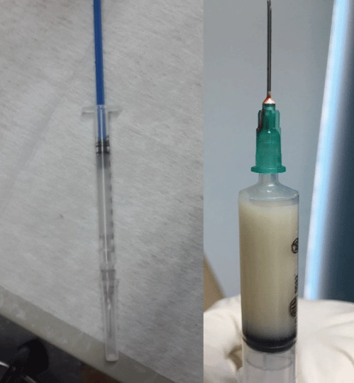 Figure 2: Multiple evacuations performed for symptomatic relief. Material obtained from fine needle aspiration (FNA) performed one week after the masses appeared: citrine liquid was obtained. Material obtained from FNA two weeks after the masses appeared: milky liquid was obtained. View Figure 2
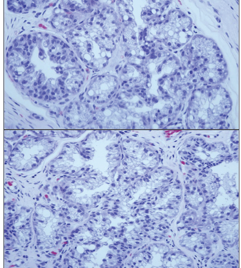 Figure 3: (Hematoxylin-eosin, original magnifications X100 [2] and X200 [1]): Histomorphological features of lactational hyperplasia in ectopic breast tissue: the markedly enlarged lobules are composed of a greatly increased number of glands. The lactating glands are larger and more irregular in shape than in usual ectopic mammary tissue. The cytoplasm has prominent apical blebs and frayed appearance at the luminal border. Nuclei are condensed and hyperchromatic. View Figure 3
Because the discomfort persisted, the patient agreed to inhibition of lactation for symptomatic relief. Soon after, discomfort improved, and the masses became smaller and softer. Five months later, the patient still complained of pain upon exercise. Surgical excision was proposed and was performed, obtaining multiple whitish nodular masses (Figure 4). Meticulous dissection and repair were used to preserve cosmesis. The surgery did not present complications and the patient was discharged 12 hours later. The pathology report informed ectopic mammary parenchyma with fibrocystic changes and adenosis as well as a vulvar intracanalicular fibroadenoma (Figure 5).
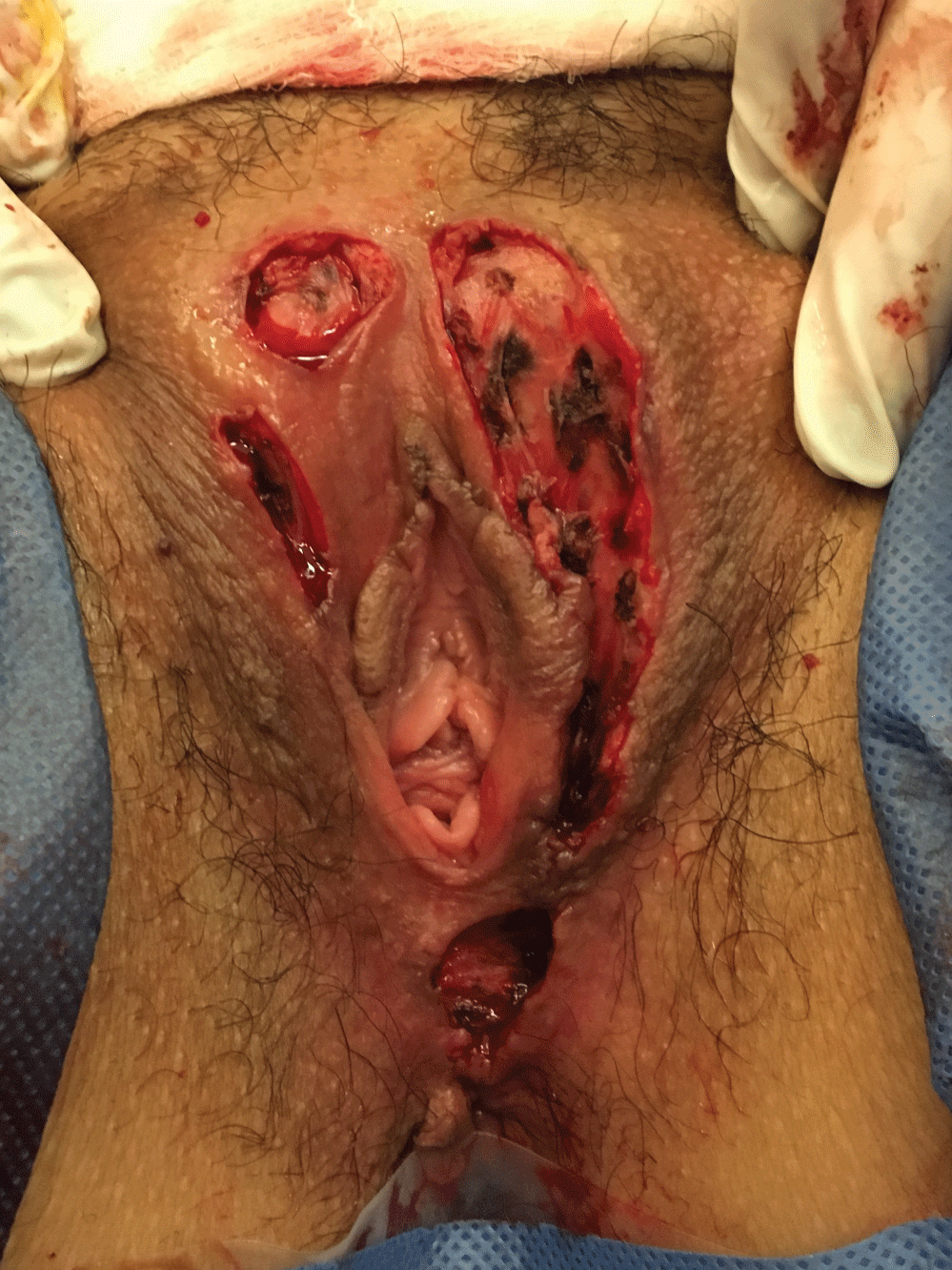 Figure 4: Surgical procedure performed under general anesthesia, working together with the plastic surgery team in order to obtain a better cosmetic result. View Figure 4
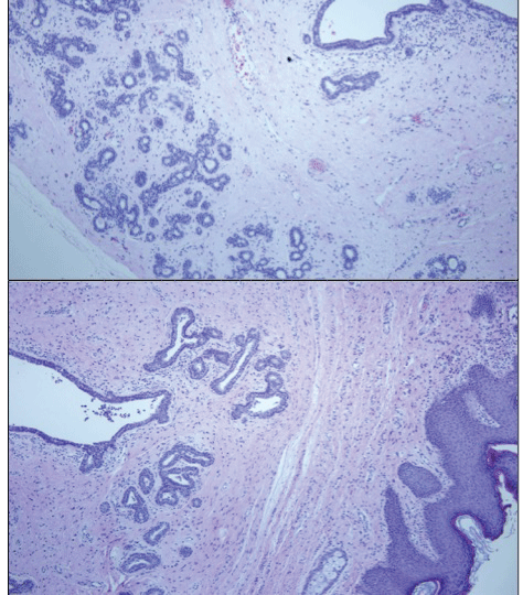 Figure 5: (Hematoxylin-eosin, original magnifications X100 [2] and X200 [1]): Histomorphological features of fibroadenoma in ectopic breast tissue: Microphotograph showing encapsulated growth consisting of compressed glands and branching ducts within a dense fibrocollagenous stroma. View Figure 5
Postoperative follow up was without complications and the patient resumed her normal activity fifteen days after surgery. She remains without recurrence to date (Figure 6).
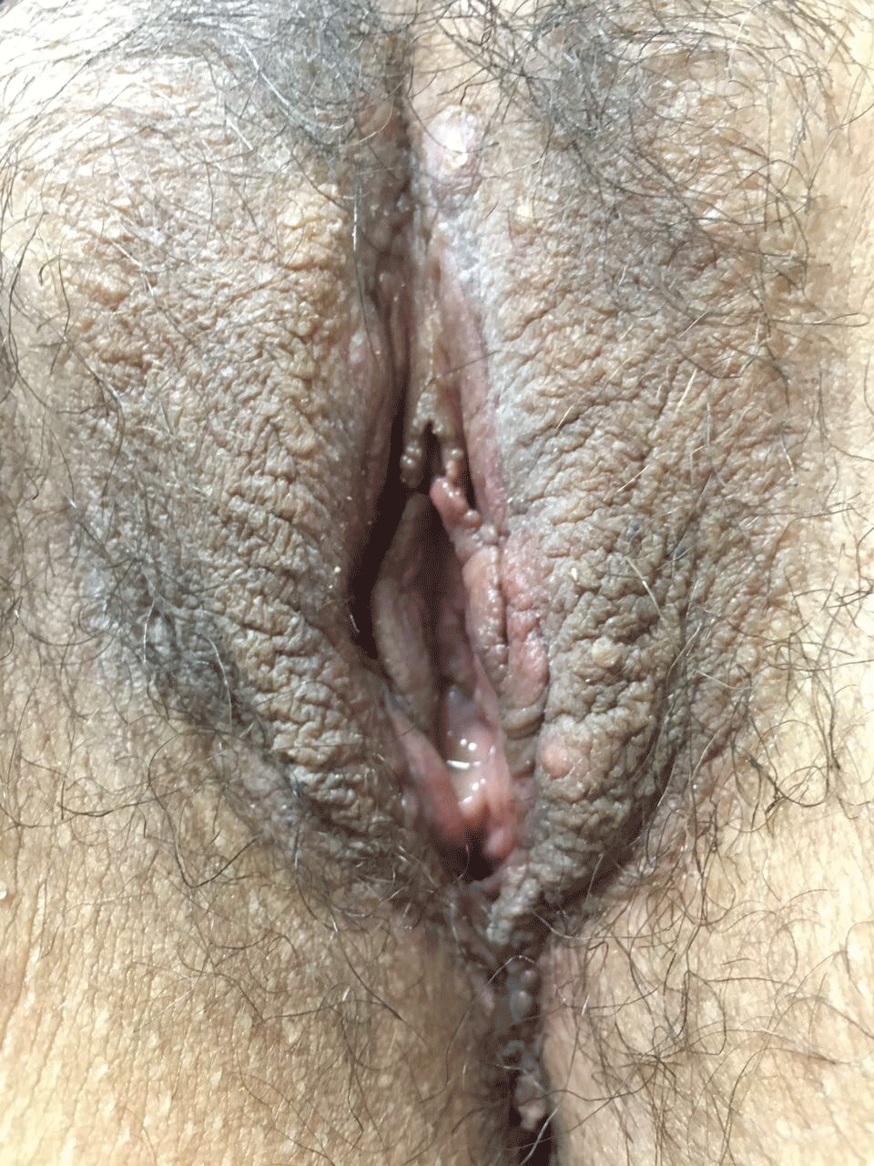 Figure 6: Six months after surgery, without evidence of new lesions. View Figure 6
Ectopic breast tissue is used to describe all types of breast tissue found outside its normal location in the thoracic mammary glands [4]. This tissue is hormonally responsive and may develop benign and malignant pathologic processes similar to those seen in normally located breast tissue, such as fibroadenoma, intraductal papilloma, fibrocystic disease, lactating adenoma and carcinoma [1,4].
There are two hypotheses to explain the embryogenesis of ectopic breast tissue in the vulva: the first one is the traditional theory based on the embryonic milk lines and its incomplete regression in the genital region. The second and more recent hypothesis subscribes to the notion that mammary-like anogenital glands are a normal constituent of the vulva. These anogenital glands have the potential to evolve into benign lesions like fibroadenoma or neoplastic lesions like extramammary Paget's and invasive adenocarcinoma. This theory dismisses the milk line hypothesis by suggesting that the distance between the breast and the vulva is too great to allow for migration of mammary primordial cells [2,5].
The differential diagnosis of a mass in the vulva includes both benign and malignant neoplastic processes. Most common benign masses in this region are cystic lesions such as Bartholin cyst and sebaceous cysts, lipomas, lymphangiomas and hidradenoma papilliferum. Malignant diseases include squamous cell carcinoma, melanoma, Paget's disease, adenocarcinoma arising from Bartholin glands and metastatic disease. The definitive diagnosis is confirmed by histology [6].
During pregnancy, high levels of estrogen, progesterone and prolactin induce growth and proliferation of breast tissue, regardless of its location. After delivery, elevated levels of prolactin and withdrawal of estrogen and progesterone result in the onset of milk secretion. If the woman is breastfeeding, the levels of prolactin remain high, stimulating lactogenesis and growth of the ectopic breast tissue [6]. Our patient initiated breastfeeding on the same day of delivery, therefore the rapid growth in size and beginning of discomfort of these labial masses suggest prolactin-induced growth.
Although there is no scientific evidence that supports inhibition of lactation as an alternative treatment to relieve pain, in this case it was considered a valid option to immediately relieve discomfort until surgery could be performed. Complete surgical excision of the ectopic breast tissue is the recommended treatment [3]. Unfortunately, there are no pathognomonic clinical signs for this condition, which is why most diagnoses are made after histopathological examination. Therefore, it is important to elaborate a detailed clinical history and always include ectopic breast tissue as a differential diagnosis if swelling occurs along the milk line [7].
The challenge of this case was significant, due to both the large size and bilateral presentation and distortion of the normal vulvar anatomy. The ectopic breast tissue was completely excised, and histology was consistent with benign changes (fibroadenoma). There is still no evidence of recurrence at follow-up.
Though ectopic breast lesions of the vulva are rare, clinicians still should be aware of their existence when formulating the differential diagnosis for labial masses. Ectopic breast tissue of the vulva is vulnerable to the same benign and malignant changes as the normal breast. Since the diagnosis is confirmed by histology, complete surgical excision with a cosmetic approach is the recommended treatment [2,8].
No financial support was received for this study. No conflict of interest.