Background: Adverse complications of anti-diabetic drugs demand for alternative novel therapy. This study investigated efficacy of Anacardium occidentale nuts methanolic extract on oxidative stress, inflammation and apoptosis in pancreas of high-fat diet (HDF)/streptozotocin (STZ)-induced diabetic rats.
Methods: Forty matured male rats were used. The animals were fed with HFD and injected intraperitoneally with freshly prepared STZ (35 mg/kgb.wt) single dose to induce diabetes. The animals were grouped into five groups, eight rats/group. Group I: control; Group II: diabetic rats; Groups III & IV: diabetic rats + 100 mg/kgb.wt & 200 mg/kgb.wt A. occidentale nuts methanolic extract; Group V: diabetic rats + 200 mg/kgb.wt metformin. The animals were sacrificed by the end of the experiments. Blood samples were collected and pancreases were excised for homogenization and histological examination. Blood samples and pancreas homogenates were centrifuged and supernatants retrieved were used for biochemical assay.
Results: Feed intake, fasting blood glucose (FBG), insulin, pancreatic malondialdehydes (MDA), tumor necrosis factor-alpha (TNF-α) and caspase-3 levels significantly (p < 0.05) elevated in diabetic rats with diminished body weight, pancreatic superoxide dismutase (SOD), catalase (CAT), reduced glutathione (GSH), interleukins-10 (IL-10) and B cell lymphoma-(Bcl-2). Administration of A. occidentale nuts methanolic extract significantly improved the body weight, pancreatic antioxidants, IL-10 and Bcl-2 levels and suppressed the food intake, FBG, insulin and pancreatic MDA, TNF-α and caspase-3 levels.
Conclusion: A. occidentale nuts ameliorates pancreatic oxidative stress, inflammation and apoptosis. Therefore, safe medication could be developed from this nuts to manage diabetes and its related organ complications.
A. occidentale nuts, Pancreas, Oxidative stress, Inflammation, Apoptosis
Diabetes mellitus (DM) is considered one of the four most common non-communicable diseases due to its high frequency and increasing in its prevalence yearly. According to the International Diabetes Federation (IDF), in 2021, it's estimated that diabetes affects approximately 537 million adults (aged 20-79) and projected that by the year 2045, the number of people that will be living with diabetes will rise to 783 million globally with a higher incidence in developing countries [1].
DM is a metabolic disorder characterized by chronic hyperglycemia with disruptions in the metabolism of carbohydrates, proteins, and lipids resulting from dysfunction of insulin secretion, impaired insulin action, or a combination of both [2]. These metabolic alterations cause many diabetic-related macro and micro-vascular complications such as diabetic nephropathy, retinopathy, neuropathy, cardiovascular disease, stroke and premature death [3]. Clinically, diabetes mellitus symptoms include long-term elevated blood sugar levels, polydipsia, polyphagia, polyuria and weight loss [4-6].
Chronic hyperglycemia in diabetes mellitus triggers the production of oxidative stress [7]. Oxidative stress is well-recognized for the stimulation of pro-inflammatory cytokines production, including tumor necrosis factor-alpha (TNF-α). Inflammation-induced by oxidative stress contributes to the progression of diabetes mellitus [8]. Hyperglycemia-induced oxidative stress and inflammation appear as the key event in the apoptosis of pancreatic beta-cells in diabetes conditions [9]. In addition, it has been well known that an imbalanced balance between excessive ROS production and the body's antioxidant defence system generated by oxidative stress in chronic hyperglycemia critically contributes to the pathogenesis of diabetes mellitus cellular damage-related complications [10].
Though, there is a wide variety of hypoglycemic agents available globally to effectively manage diabetes mellitus [11,12]. However, many adverse effects associated with these therapies include weight gain, hypoglycemia, hypersensitivity, heart failure, hepatic damage and gastrointestinal disturbances [13], and there's a necessity for scientists to source promising alternatives therapies with efficient blood glucose-lowering potentials without adverse toxicity [14]. World Health Organization (WHO) has propounded those traditional medicines offered some promising alternate therapies for chronic diseases including diabetes mellitus [15].
Extracts from numerous medicinal plants have been extensively studied and documented for their remarkable effectiveness in diabetes management [16]. Extracts of these medicinal plants possess divers of chemical constituents such as flavonoids, phenolic compounds, terpenoids, alkaloids, glycosides and coumarins, which can lead to the development of novel antidiabetic drugs [17].
Anacardium occidentale L. (A. occidentale) popularly known as cashew tree originated in Brazil and has become extensively cultivated across the globe [18]. A. occidentale is a medicinal plant with remarkable therapeutic effects [19]. A. occidentale stem, leaves, fruit and flowers have been reported for their different pharmacological activity [20]. The cashew nuts and its derivatives possess a diverse array of biological properties including free radicals scavenging and anti-bacterial activities [21]. A. occidentale nuts are rich sources of various chemical compounds such as unsaturated fatty acids (UFAs) oleic acid (-9) and linoleic acid (-6), flavonoids, anthocyanins, tannins, tocopherols, fiber and folate which are potent antioxidants that protect cells from damage caused by free radicals and reduce chronic diseases risk [22]. Previously, the attenuation of inflammatory mediators by cashew nuts has been demonstrated in animal models [23]. Nevertheless, research on A. occidentale nuts extract on chronic hyperglycemia-induced oxidative, inflammation and apoptosis in the pancreas is still univestigated.
The present experimental study investigates A. occidentale nuts methanolic extract (AONME) on pancreatic oxidative stress, inflammatory and apoptotic markers in diabetic-induced animal model.
Streptozotocin, formalin, phosphate buffer, ketamine, xylazine, methanol, metformin.
Freshly harvested Anacardium occidentale nuts was obtained from the Agricultural Research Farm at Ladoke Akintola University of Technology in Ogbomosho, Oyo State, Nigeria. The nuts was identified, authenticated, and assigned the voucher specimen number LH0533 by Dr. A. T. J. Ogunkunle from the Biology Department at Ladoke Akintola University of Technology, Ogbomoso, Oyo State, Nigeria.
Freshly harvested cashew nuts ( Anacardium occidentale ) were washed thoroughly and then air-dried at room temperature. After drying, the outer coat was removed and the nuts were ground by an electric blender into a fine powder form and stored in an air-tight container. 500g of the finely powdered cashew nut was subjected to extraction in a Soxhlet apparatus using methanol (95%) as the solvent. A semi-solid methanolic extract was collected and stored in a separate air-tight container at 4 °C until needed.
A total of forty matured male albino Wistar rats of average weight (180 ± 20g) were obtained and housed at the Animal Research House of the Physiology Department, Ladoke Akintola University of Technology, Ogbomoso, Oyo State, Nigeria. The animals were kept in clean polypropylene cages, eight rats/cage, and were provided with standard feed and water ad libitum under a ventilated, pathogen-free and hygienic environment with a constant temperature of 25 ± 2 °C, relative humidity 45% ± 5% and a natural 12:12 hrs light/dark cycle. Before the commencement of the experiment, the rats were allowed to acclimate to laboratory conditions for a week.
The experimental procedures adhered to the guidelines outlined in the National Institutes of Health (NIH) Guide for the Care and Use of Laboratory Animals. The research protocols were duly approved by the Research Ethical Committee of Ladoke Akintola University of Technology.
Groups II-V animals were subjected to a high-fat diet (HFD) for six weeks following an acclimatization period. After the HFD feeding, the animals were fasted overnight (12 hours) and diabetes was induced by intraperitoneal injection of repeated doses of freshly prepared streptozotocin (STZ) (35 mg/kgb.wt) dissolved in 0.1 M citrate buffer (pH 4.5). To prevent drug-induced hypoglycemic death, the animals were administered a 20% glucose solution overnight.
Diabetes induction was confirmed after 72 hours of STZ injection by obtaining fasting blood samples from the rats' tail veins and measuring the blood glucose levels using a digital glucometer (Accu-Chek) with test strips. Animals with fasting blood glucose levels ≥ 250 mg/dL were considered diabetic and selected for the experiments.
The forty (40) experimental animals were randomly divided into five groups, 8 rats/group and administered with different treatments as follows:
Group I: Normal control rats + 0.5 ml/kgb.wt distilled water.
Group II: Diabetic (untreated) rats + 0.5 ml/kgb.wt distilled water.
Group III: Diabetic rats + 100 mg/kgb.wt A. occidentale nuts methanolic extract.
Group IV: Diabetic rats + 200 mg/kgb.wt A. occidentale nuts methanolic extract.
Group V: Diabetic rats + 200 mg/kgb.wt metformin.
During the treatment period, the rats were fed with normal standard feed and treated once a day for consecutive 21 days. The body weight and blood glucose levels were recorded on the 1 st , 7 th and 14 th days. Daily food and water intake were also recorded on a daily basis throughout the study.
A day after completing the treatment period, the animals were anesthetized using ketamine (45 mg/kg) and xylazine (5 mg/kg), and then euthanized by cervical dislocation. Blood was collected via cardiac puncture and the pancreas was excised, washed in ice-cold saline to remove blood and sliced into pieces. The blood was centrifuged at 3500 rpm for 15 minutes at 4 °C. The pancreatic sliced tissues were homogenized in phosphate-buffered saline (pH 7.4) and subsequently centrifuged at 6000 rpm for 15 minutes at 4 °C. The retrieved supernatants from the blood and pancreatic tissues were used for the assessment of pancreatic biochemical changes.
Glucose oxidase-peroxidase (GOD-POD) method was used to determine the plasma fasting blood levels using an active digital glucometer (Accu-chek) via vein blood obtained from the rats' tail.
The levels of plasma insulin concentration and pancreatic parameters, superoxide dismutase (SOD), catalase (CAT), pro-inflammatory cytokines tumor necrosis factor-alpha (TNF-α), anti-inflammatory interleukins-10 (IL-10), caspase-3 (apoptotic marker), B cell lymphoma-2 (Bcl-2, anti-apoptotic marker) and oxidative stress marker malondialdehyde (MDA) levels were determined by enzyme-linked immunosorbent assay (ELISA) method using each assay specific ultrasensitive rats ELISA kits following manufacturer's protocols. Gupta and Gupta method was used to determine pancreatic reduced glutathione levels [24].
The pancreases were fixated in 10% formalin solution for a week at room temperature. After fixation, the pancreases were embedded in paraffin, sections 5-6 μm were cut by microtome and stained using the hematoxylin-eosin (H&E) staining. Pancreatic tissue histopathological alteration was observed under a light microscope with magnification (X400).
The data were analyzed with Statistical Package for Social Science (SPSS version 20.0), and the results are presented as the mean ± standard error of the mean (Mean ± SEM). One-way analysis of variance (ANOVA) was used to determine the statistical significance between groups, followed by Tukey's post-hoc test. A P < 0.05 was considered statistical significance.
In the HFD/STZ-induced diabetic rats, the body weight and food intake reduced significantly (p < 0.05) with water intake increased significantly as compared with non-diabetic rats. Administration of low dose (100 mg/kgb.wt) and high dose (200 mg/kgb.wt) of A. occidentale nuts methanolic extract improved the body weight and food intake and lessen the water intake in comparison with the untreated HFD/STZ-induced diabetic rats (Table 1).
Table 1: Effect of A. occidentale nuts methanolic extract on body weight, food intake and water intake in HFD/STZ-induced diabetic rats. View Table 1
As compared with the non-diabetic rats, plasma insulin and fasting blood glucose levels of HFD/STZ-induced diabetic rats dramatically elevated (p < 0.05). Low dose (100 mg/kgb.wt) and high dose (200 mg/kgb.wt) of A. occidentale nuts methanolic extract administration significantly lowered the insulin and fasting blood glucose levels compared with the untreated HFD/STZ-induced diabetic rats (Figure 1A and Figure 1B).
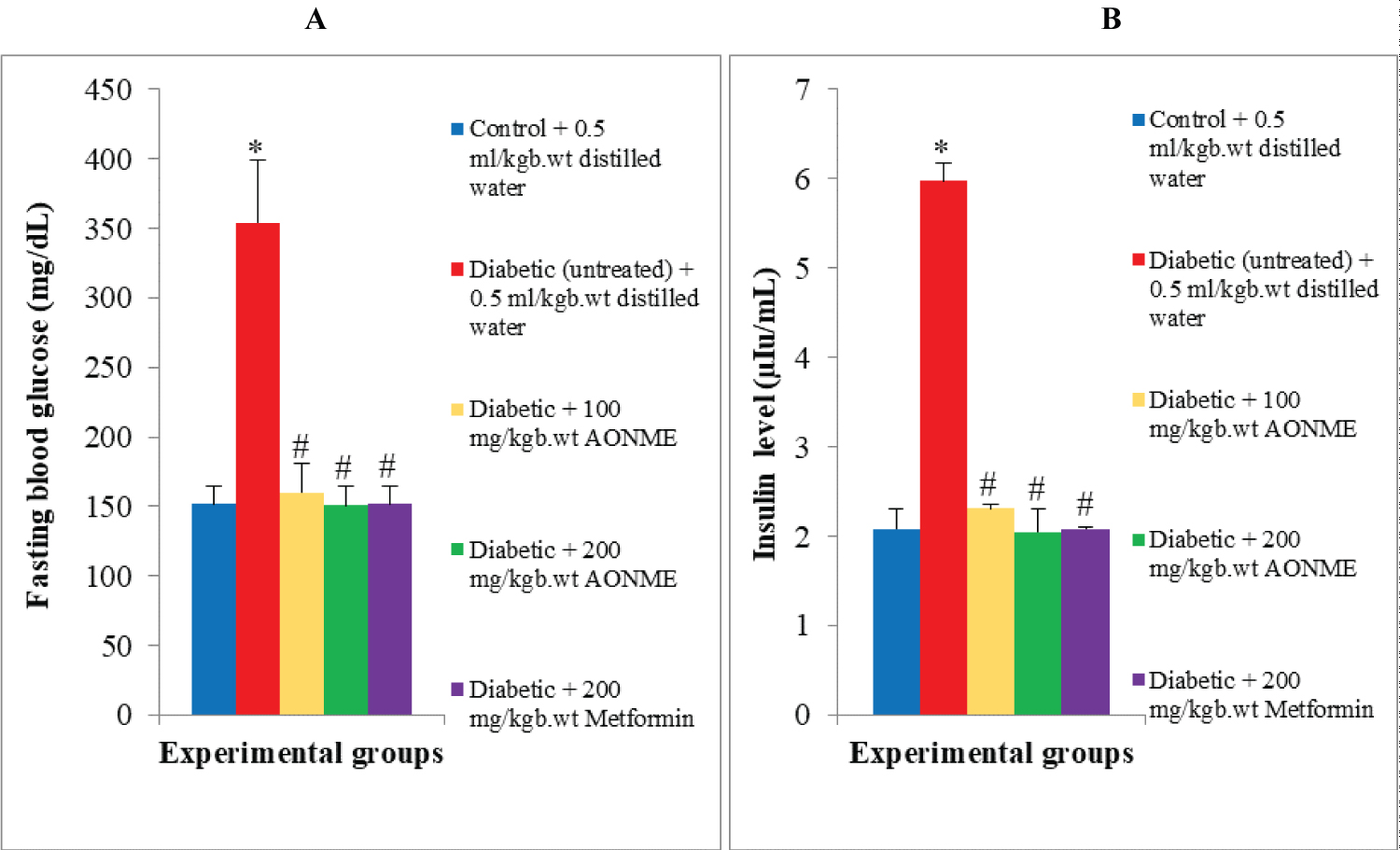 Figure 1: Effects of A. occidentale nuts methanolic extract on (A) fasting blood glucose, (B) insulin levels in HFD/STZ-induced diabetic rats.
Values are expressed as mean ± SEM (n = 8). *significant at p < 0.05 compared with control; #significant at p < 0.05 compared with untreated diabetic group.
View Figure 1
In the pancreas of HFD/STZ-induced diabetic rats, antioxidant superoxide dismutase (SOD), catalase (CAT), and reduced glutathione (GSH) levels reduced (p < 0.05) significantly, and oxidative stress marker malondialdehyde (MDA) higher significantly compared with non-diabetic rats. Administration of low dose (100 mg/kgb.wt) and high dose (200 mg/kgb.wt) of A. occidentale nuts methanolic extract increased the pancreatic antioxidant SOD, CAT, and GSH and reduced pancreatic MDA in comparison with untreated HFD/STZ-induced diabetic rats (Figure 2A and Figure 2B).
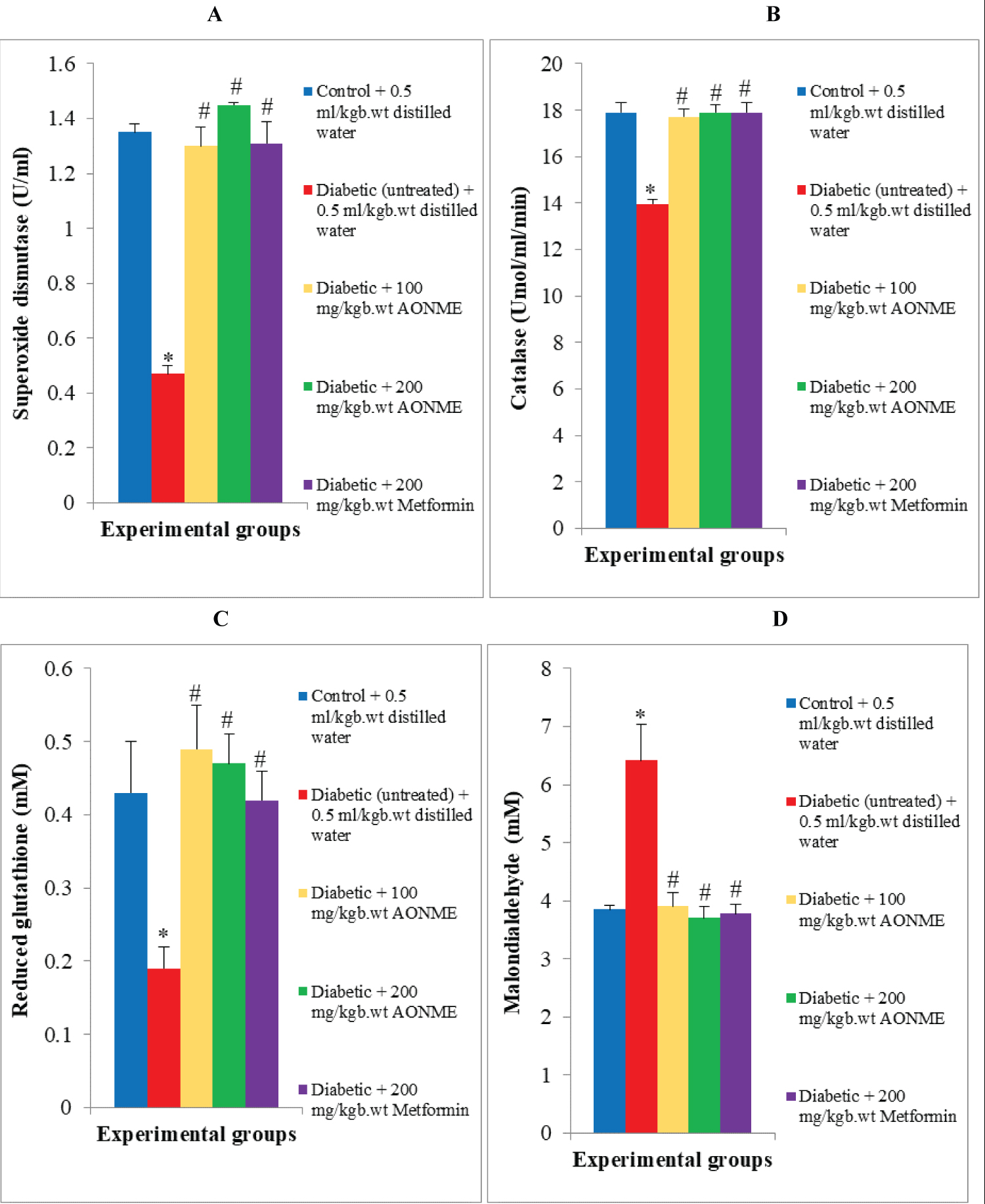 Figure 2: Effects of A. occidentale nuts methanolic extract on pancreatic (A) superoxide dismutase (SOD), (B) catalase (CAT), (C) reduced glutathione (GSH), (D) malondialdehyde (MDA) levels in HFD/STZ-induced diabetic rats.
Values are expressed as mean ± SEM (n = 8). *significant at p < 0.05 compared with control; #significant at p < 0.05 compared with untreated diabetic group.
View Figure 2
In comparison with non-diabetic rats, anti-inflammatory cytokine interleukins-10 (IL-10) in the pancreas of HFD/STZ-induced diabetic rats significantly (p < 0.05) reduced, with a significant increase in pro-inflammatory cytokine tumor necrosis factor-alpha (TNF-α). Low dose (100 mg/kgb.wt) and high dose (200 mg/kgb.wt) of A. occidentale nuts methanolic extract administration significantly elevate the pancreatic anti-inflammatory cytokine IL-10 and decreased the pro-inflammatory cytokine TNF-α compared with untreated HFD/STZ-induced diabetic rats (Figure 3A and Figure 3B).
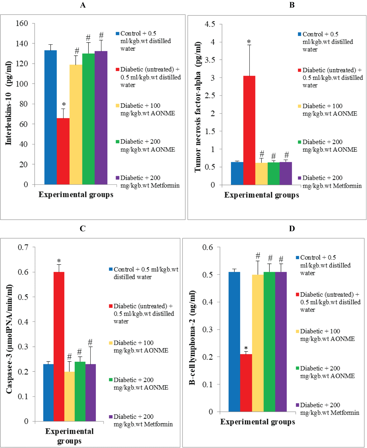 Figure 3: Effects of A. occidentale nuts methanolic extract on pancreatic (A) interleukin -10 (IL-10), (B) tumor necrosis factor-α (TNF-α), (C) caspase-3 (D) B-cell lymphoma-2 (Bcl-2) levels in HFD/STZ-induced diabetic rats.
Values are expressed as mean ± SEM (n = 8). *significant at p < 0.05 compared with control; #significant at p < 0.05 compared with untreated diabetic group.
View Figure 3
Diabetic (untreated) rats exhibited a significant (p < 0.05) increase in pancreatic apoptotic marker caspase-3 and pancreatic anti-apoptotic marker B-cell lymphoma-2 (Bcl-2) reduced significantly compared with non-diabetic rats. The administration of A. occidentale nuts methanolic extract at a low dose (100 mg/kgb.wt) and high dose (200 mg/kgb.wt) lowered the pancreatic caspase-3 and raised the pancreatic Bcl-2 makers significantly in diabetic-treated rats in comparison with untreated diabetic rats (Figure 3C and Figure 3D).
Figure 4A, Figure 4B, Figure 4C, Figure 4D and Figure 4E.
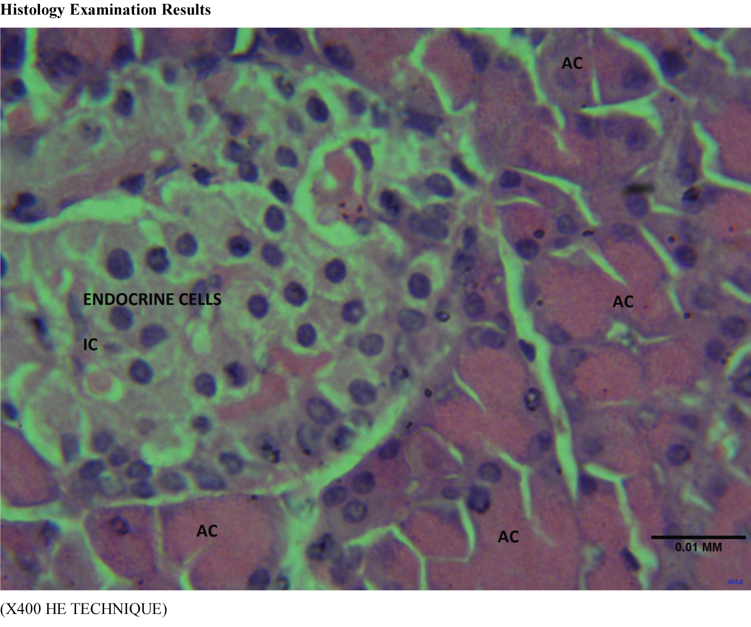 Figure 4a: Pancreatic histological examination of control rats. Section shows the pancreatic tissue composed of the endocrine unit made up of the islet cells (IC), the exocrine unit made up of the acinar cells (AC) lined by regular epithelium and the pancreatic duct, all dispersed with a loose connective tissue and separated by the interstitium. The pancreatic vessel and ducts appears unremarkable. Features consistent with normal pancreatic histomorphology.
View Figure 4a
Figure 4a: Pancreatic histological examination of control rats. Section shows the pancreatic tissue composed of the endocrine unit made up of the islet cells (IC), the exocrine unit made up of the acinar cells (AC) lined by regular epithelium and the pancreatic duct, all dispersed with a loose connective tissue and separated by the interstitium. The pancreatic vessel and ducts appears unremarkable. Features consistent with normal pancreatic histomorphology.
View Figure 4a
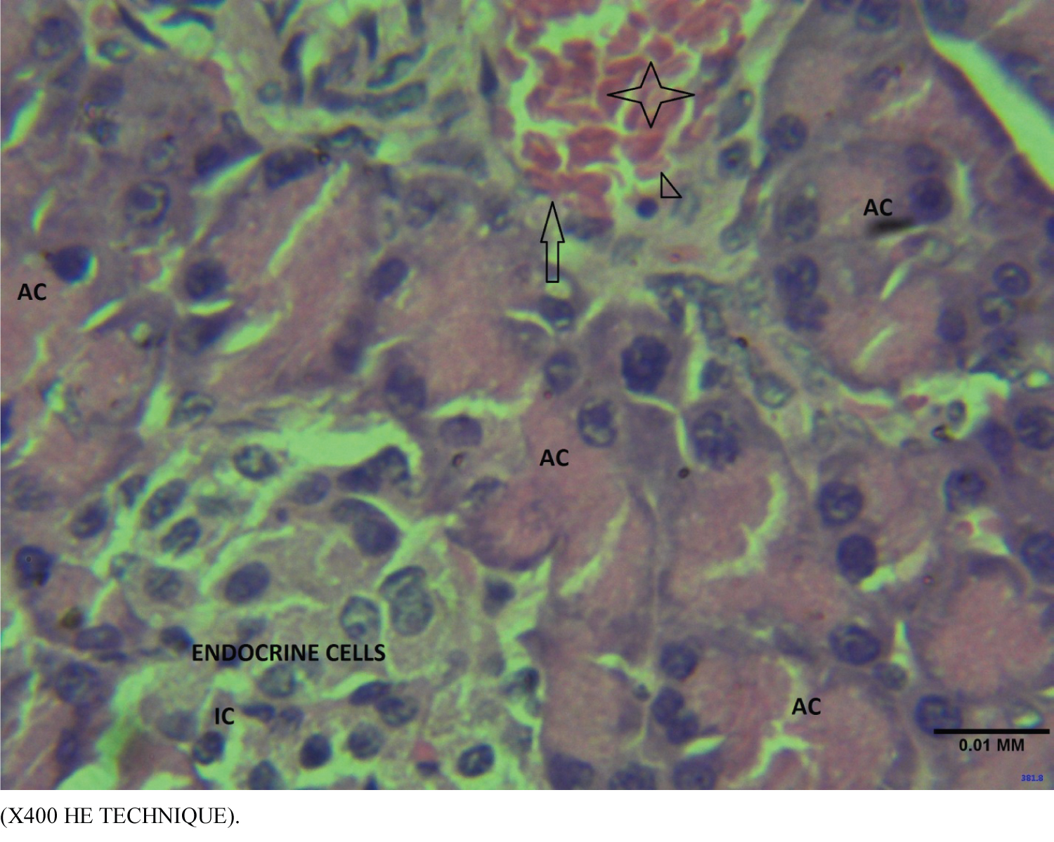 Figure 4b: Pancreatic histological examination of diabetic rats Section shows the pancreatic tissue composed of the endocrine unit made up of the islet cells (IC), the exocrine unit made up of the acinar cells (AC) lined by regular epithelium and the pancreatic duct, all dispersed with a loose connective tissue and separated by the interstitium. The pancreatic vessel (arrow) appears congested (Star) with surveilling white blood cell (arrow head). Features suggestive of vascular response to inflammation.
View Figure 4b
Figure 4b: Pancreatic histological examination of diabetic rats Section shows the pancreatic tissue composed of the endocrine unit made up of the islet cells (IC), the exocrine unit made up of the acinar cells (AC) lined by regular epithelium and the pancreatic duct, all dispersed with a loose connective tissue and separated by the interstitium. The pancreatic vessel (arrow) appears congested (Star) with surveilling white blood cell (arrow head). Features suggestive of vascular response to inflammation.
View Figure 4b
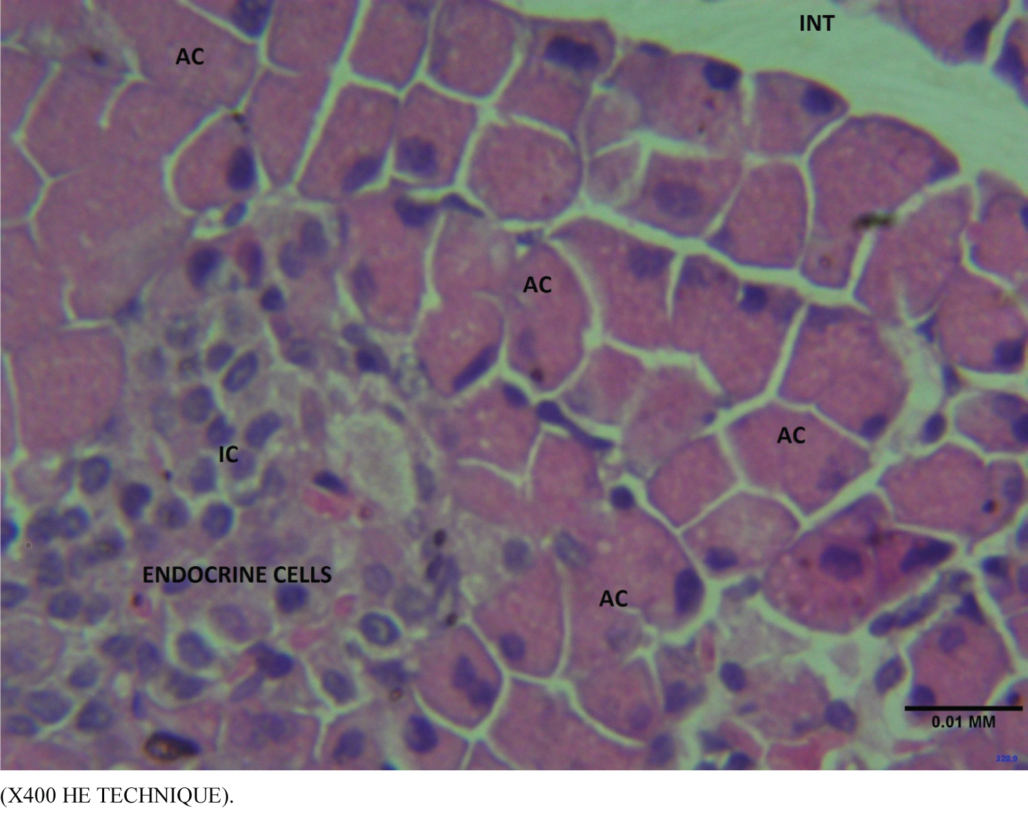 Figure 4c: Pancreatic histological examination of diabetic rats treated with A. occidentale nuts low dose (100 mg/kgb.wt) methanolic extract. The section shows the pancreatic tissue composed of the endocrine unit made up of the islet cells (IC), the exocrine unit made up of the acinar cells (AC) lined by regular epithelium and the pancreatic duct, all dispersed with a loose connective tissue and separated by the interstitium (INT). The pancreatic vessel and ducts appear unremarkable. Features consistent with normal pancreatic histomorphology.
View Figure 4c
Figure 4c: Pancreatic histological examination of diabetic rats treated with A. occidentale nuts low dose (100 mg/kgb.wt) methanolic extract. The section shows the pancreatic tissue composed of the endocrine unit made up of the islet cells (IC), the exocrine unit made up of the acinar cells (AC) lined by regular epithelium and the pancreatic duct, all dispersed with a loose connective tissue and separated by the interstitium (INT). The pancreatic vessel and ducts appear unremarkable. Features consistent with normal pancreatic histomorphology.
View Figure 4c
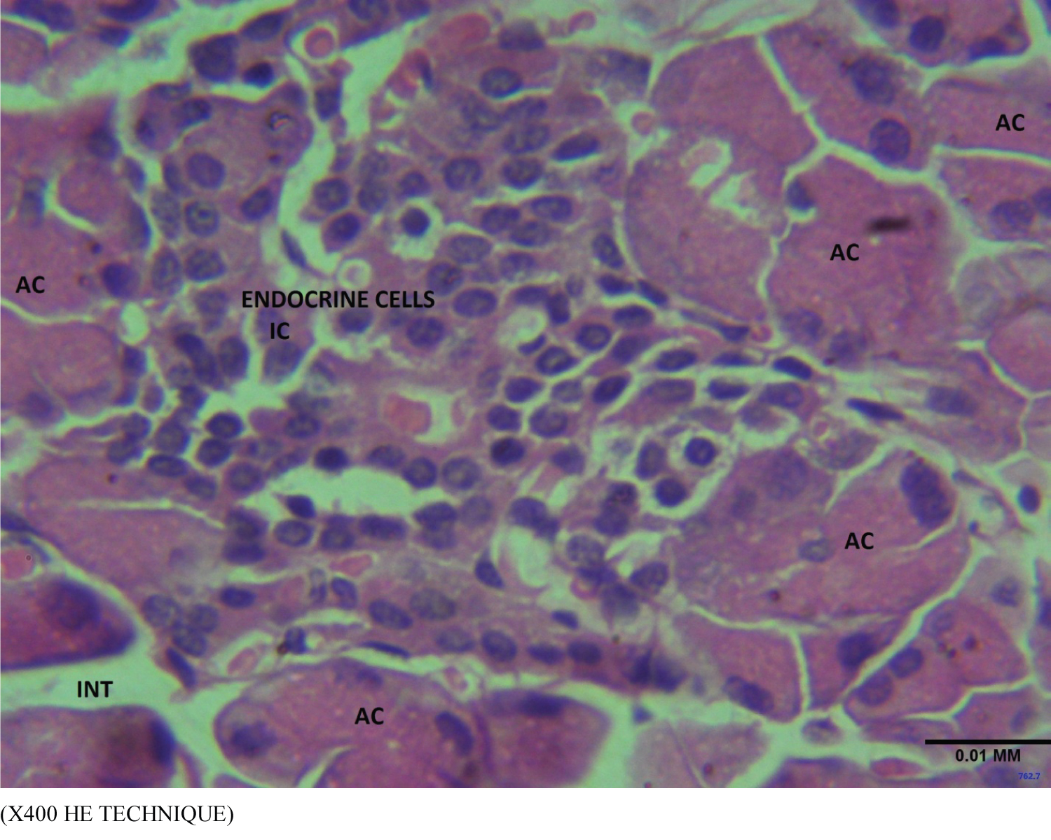 Figure 4d: Pancreatic histological examination of diabetic rats treated with A. occidentale nuts low dose (200 mg/kgb.wt) methanolic extract. The section shows the pancreatic tissue composed of the endocrine unit made up of the islet cells (IC), the exocrine unit made up of the acinar cells (AC) lined by regular epithelium and the pancreatic duct, all dispersed with a loose connective tissue and separated by the interstitium (INT). The pancreatic vessel and ducts appears unremarkable. Features consistent with normal pancreatic histomorphology.
View Figure 4d
Figure 4d: Pancreatic histological examination of diabetic rats treated with A. occidentale nuts low dose (200 mg/kgb.wt) methanolic extract. The section shows the pancreatic tissue composed of the endocrine unit made up of the islet cells (IC), the exocrine unit made up of the acinar cells (AC) lined by regular epithelium and the pancreatic duct, all dispersed with a loose connective tissue and separated by the interstitium (INT). The pancreatic vessel and ducts appears unremarkable. Features consistent with normal pancreatic histomorphology.
View Figure 4d
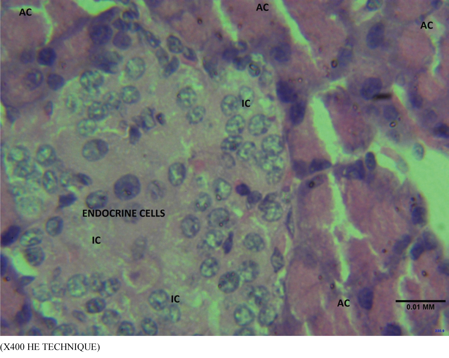 Figure 4e: Pancreatic histological examination of diabetic rats treated with metformin (reference drug). The section shows the pancreatic tissue composed of the endocrine unit made up of the islet cells (IC), the exocrine unit made up of the acinar cells (AC) lined by regular epithelium and the pancreatic duct, all dispersed with a loose connective tissue and separated by the interstitium (INT). The pancreatic vessel and ducts appear unremarkable. Features consistent with normal pancreatic histomorphology.
View Figure 4e
Figure 4e: Pancreatic histological examination of diabetic rats treated with metformin (reference drug). The section shows the pancreatic tissue composed of the endocrine unit made up of the islet cells (IC), the exocrine unit made up of the acinar cells (AC) lined by regular epithelium and the pancreatic duct, all dispersed with a loose connective tissue and separated by the interstitium (INT). The pancreatic vessel and ducts appear unremarkable. Features consistent with normal pancreatic histomorphology.
View Figure 4e
Diabetes mellitus (DM) is a prevalent global health concern and patients affected frequently experience many complications such as injury to pancreatic cells, liver, kidney and heart disease. These complications pose significant challenges to managing diabetes mellitus effectively [25]. The current study demonstrated the promising effects of A. occidentale nuts extract in improving hyperglycemia-induced oxidative stress, inflammation and apoptosis in the pancreas of diabetic rats.
Polyphagia, polydipsia, polyuria and body weight loss are common symptoms for diagnosis of diabetes mellitus [26]. Experimental-induced diabetic rats in the current investigation also exhibited polydipsia and loss of body weight, which is an indication of diabetes induction by streptozotocin. The finding is in agreement with reports on body weight loss in uncontrolled hyperglycemia [27]. The loss of body weight may be attributed to altered energy metabolism and excessive structural protein lysis, a consequence of insulin deficiency in hyperglycemia [28]. However, the supplement of A. occidentale nuts methanolic extract at a low dose (100 mg/kgb.wt) and high dose (200 mg/kgb.wt) effectively restored the body weight and calm the excessive water intake in diabetic rats, this corroborates with the report of Oyebode, et al. [29]. This may be attributed to the normalizing of insulin secretion by the nuts extract to enhance energy metabolism, protein synthesis and inhibit structural protein lysis.
Hyperglycemia is a prominent clinical feature observed in individuals with inadequately managed diabetes mellitus [30]. Insulin is a vital hormone majorly responsible for regulating blood glucose levels [31,32]. In diabetes, insulin secretion and responsiveness at target organs are impaired; leading to hyperglycemia condition [33]. Hyperglycemia-hyperinsulinemia was observed in this study, which revealed the establishment of type-2 diabetes mellitus and damage to pancreatic islets confirmed by histological examination in diabetic rats. This contradicts the finding of Altında˘g, et al. [34], who reported type -1 diabetes mellitus in streptozotocin-induced diabetic rats, noticed by hyperglycemia-hypoinsulinemia . A. occidentale nuts methanolic extract low dose (100 mg/kgb.wt) and high dose (200 mg/kgb.wt) administration normalized the islets insulin secretion, attenuates the elevated blood glucose level and restored the cellularity of pancreatic islets cell (acini and beta cells) integrity supported by the histological analysis. This anti-hyperglycemia potential of the extract could mainly stem from the bioactive compounds present in A. occidentale nuts which might increase insulin sensitivity at the target for glucose uptake and inhibit gluconeogenesis. The findings of this study support other previous reports on the attenuation of hyperglycemia by the consumption of fresh fruit [35].
Oxidative stress arises as a pathological consequence of an imbalance between the increased production of free radicals (oxidants) production and a reduction in the antioxidant free radical scavenging system effectiveness. Persistent chronic hyperglycemia can trigger oxidative stress in pancreatic cells. Pancreatic beta cells are highly vulnerable to the toxic effects of oxidative stress due to low antioxidant enzyme content than other tissues. ROS can directly inflict damage on DNA, proteins, and lipids, leading to dysfunction and apoptosis of beta cells [36]. In line with similar reports in animal experimental diabetes [37], this present study revealed reduced activities of endogenous antioxidant enzymes, SOD and CAT, which are vital in countering ROS. Additionally, a decrease in GSH levels, along with an elevation in MDA concentration (an indicator of lipid peroxidation) was observed in the pancreas of diabetic rats. The disrupted activity of the antioxidant defense system in the pancreas of diabetic rats was improved after the administration of A. occidentale nuts methanolic extract low dose (100 mg/kgb.wt) and high dose (200 mg/kgb.wt), noticeably by enhanced SOD, CAT and GSH with concomitant depleted MDA (oxidative stress maker) level, parallel with the report of Chandirasegaran, et al. [38]. Phenolic compounds have received significant attention in recent years due to their remarkable antioxidant properties. Flavonoids, another group of compounds, are also well-known for their antioxidant properties and their ability to stimulate insulin release, leading to beneficial anti-hyperglycemic effects [39]. The observed enhancement in STZ-mediated oxidative stress improvement by the A. occidentale nuts methanolic extract could be attributed to the presence of phenolic compounds and flavonoids. These bioactive constituents are known for their potent antioxidant properties.
Inflammatory responses play a crucial role in the development of diabetes and are closely associated with increased insulin resistance and reduced responsiveness of insulin target organs [40]. Elevated levels of inflammatory mediators such as TNF-α in blood, have been linked to the consequence of chronic hyperglycemia [41]. These pro-inflammatory cytokines are found at higher concentrations in individuals with diabetes, ultimately leading to cellular apoptosis [42,43]. In accordance with the findings of Yosra Raziani, et al. [44], the up-regulation of pro-inflammatory cytokines TNF-α and down-regulation of anti-inflammatory IL-10 were manifested in the pancreas of experimental diabetic rats in this study. Also, histology examinations revealed vascular response to inflammation in pancreas with a congestive pancreatic vessel and clump of white blood cells. Administration of A. occidentale nuts methanolic extract at either 100 or 200 mg/kgb.wt doses resulted in a notable down-regulation of TNF-α and up-regulation of IL-10 levels in the pancreas and suppresses the inflammatory response in pancreas, also confirmed from the histology studied. This down-regulation in inflammatory cytokines indicates that A. occidentale nuts extract effectively alleviated inflammation, suggesting its anti-inflammatory activity against hyperglycemia-induced pancreatic damage. In agreement with Alabi, et al. findings [45], this anti-inflammatory efficacy might stem from its antioxidant efficacy in repressing the consequence of oxidative stress in the pancreas.
Apoptosis, also known as programmed cell death, is a vital physiological and biological process essential for maintaining cellular homeostasis. Hyperglycemia-induced oxidative stress and inflammation can be triggered by various pathways that lead to cell apoptosis. Hyperglycemia disrupts the balance in the apoptotic process by increasing the pro-apoptotic protein caspase-3 and declining the anti-apoptotic protein Bcl-2 [46]. In the pancreas of diabetic rats, an upsurge in caspase 3 levels (apoptotic marker) with a concurrently diminishing in the levels of Bcl-2 (anti-apoptotic marker) was observed. Various studies have documented comparable expression patterns in the pancreas and liver of streptozotocin-induced diabetic animal models [47,48]. Diabetic rats administered with low dose (100 mg/kgb.wt) and high dose (200 mg/kgb.wt) A. occidentale methanolic nuts extract exhibited a remarkable diminution in the level of pro-apoptotic caspase 3 protein expression, accompanied by elevated anti-apoptotic Bcl-2 protein expression levels in the pancreas. The restoration of the apoptotic process in the pancreas is evidence that A. occidentale nuts extract has apoptosis inhibition potential and this agreed with the findings of Kim, et al. [49], who also reported balancing in the pancreas apoptotic process after administration of berberine chloride to diabetic rats. Numerous antioxidants compounds possessed by A. occidentale nuts could be responsible for this anti-apoptotic potential in pancreas.
Findings of this study showed that A.occidentale nuts exhibited anti-oxidative, anti-inflammatory and anti-apoptotic efficacy. It suppresses the consequence of hyerglycemia-induced oxidative, inflammation and apoptosis in pancreas. A. occidentale nuts could be explored to develop alternative promising therapeutic agent for diabetes mellitus-related complications.
FO conceived the original idea, designed and supervised the research. OA, NO, LA performed the experiments with the support of FO. OA, NO, LA collected the data. FO, MO analyzed the data and prepared the manuscript. FO, MO reviewed the manuscript. All authors’ have read and approved the final manuscript.
All data generated and analyzed during this study are included in this article.
No competing interests.
No funding.