The superficial temporal artery aneurysm is a rare pathology. We report the case of a 7-year-old child who consulted for a beating mass on the left forehead. The doppler ultrasound and The CT angiography confirmed the diagnosis of a superficial temporal artery aneurysm. The treatment was surgical and the postoperative course was simple.
Superficial temporal artery, Aneurysm, Surgery
An aneurysm is defined as an increase in the diameter of a vessel than 1.5 times. It can be fusiform involving the body of the artery or sacciform. A true aneurysm involves all the layers of the vessel wall unlike the pseudoaneurysm in which the expansion involves only the outermost layer of the arterial wall [1].
The temporal artery aneurysms represent 1% of arterial aneurysms. The pseudoaneurysms rank first (95%) while true aneurysms are extremely rare (5%) [2].
A 7-year-old child with no pathological history consulted for a swelling on the left forehead evolving for 3 weeks (Figure 1). On examination, it is a beating and expansive mass. The CT angiography confirmed the diagnosis of a left superficial temporal artery aneurysm measuring 1.5 cm in diameter (Figure 2). He was operated under general anesthesia. He had an aneurysm excision by ligature section. The anatomopathological examination of the specimen showed a muscular artery presenting on one side of its wall a sacciform aneurysmal dilation marked by the total destruction of the media with interruption of the internal elastic lamina. The cavity is filled with a fibrino-cruoric thrombus adhering to a largely abraded intima.
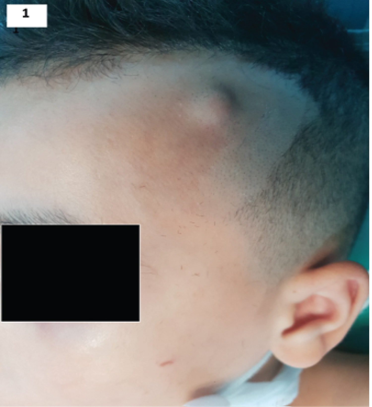 Figure 1: Left forehead swelling.
View Figure 1
Figure 1: Left forehead swelling.
View Figure 1
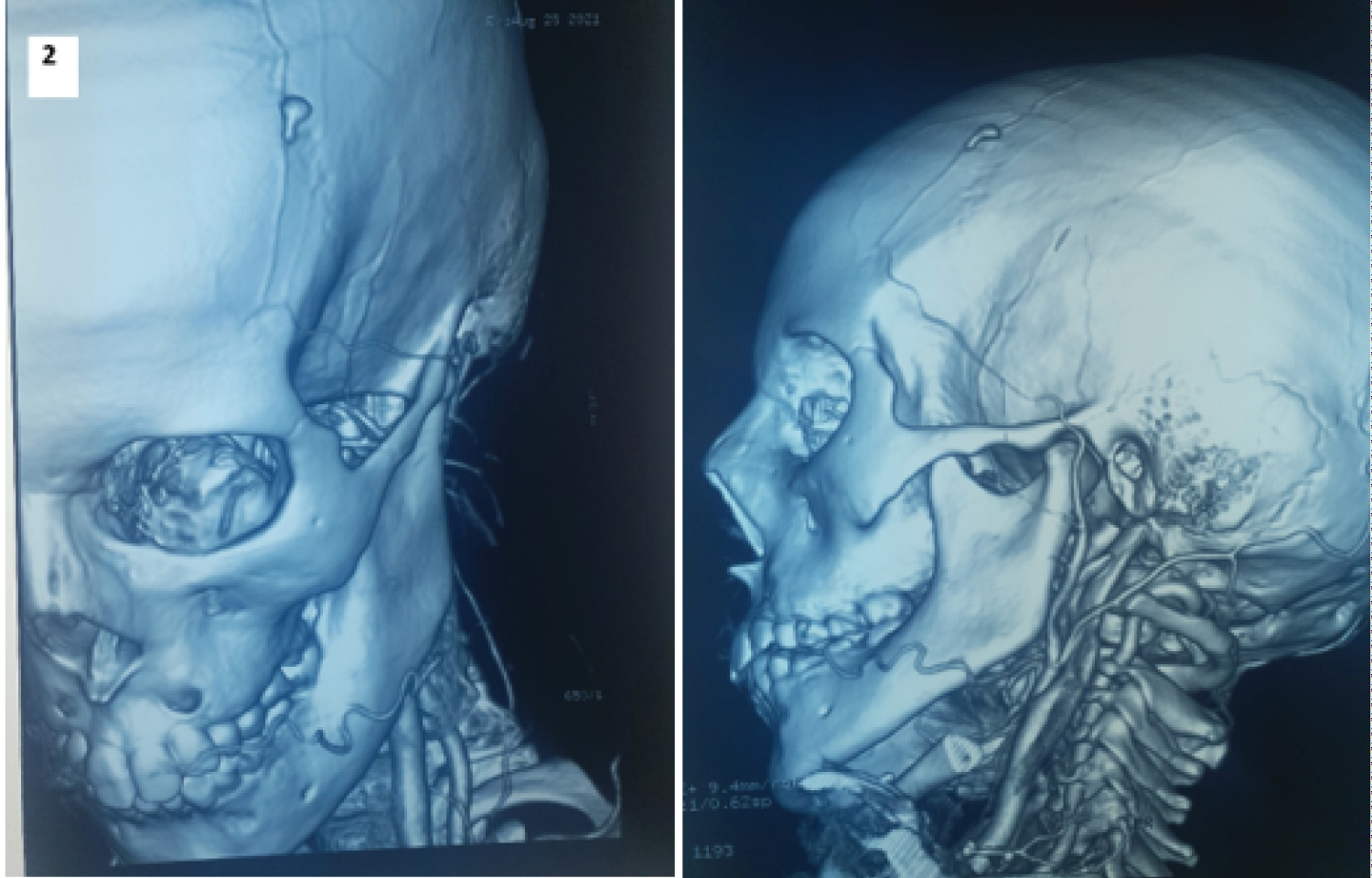 Figure 2: The CT angiography showed a left superficial temporal artery aneurysm measuring 1.5 cm in diameter.
View Figure 2
Figure 2: The CT angiography showed a left superficial temporal artery aneurysm measuring 1.5 cm in diameter.
View Figure 2
The media and the intima are replaced by hemorrhagic and inflammatory connective tissue. The adventitia shows a rich capillary neovascularization. The wall resumes a normal morphology and caliber on either side of this aneurysm.
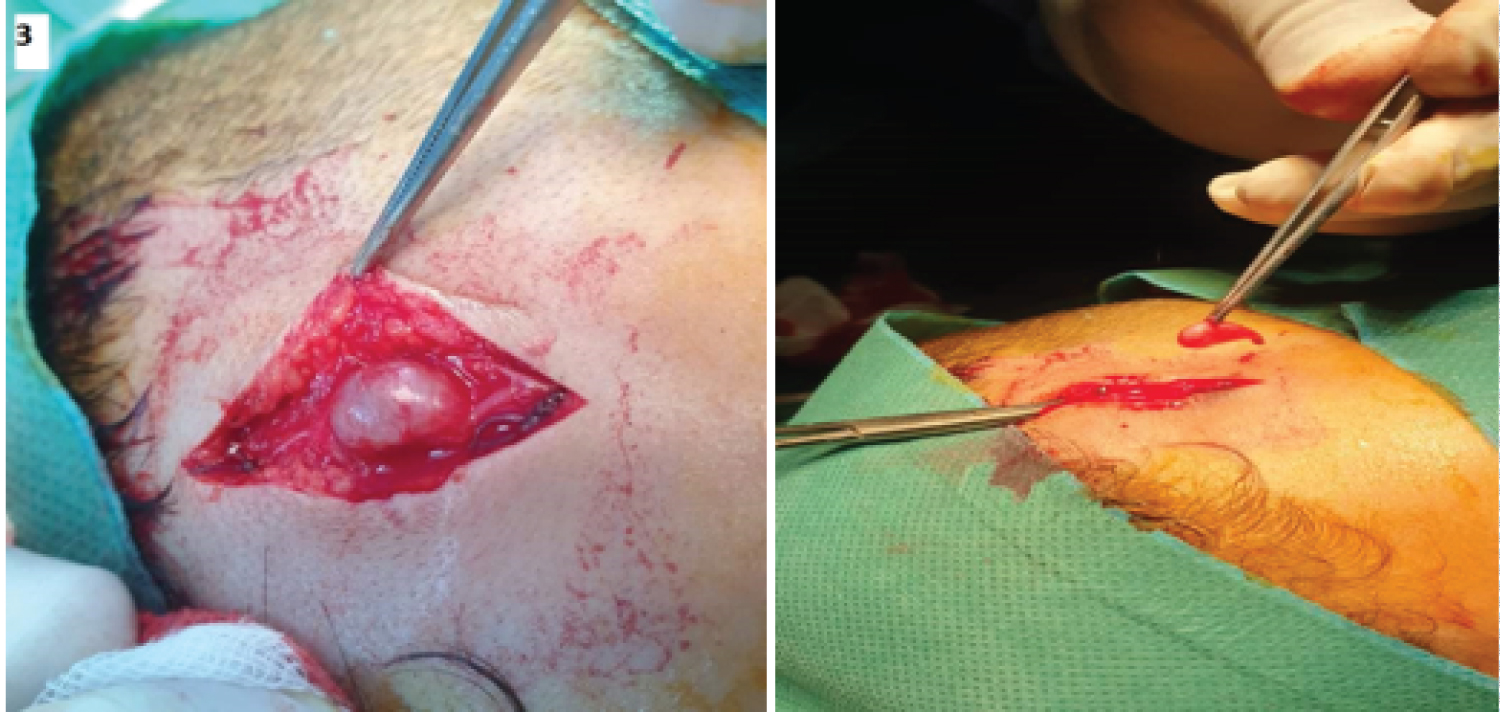 Figure 3: An intraoperative view of the aneurysm of the left superficial temporal artery.
View Figure 3
Figure 3: An intraoperative view of the aneurysm of the left superficial temporal artery.
View Figure 3
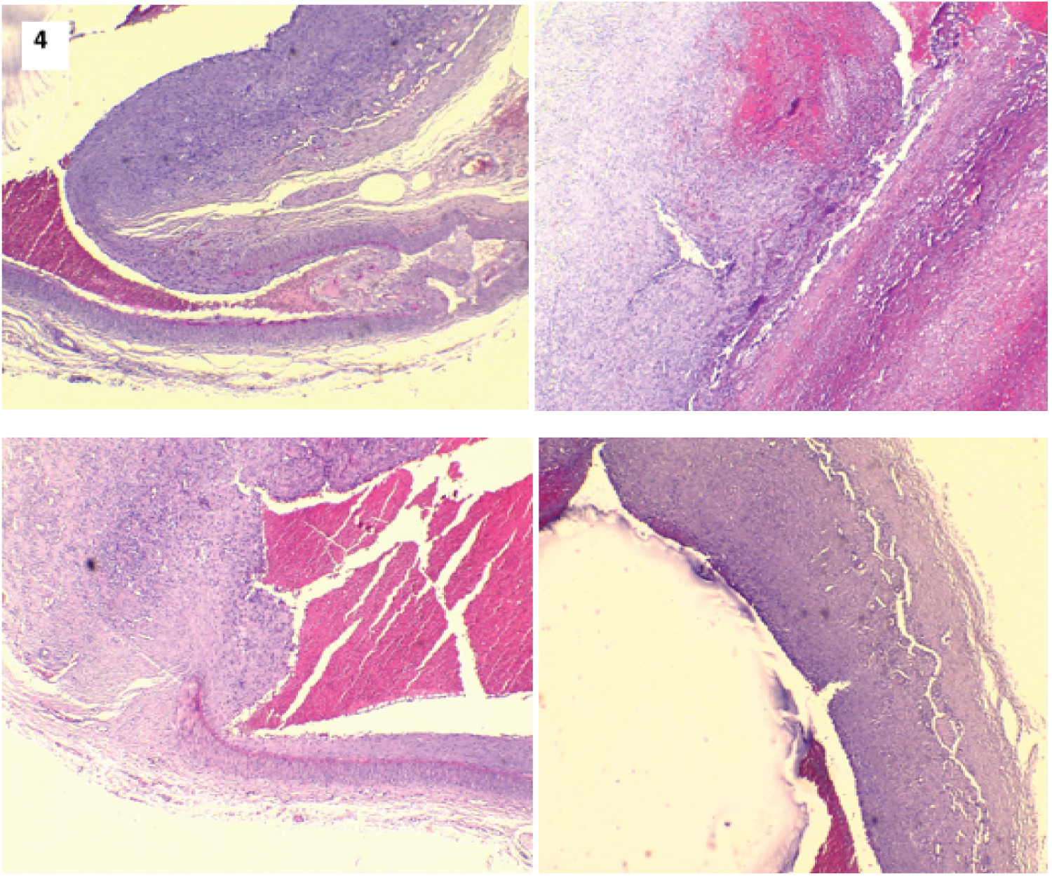 Figure 4: Histological examination of the aneurysm which showed: the appearance of the neck of the aneurysm and the interruption of the internal elastic limit and of the media.
View Figure 4
Figure 4: Histological examination of the aneurysm which showed: the appearance of the neck of the aneurysm and the interruption of the internal elastic limit and of the media.
View Figure 4
The first case of superficial temporal artery aneurysm was described by Thomas Bartholin in 1740 [3].
The clinical expression is a beating swelling on the forehead. The main differentials diagnosis include arteriovenous fistula, abscess, parotid gland lesion or soft tissue tumor [4].
The radiological explorations include Doppler ultrasound and CT angiography. These examinations are not mandatory for diagnosis but may reveal other synchronous lesions [5].
The treatment of a temporal artery aneurysm is often surgical [6]. It consists in the resection of the aneurysmal sac with double ligation of the proximal and the distal ends [7,8]. The endovascular techniques have been described either by ultrasound-guided direct injection of thrombin [9] or embolization [10]. The endovascular intervention is preferable when the facial nerve is close to the parotid gland [4].
The diagnosis of an aneurysm of the superficial temporal artery is clinical. It must be evoked in front of a beating mass of the forehead. The treatment is in most cases surgical.