Robotic surgery, Complications, Surgical subcutaneous emphysema
As minimally invasive surgery becomes more prevalent, it is important to keep in mind the unique complications that can be associated with the use of carbon dioxide insufflation. Postoperative patients presenting with lower extremity pain and swelling should have the diagnosis of iatrogenic subcutaneous emphysema on the differential, alongside deep venous thrombosis and necrotizing fasciitis. A careful history and physical examination can help guide the appropriate management and avoid unnecessary morbidity and mortality.
Complications of Robotic surgery are well known and have been mentioned in the literature. These problems occur in a manner either directly or indirectly related to the robotic system. The use of carbon dioxide insufflation to create pneumoperitoneum is common during robotic-assisted laparoscopic procedures in the fields of urology, gynecology, and general surgery. Known complications of such procedures include pneumothorax, pneumomediastinum, and hypercarbia.
We report a case of a 65-year-old G7P4034 female who presented with tender crepitus and swelling of her bilateral lower extremities on postoperative day one, status post a robotic-assisted laparoscopic supracervical hysterectomy with bilateral salpingo-oophorectomy, sacrocolpopexy and posterior colporrhaphy. Computed tomography of her abdomen and pelvis confirmed the presence of subcutaneous emphysema within the abdominal wall and in both thighs. Her condition was self-limited, and her symptoms were managed conservatively with the patient being discharged home on postoperative day two.
A 65-year-old G7P4034 female underwent robotic-assisted laparoscopic supracervical hysterectomy with bilateral salpingo-oophorectomy, sacrocolpopexy, and posterior colporrhaphy for abnormal uterine bleeding and stage 4 pelvic organ prolapse. Her preoperative symptoms included unexplained weight loss and a bothersome vaginal bulge with occasional leakage of urine while coughing or sneezing. She had failed pessary use and desired to proceed with definitive surgical management. Hey gynecologic history was significant for four vaginal deliveries, dilation and curettage three times, and menopause at the age of 51. Her medical history was with controlled hypertension but with significant for recurrent urinary tract infections, constipation, interstitial cystitis, and atrophic vaginitis managed previously with Premarin vaginal cream. She had a significant family history of colon cancer. Her body mass index was 24.6 at the time of surgery. A fluoroscopic urodynamic study ruled out stress urinary incontinence. A pelvic ultrasound revealed uterine fibroids and a Pap smear showed no malignant cells. She had been started on a three-month preoperative course of antibiotics and her urine culture was negative prior to her surgery. Preoperative assessment was done with the anesthesia team and has ASA-2.
The total operative time was 240 minutes. A Veress needle was used for initial carbon dioxide insufflation. Port placement was as follows: A 12 mm robotic camera port in the umbilicus, three 8 mm robotic arm ports (two in the left abdomen and one in the medial aspect of right abdomen), and a 12 mm AirSeal port used as an assistant channel for suture insertion. The AirSeal was placed in the lateral aspect of the right abdomen with pneumoperitoneum maintained at 15 mmHg for a total of 160 minutes. The patient was placed in steep Trendelenburg throughout the procedure. The case proceeded without any intraoperative surgical or anesthetic complications. After the patient was extubated, she was taken to the post-anesthesia care unit.
She was observed overnight on a surgical recovery unit. There were no acute events through the night. On postoperative day one, she complained of mild pain in her right flank and thigh and reported nausea and vomiting. Her blood pressure was low, but responsive to a 1 L bolus of normal saline. She was afebrile, had generalized tenderness to palpation in her abdomen, and had swelling and tender crepitus in both thighs, noticeably worse on the right.
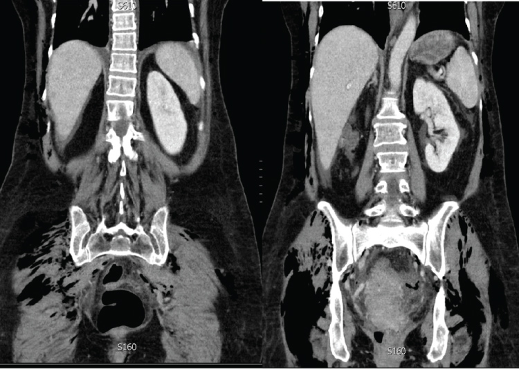
A complete blood count revealed mild leukocytosis with a white blood cell count of 10.8 × 10⁹ cells/L. The basic metabolic panel was within normal limits. Doppler ultrasound examination of her lower extremities did not reveal any underlying pathology. Computed tomography of her abdomen and pelvis with intravenous contrast revealed extensive subcutaneous emphysema along the abdominopelvic wall and extending into her thighs, more so on the right (Figure 1).
 Figure 1: CT abdomen and pelvis showed extensive retroperitoneum and subcutaneous emphysema on both thighs. View Figure 1
Given the findings on imaging, our team decided that the subcutaneous emphysema was likely postsurgical in nature. However, we still had a high suspicion for deep venous thrombosis, as it had not been completely ruled out by ultrasound examination. In addition, she had several deep venous thrombosis risk factors, including prolonged immobilization, history of transvaginal estrogen use, and recent surgery. Lower on the differential was necrotizing fasciitis, given the findings of tender crepitus and leukocytosis.
The patient was encouraged to ambulate and apply cold compresses to her thighs. She was also given a 30 mg dose of ketorolac intramuscularly every six hours for the pain and inflammation.
At her two-week postoperative follow-up, there was complete resolution of her pain and swelling. Final pathology on the surgical specimens was benign.
There have been three previously reported cases of lower extremity subcutaneous emphysema after a robotic-assisted laparoscopic hysterectomy. Only one of these cases involved a patient with isolated subcutaneous emphysema of the bilateral lower extremities, in a similar fashion to our case reported here. There are many causes for the development of such complication and theories behind it; But our experiences recommended some important steps that can be taken to reduce the incidence of iatrogenic subcutaneous emphysema which include decreasing operative time, reducing the number of laparoscopic ports, maintaining low pneumoperitoneum pressure, and minimizing dissection of the infundibulopelvic ligament (during hysterectomy and salpingo-oophorectomy).
McLennan and Lefebvre [1] reported a case of a 71-year-old female who presented to the emergency department on postoperative day two status post a robotic-assisted laparoscopic supracervical hysterectomy with sacrocolpopexy. Her postoperative course was otherwise uneventful. Her chief complaint in the emergency department was bilateral lower extremity pain and swelling, beginning in her upper thighs and extending down to her ankles. On physical examination, her vitals were within normal limits and the only significant finding was diffuse tender crepitus in her bilateral lower extremities. Doppler ultrasound examination failed to identify any deep venous thrombosis. A diagnosis of iatrogenic subcutaneous emphysema was made after consulting with the patient's surgical team. They suggested that retained carbon dioxide gas within the peritoneum could have traveled inferiorly through the femoral canal and collected in the lower extremities. The patient was discharged home and instructed to apply warm compresses. At her two-week postoperative follow-up, her symptoms had completed resolved.
Vetter, et al. [2] reported a case of a 89-year-old female who underwent a robotic-assisted laparoscopic hysterectomy with bilateral salpingo-oophorectomy, omentectomy, pelvic lymph node dissection, and anterior/posterior colporrhaphy for uterine carcinosarcoma and pelvic organ prolapse. The total operative time was 260 minutes, including 150 minutes of pneumoperitoneum at 14 mmHg with the patient in steep Trendelenburg. Five laparoscopic ports were used. On postoperative day one, she presented with right lower extremity pain. On physical examination, her blood pressure was hypertensive at 148/92, but her vitals were otherwise within normal limits. Crepitus was noted at the right lateral malleolus and extended into her knee. Radiography of the right ankle revealed subcutaneous emphysema posterior to the tibia. Doppler ultrasound examination of her lower extremity was negative for deep venous thrombosis. Further examinations of the patient revealed an incisional hernia at the site of a prior appendectomy. It was proposed that this disruption of the fascial planes provided a means for retained carbon dioxide in the peritoneum to diffuse into the right lower extremity. She was diagnosed with iatrogenic subcutaneous emphysema and treated with compression stockings and cold compresses. The remainder of her postoperative course was uneventful, and she was discharged home on postoperative day three. At her three-week postoperative follow-up, there was complete resolution of her symptoms.
Patti, et al. [3] reported a case of a 62-year-old female who presented to the emergency department on postoperative day two status post a robotic-assisted laparoscopic supracervical hysterectomy with sacrocolpopexy. Her postoperative course was otherwise unremarkable. Her chief complaint in the emergency department was substernal pleuritic chest pain radiating to both shoulders, along with a subcutaneous "crunching" sensation on her neck and torso. On physical examination, her vital signs were within normal limits and the only significant finding was diffuse crepitus of the mandible, neck, anterior chest, abdomen, and bilateral thighs. Computed tomography angiography of the chest, abdomen, and pelvis revealed subcutaneous emphysema of the thoraco-abdominopelvic wall, in addition to pneumoperitoneum, pneumomediastinum, and a right apical pneumothorax. Doppler ultrasound examination did not reveal any underlying thrombosis of the distal deep veins. She had admitted for observation, treated with oxygen via nasal cannula for the pneumothorax, and discharged without any complications on hospital day two.
1. Carbon dioxide insufflation is used to create pneumoperitoneum during robotic-assisted laparoscopic procedures.
2. Consider iatrogenic subcutaneous emphysema as part of your differential in a patient presenting with lower extremity pain and swelling after a recent robotic-assisted laparoscopic hysterectomy.
3. Use conservative management for iatrogenic subcutaneous emphysema, as the condition is self-limited.
4. Steps that can be taken to reduce the incidence of iatrogenic subcutaneous emphysema include decreasing operative time, reducing the number of laparoscopic ports, maintaining low pneumoperitoneum pressure, and minimizing dissection of the infundibulopelvic ligament (during hysterectomy and salpingo-oophorectomy).