Mycotic keratitis has been reported from many different parts of the world, with a worldwide incidence varying from 17% to 36% in various studies [1]. It is particularly common in tropical areas, where the incidence ranges between 14%-40% of all ocular mycoses [2]. Early diagnosis is highly important in every patient with the corneal lesion because infectious keratitis caused by various organisms such as bacteria, viruses, fungi, or Acanthamoeba the leading cause of monocular blindness. Rapid and aggressive treatment is needed to halt disease progression and prevent vision loss. More than 50% of infectious keratitis cases are fungal keratitis (FK), especially in the developing countries [3]. FK develops very rapidly, and easily induces corneal perforation, which usually requires surgical intervention. Therefore, accurate diagnosis in the early stages is important for better treatment of infectious keratitis [4].
Among the important predisposing factors related to corneal ulcer are trauma (generally with plant materials), chronic ocular surface disease, contact lens usage, ocular surgery, corneal anesthetics abuse, diabetes mellitus, vitamin deficiency and immune deficiencies. Treatment remains limited by the scarcity of effective antifungal drugs and the emerging resistant organisms. Therefore, establishing a rapid, reliable method to diagnose fungal keratitis, as well as understanding the etiological and epidemiological characteristics of keratitis, is significant for early diagnosis, effective treatment, and prognosis. An effective DNA extraction method from corneal scrapings and multiplex polymerase chain reaction (PCR) should be established to identify the predominant pathogens [5].
We describe a case of poly-microbial keratitis caused by Rhizopus and Morganella precipitated by trauma in a 64-year-old hypertensive female patient. She was presented with hypopyon and was treated with six week course of oral Isavuconazole and topical tobramycin with resolution of hypopyon. Because of the delay of seeking medical care immediately, corneal scarring was observed and corneal grafting was required to restore vision.
64-year-old female with history of hypertension presented with two weeks duration of right eye blurry vision, watery discharge and foreign body sensation preceded by eye trauma. Three weeks prior to the admission, while sitting outside of her house, watching her grandson playing basketball, a tiny invisible particle got into her right eye and she developed irritation of eye and watery discharge. She presented to our hospital after one week when her right eye irritation persisted, developed redness and blurry vision. Patient denied history of runny nose, pain on eye movements, dental problems, dry eye or dry mouth. She denied of contact lens use or swimming. She also denied having headaches, fever, night sweats, cough, dyspnea, chest pain, abdominal pain, rash, diarrhea or joint pains. She was afebrile and hemodynamically stable. On exam, right periorbital swelling was present with active watery discharge from the right eye. Cornea was congested and pus is seen in the anterior chamber. There was no pain on ocular movements and no nystagmus. Physical exam findings on admission were described in Table 1 and Table 2.
Table 1: External Exam. External eye exam findings on admission. View Table 1
Table 2: Slit lamp exam findings on admission. View Table 2
External Exam: Table 1
Slit Lamp Exam: Table 2
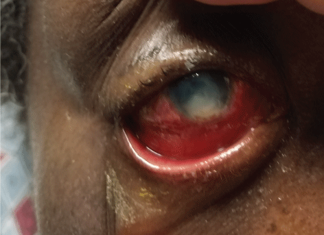
Direct microscopy showed fungal hyphal elements consistent with Rhizopus and cultures grew Rhizopus species and Morganella morganii. She was diagnosed with traumatic poly-microbial keratitis likely primary Rhizopus infection, secondarily infected with Morganella by rubbing of her eyes. CT sinuses was not done as the patient did not complain of sinus pain, runny nose or seasonal allergies. She was started on vancomycin 25 mg/ml, Tobramycin 14 mg/ml, Amphotericin 0.25% eye drops, one drop of each antibiotic every hour with 5 minute interval while awake and every two hours while asleep. She was also given prednisone 60 mg for 5 days for hypopyon. There was no improvement in redness with topical treatment for 10 days and her eye looked still congested and hypopyon was not resolved. We started on Isavuconazole orally 372 mg three time daily for 48 hours then 372 mg daily and continued on Amphotericin eye drops (Figure 1).
 Figure 1: Severe chemosis and hypopyon on presentation. View Figure 1
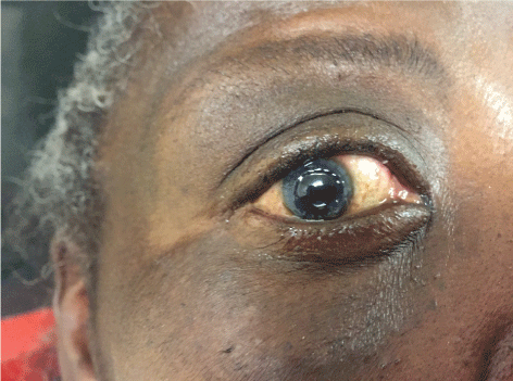
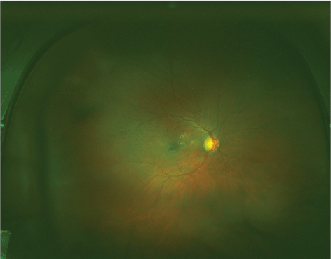
After 10 days of treatment, Patient's ulcer was slowly improved. On slit lamp exam, 10% hypopyon was seen and 1+ chemosis (Table 3 and Table 4). She was discharged with topical tobramycin and topical amphotericin to continue 4 times a day and Isavuconazole 372 mg daily to continue for 6 weeks. She was followed up by ophthalmology at outpatient clinic. At 3 month follow up, eye exam was as follows and she was microbiologically cured. However, since in the delay of seeking medical care initially, corneal scarring was observed and corneal grafting was required to restore vision (Figure 2, Figure 3).
 Figure 2: After corneal graft placement. View Figure 2
 Figure 3: No significant abnormality present on funduscopic exam. View Figure 3
Table 3: External eye exam findings on discharge. View Table 3
Table 4: Slit lamp exam findings on discharge. View Table 4
Mycotic keratitis since its recognition in 1879 has emerged to be a major ophthalmic problem and important cause of ocular morbidity. The risk factors for FK are trauma, and topical corticosteroid use which facilitates the penetration of pathogens, and the popularization of topical antibiotics, which create an environment of lower competition among microorganisms on the ocular surface [6]. In a study done by Dan He, et al. 275 patients with fungal keratitis were examined. In those, 198 patients were diagnosed by positive fungal cultures. The sensitivity of fungal culture was 72.0% in this study. The most common isolated were Fusaerium (49.5%) followed by Aspergillus (18.6%), and Candida (12.4%). Only one case of Rhizopus was reported in this study, representing 0.5% of all diseases. A similar study is done to assess the prevalence of fungal keratitis by Anup K. Ghosh, in a retrospective study over 7 years (2005 through 2011) at a tertiary care center in North India, Rhizopus was reported to be present in 0.3% of all cases [6,7].
Most patients with FK present with pain and redness in the eye, blurred vision, and sensitivity to light, and discharge from eyes. Complications of FK include corneal perforation, hypopyon formation, limbus or scleral involvement [8]. The disadvantages of fungal cultures are; low sensitivity and it is a time-consuming process, often takes a few weeks to grow fungi on solid media. The fastest way to get FK diagnosis is PCR testing which takes 2-3 hours. However it requires a specialized equipment that is not available in all centers. Direct microscopic detection of fungal structures by methylene blue staining in corneal scrapes is another fast and effective method for early diagnosis of fungal keratitis [9,10].
Current literature suggests a promising role of voriconazole in the treatment of fungal keratitis, and given its better penetration, this drug is purported to be a superior alternative to natamycin. A high microbiological cure rate can be achieved in eyes with fungal keratitis; however, therapeutic keratoplasty is often needed to achieve this objective [11].
On our through literature review, we found only three other Rhizopus keratitis cases. The first one was a 24-year-old man following a perforating injury of the cornea with a soil-contaminated screwdriver. The infection was documented by positive cultures from the corneal ulcer, anterior chamber and by histopathologic studies [12]. Second case of Rhizopus keratitis was a 48-year-old male presented after trauma from a metal wire. The patient was treated successfully with oral and topical voriconazole [13]. Third case was associated with contact lens wear. Although staining and cultures of the corneal scraping were negative for fungi, he was diagnosed with fungal keratitis based upon clinical presentation coupled with contact lens culture and response to antifungal therapy. Our case is also associated with minor ocular trauma similar to these three previous Rhizopus keratitis. However, it is unique, in our case that Morganella also grew in the culture, made it a polymicrobial keratitis. Authors believe that it is a case of Rhizopus keratitis secondarily infected by a gram negative Morganella probably by feco-ocular transmission by rubbing of eyes. We successfully used isavuconazole, which is a drug that has not been used before. Isavuconazole (prodrug Isavuconazonium sulfate) is approved for the treatment of invasive mucor infection based on historical controls [14].
While Rhizopus is known to cause a rapid and severe destruction of the extraocular and rhinocerebral soft tissues, the observation was that it behaves less aggressively in the cornea, perhaps due to a vascularity of this tissue [15]. Fungal cultures are the gold standard test for FK. If available, PCR testing is strongly recommended.