A 56-year-old female came into our institution due to dull, non-radiating, 8/10 pain scale, persistent bifrontal headache. Associated with drowsiness, nausea, and 3 episodes of non-projectile vomiting. Work up revealed a frond-like mass with small enhancing bands within the distal body extending to the right atrium of the right lateral ventricle. It was measured to be 4.5 × 2.2 × 2.6 cm. Patient underwent posterior parietal approach craniotomy, excision of tumor with ventriculostomy tube placement on August 16, 2023. Histopathologic findings revealed that it is a Metastatic Colonic Adenocarcinoma. This is currently an unreported case in the Philippines based on HERDIN.
In the world, intraventricular metastasis secondary to colorectal cancer are extremely rare with only four published cases. Our goal for this study is to report this rare occurrence of having a completely resected Intraventricular Metastatic tumor in the hopes of raising awareness and providing options for our future patients.
Metastasis, Intraventricular, Colonic adenocarcinoma
Brain metastases are the most common intracranial tumor, with a staggering number of cases each year. Cases have been steadily increasing as newer treatments prolong the lives of cancer patients. Despite this, finding metastasis in an intraventricular location proves to be a rarity. Comprising only isolated case reports which confirm their existence [1]. In particular, ventricular metastasis are computed to comprise of 0.52%-0.9% of all intracranial metastases reported [2]. The primary of these metastasis could vary from lung, breast, melanoma, thyroid, bladder carcinoma gastric carcinoma or the gastrointestinal system [2,3]. Given that these tumors are situated at precarious locations (deep within the ventricles), neurosurgical interventions such as biopsy and en bloc resection have not always been possible. Attempts at resections have the possibility of life-altering morbidity and even mortality. Biopsy is necessary, as these intraventricular tumors’ differentials include choroid plexus carcinomas (CPCs), subependymal giant cell astrocytoma’s (SEGAs), ependymomas, sub ependymomas, central neurocytomas, and meningiomas [4]. In addition, should there be concurrent masses that could represent other intracranial metastases, patients are directed towards a systemic treatment multidisciplinary approach. Patient or relatives would opt to undergo either stereotactic radiosurgery, chemotherapy, whole brain radiation therapy or a combination. These further lessen that chance of documenting a rare case of a true intraventricular metastatic tumor. It is vital to contribute to the local and international literature to raise awareness towards this rare case. There are no formal guidelines towards the optimal management of intraventricular metastases. In hopes of adding to the database, we report a case of a 56-year-old female, with a metastatic intraventricular colonic adenocarcinoma, managed surgically with a posterior parietal approach craniotomy, and complete excision of the tumor.
Our goal for this study is to report this rare occurrence of having a completely resected Intraventricular Metastatic tumor in the hopes of raising awareness and providing options for our future patients.
Secured written and verbal consent from the patient and husband.
A 56-year-old female was brought to our institution obtunded, with one-to-two-word utterances, did not follow commands, but with spontaneous movement of all extremities. She underwent Cranial MRI which revealed a frond like mass in the right lateral ventricle with extension to the right atrium measuring 3.4 × 3.1 × 2.1 cm. There was leftward bowing of the septum pellucidum 1 cm from midline resulting in moderate obstructive hydrocephalus with transpendymal edema. She underwent ventriculoperitoneal shunt insertion and was appraised regarding excision of the mass. Relatives refused and opted to be discharged without biopsy or excision of the tumor.
Patient returned to our institution after 1 month due to dull, non-radiating, 8/10 pain scale, persistent bifrontal headache. This was associated with drowsiness of the patient coupled with nausea and 3 episodes of non-projectile vomiting. Patient known to have hypothyroidism (> 10 years) s/p Total Thyroidectomy maintained on Levothyroxine 100 mcg/tablet once a day. Dyslipidemic is maintained on Rosuvastatin 20 mg/tabelet 1 tablet once a day. Her hyperuricemia is maintained on Febuxostat 40 mg tablet, 1 tablet once a day. Patient underwent Cranial MRI with contrast which revealed a frond-like mass with small enhancing bands within the distal body extending to the right atrium of the right lateral ventricle. It measures 4.5 × 2.2 × 2.6 cm. Patient underwent posterior parietal approach craniotomy, excision of tumor with ventriculostomy tube placement on August 16, 2023 (Figure 1, Figure 2 and Figure 3).
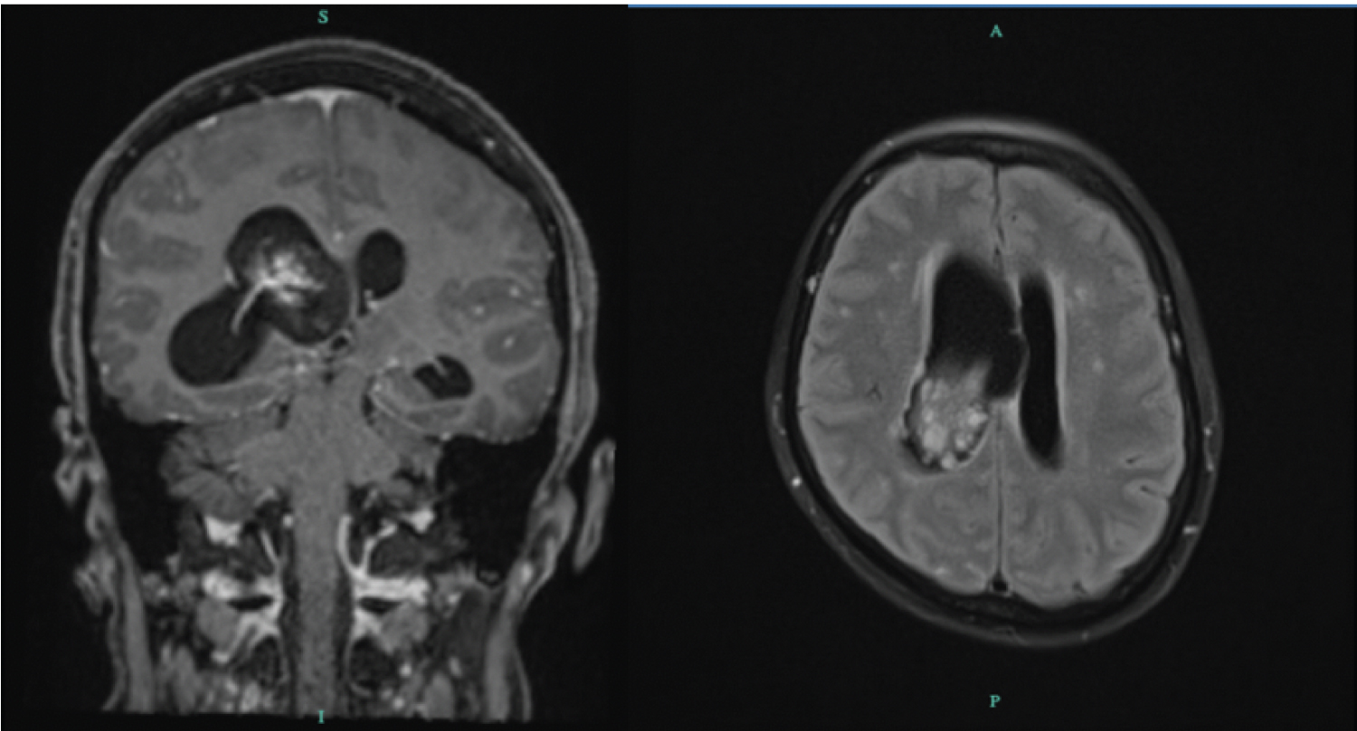 Figure 1: Cranial MRI with Contrast. Coronal view, T1 with contrast showing a frond-like mass with avid enhancing bands and nodules within, centered in the distal body of the right lateral ventricle, extending to the right atrium. It measures approximately 34 × 31 × 21 mm (AP × T × CC). Leftward bowing of the septum pellucidum 10 mm from the midline. Axial view T2 sequence shows the same Frond-like mass with avid enhancing bands and nodules within, centered in the distal body of the right lateral ventricle, extending to the right atrium with evidence of recent hemorrhage and containing hemosiderin and/or calcifications within.
View Figure 1
Figure 1: Cranial MRI with Contrast. Coronal view, T1 with contrast showing a frond-like mass with avid enhancing bands and nodules within, centered in the distal body of the right lateral ventricle, extending to the right atrium. It measures approximately 34 × 31 × 21 mm (AP × T × CC). Leftward bowing of the septum pellucidum 10 mm from the midline. Axial view T2 sequence shows the same Frond-like mass with avid enhancing bands and nodules within, centered in the distal body of the right lateral ventricle, extending to the right atrium with evidence of recent hemorrhage and containing hemosiderin and/or calcifications within.
View Figure 1
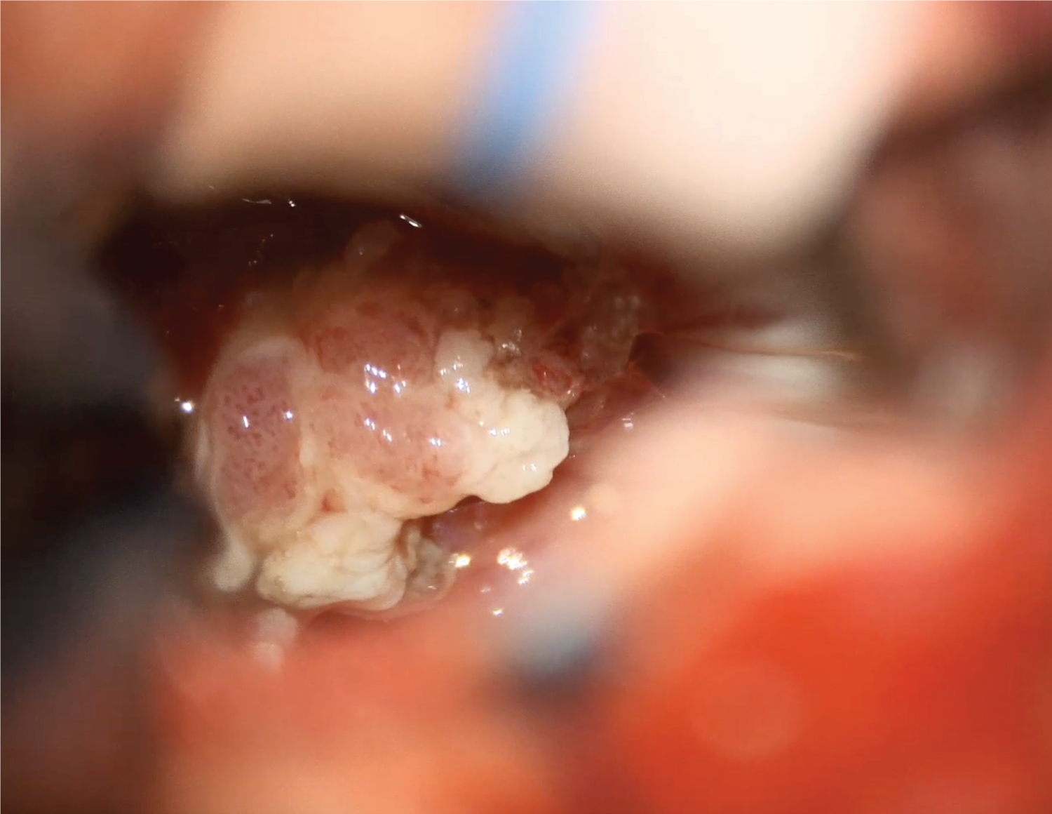 Figure 2: Patient underwent Posterior parietal approach craniotomy, excision of tumor. The intraoperative photo taken through microscope shows mid-dissection of the intraventricular tumor, still with attachments to the lining of the ventricle.
Intraoperative findings revealed an irregularly shaped cream white to tan, brown tumor that was soft to mucoid, not highly vascular. Successfully removed in toto and was measured to be 2.5 × 2.0 × 1.5 cm.
View Figure 2
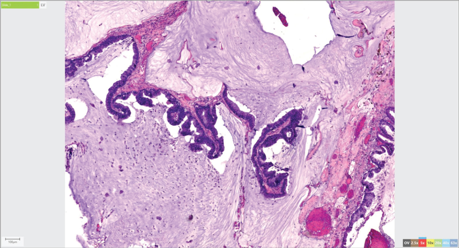 Figure 3: Biopsy on Hematoxylin-eosin staining revealed simple columnar to pseudostratified columnar with areas of dysplasia. Glandular cells are present in supposedly neural tissue, floating in large extracellular mucin lakes.
View Figure 3
Figure 3: Biopsy on Hematoxylin-eosin staining revealed simple columnar to pseudostratified columnar with areas of dysplasia. Glandular cells are present in supposedly neural tissue, floating in large extracellular mucin lakes.
View Figure 3
Patients post-operative course was unremarkable and was deemed fit for discharge accordingly. The patient was scheduled for follow up 1 month after the procedure.
Many tumors can be found in the intraventricular region. However, by definition, a true intraventricular metastases as described by Vecil, et al. is that tumor which is located within the ventricle and is attached either to the choroid plexus or to the ependymal lining or subependymal. It would be crucial to exclude tumors which only have extensions to the ventricle as these are false intraventricular metastases [2]. In addition, any leptomeningeal spread should not be considered as a true intraventricular metastasis. In a study by Bernstein L, Schreiber D, et al. they found that among 737 malignant tumors they studied, only 12.6% of their patients had metastases to the brain. While only 2.6% of the patients had intraventricular tumors. Lesser even were those that had a singular intraventricular metastasis which were found in 1/737 or 0.14%. Among those published cases of intraventricular brain metastates, it was clear that the most common was that of Renal cell carcinoma. It is still unclear why there is a tendency for renal cell carcinomas to metastasize to the ventricles. In the case of our patient, with histopathologic diagnosis of adenocarcinoma. Biopsy diagnosis of Colonic adenocarcinoma was strengthened with findings of glandular cells present in supposedly neural tissue, with characteristic simple columnar to pseudostratified columnar cells. Specimen sent for immunohistochemical staining further confirms the diagnosis with positive CK20 and CDX2. According to Berrada K, Adil H, et al. [5] (2022) there have only been four cases published that describe intraventricular metastasis secondary to colorectal cancer or adenocarcinoma. In our country, the Philippines, there has not been a single publication or report of Intraventricular Metastasis secondary to Adenocarcinoma according to the HERDIN database. This study can serve as guidance for future cases of this nature, which, as seen in our case, can be operated on successfully and safely.
Intraventricular metastases of colonic adenocarcinoma are remarkably rare. Therefore, when encountered, it is imperative that they should be properly documented and published in paper to aid in the proper management of these patients.
In our patient, it was shown that resection of intraventricular tumors via posterior parietal approach can help in facilitating the diagnosis and optimizing patient treatment and management.
Nil.
None.
Dr. Leo Anthony Acedillo - Visualization, Supervision, Investigation.