Intramedullary nailing (IMN) is a standard orthopedic procedure for treating fractures in the lower extremity. While IMNs are generally effective, rare complications, such as nail bending, can present challenges for both patients and surgeons. This article presents a case report of a patient with a bent intramedullary nail in the lower extremity and provides a comprehensive literature review on the management of this unusual complication. By examining the reported cases and available literature, we aim to enhance the understanding of bent IMNs and offer insights into the most effective removal techniques and postoperative considerations.
Intramedullary nailing, Femur, Tibia, Bent, Broken
Intramedullary nailing has become the gold standard for stabilizing fractures in the lower extremity, promoting early mobilization and faster healing. However, a small subset of patients may experience complications, such as a bent intramedullary nail [1].
The removal of a bent intramedullary nail (IMN) is a rare but challenging issue in orthopedics. While various removal techniques have been described in the current literature, there is a lack of comprehensive reviews or established algorithms to guide surgeons in managing such cases effectively [2].
To address this gap, the purpose of this review is threefold. Firstly, it aims to describe the most commonly used removal techniques for bent IMN. These techniques may vary in complexity, invasiveness, and success rates, and understanding their differences is essential for choosing the most appropriate approach for each patient.
Secondly, the review aims to discuss the advantages and disadvantages of these removal techniques. By providing a comprehensive analysis of the pros and cons of each method, surgeons can make informed decisions based on the specific characteristics of the patient's case, such as the degree of angulation, localization of the bending, and implant material.
Finally, the review aims to assist surgeons in formulating a proper management strategy based on the available literature. By synthesizing the existing knowledge on bent IMN removal techniques, the review provides valuable insights to guide surgeons in making the best decisions for their patients.
A 21-year-old male involved in a motorcycle accident sustained a left open tibial fracture and was treated with an IMN in another medical center. During the postoperative period and follow-up, the fracture healed and patient returned back to his previous level of activity within 6 months. However, the patient had a traffic accident again and applied to the emergency department with this complaint. On physical examination, there was a fixed varus deformity of his leg. Both knee and ankle range of motion were normal, and neurovascular status was intact. Plain radiographs showed a re-fracture of the tibia and 30 of angulation of the nail in the coronal plane. Under spinal anesthesia and tourniquet control, patient in supine position, a manual straightening of the nail was first attempted under fluoroscopic control but resulted with failure. An incision was made from the fracture line, then the nail was exposed, the screws in the distal and proximal sections were removed. Then it was cut from the bending line with a metal drill (with saw) and then removed in two parts (Figure 1). Then the fracture was reduced and an 11 mm titanium nail was inserted.
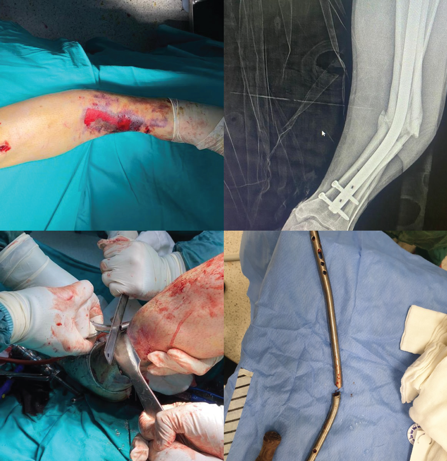 Figure 1: When removing the bent nail.
View Figure 1
Figure 1: When removing the bent nail.
View Figure 1
The literature search was conducted using electronic databases, including PubMed, Clinical Key/Elsevier, EBSCO Discovery Service, MD Consult Science Direct, Scopus, EMBASE, Medscape, and Google Scholar. The search encompassed publications in any language and utilized the following terms: "intramedullary nail," "removal," "bent," "nail removal," and "removal technique." This comprehensive search yielded a total of 30 published case reports.
Upon retrieval of the relevant texts, a detailed examination was performed to evaluate all removal techniques used from 1970 to the present. While various authors claimed to introduce novel techniques, it was observed that most of these techniques shared similar features and steps, often representing modifications of previously described methods [3-35].
In essence, five primary methods for the removal of bent intramedullary nails (IMN) were identified:
1. No additional intervention is required during standard extirpation.
2. The straightening in situ is followed by the standard extirpation.
3. Afterwards, the nail is manually straightened after partial weakening.
4. Extraction of the nail in two pieces after fully cutting it.
5. An axial bone window is created.
In this article, these techniques will be described in sequence, providing valuable insights into their respective procedures and applications.
This technique capitalizes on the inherent flexibility of intramedullary nails and is based on the assumption that bent nails will conform to the shape of the intramedullary canal during standard removal without causing any harm or additional fractures. Yip and Leung pointed out that the anatomic anterolateral bowing of the femur and the larger proximal intramedullary canal of the tibia further facilitate the removal of a bent nail [3]. Additionally, Biert and Edwards suggested that bent nails, especially those made of titanium, tend to lose their original strength and adapt more easily to the medullary canal than expected [4]. A significant advantage of this technique is that it protects the bone and soft tissues by employing external correction maneuvers and open surgery, without requiring any specialized tools or equipment beyond the standard nail extirpation set. However, it is crucial to carefully evaluate the strength and stiffness of the nail when considering this technique. In cases where the nail is thick and composed of stainless steel, the anticipated rebending to its original shape may not occur, potentially leading to additional fractures. Moreover, if the degree of angulation is high (≥ 20 degrees), this technique may also cause further fracture, as the nail can become firmly stuck in the canal [5]. Therefore, the indication for this technique is primarily thin, titanium nails, associated with a simple fracture, and preferably featuring anterolateral bending of the femoral nail and apex posterior bending of the tibia, with an angulation of less than 20 degrees [3].
In this technique, the bent nail is corrected using external reduction maneuvers and subsequently removed through standard extirpation. External reduction of a bent nail can be achieved either manually or with the assistance of a specialized tool. Some authors have attempted manual reduction maneuvers as the initial correction technique, but they have reported limited success in achieving complete correction. Patterson and Ramser utilized a perineal post as a fulcrum to provide the necessary reduction force [6]. On the other hand, Haffernan, et al. argued that while varus-valgus angulations can be corrected through manual reduction, anteroposterior angulations are more challenging, and attempting the reduction manually may lead to extensive soft tissue injuries. Therefore, they advocated for the use of the F-tool, a specially designed instrument for the reduction of long bones, to correct the nail [7].
The F-Tool, manufactured by Synthes, West Chester, PA, comprises three arms and is designed to facilitate the manual reduction of long bone fractures. By clamping the extremity between two arms and applying force in opposite directions (two-point bending) with the third arm, reduction is achieved. However, this closed reduction technique may be associated with certain complications. For instance, excessive force might be required to correct thick nails, potentially leading to soft tissue injuries, including damage to collateral ligaments around the knee. Additionally, the location of the angulations on the nail is crucial because if the nail is bent too proximally or too distally, more powerful reduction maneuvers may be necessary due to the shorter moment arm. In some cases, secondary fractures may occur, especially in osteoporotic patients or in those with longitudinal cracks.
Bek, et al. reported an instance of an additional fracture line appearing after attempting manual reduction of a bent inflatable nail. This highlights the importance of careful consideration and assessment of the appropriateness of the manual reduction technique in specific cases, especially when dealing with challenging nail deformities or certain patient factors that may increase the risk of complications [8].
This technique represents one of the most commonly employed methods found in the existing literature. It revolves around the concept of weakening the nail before an external reduction can be easily achieved through manual strength. The process of partial resection or transection of the nail can be carried out using either an open or closed approach [9].
During this technique, the partial weakening or resection of the nail should be focused on the apex or tip to attain a straightened nail. If surgical exposure of the apex is feasible, this approach can be effectively utilized [10].
In the study conducted by Apivatthakakul and Chiewchantanakit, they described a percutaneous approach to weaken the nail [11]. In this method, they performed a partial resection or transection using a metal cutting drill bit and trochar sleeve through a small incision. This percutaneous technique offers the advantage of minimal invasiveness, potentially reducing the risk of complications and facilitating a quicker recovery.
The choice between open and closed approaches for partial resection or transection depends on the specific case and the location of the deformity apex. Surgeons must carefully evaluate each patient's condition to determine the most appropriate and feasible method for weakening the nail before proceeding with external reduction maneuvers [12].
This technique involves an open surgical approach, requiring complete exposure of the nail at the site of angulation. Specialized instruments are used to cut the nail during the procedure. There are various options for cutting the nail, including motorized or manual metal cutting saws. Some of the tools used in the literature include the dental drill, Midas Rex high-speed burr, Ansbach metal-cutting oscillating circular saw, simple manual metal cutting saw, and jumbo pin cutters [13].
One crucial consideration during the procedure is to minimize the risk of soft tissue damage and necrosis caused by extensive heat generation. To achieve this, continuous irrigation with saline should be employed during the transection of the nail [14]. Additionally, copious debridement should be performed to remove any metal debris that may be produced during the cutting process. Failure to remove this metal debris could potentially lead to complications such as nonunion and infection [16].
The extraction of the bent intramedullary nail is typically carried out in two steps. The proximal part of the nail is extracted using standard techniques, while the distal part is removed through the fracture site. However, in some cases, the nail may become lodged in the intramedullary canal, making its extraction challenging. In such instances, a longitudinal cortical osteotomy may be necessary to free the nail and facilitate its complete removal [15].
As with any surgical procedure, careful attention must be given to patient-specific factors and the nature of the nail deformity to ensure the success and safety of the open surgical technique for nail removal. Proper execution of the procedure, along with meticulous postoperative care, is essential to achieve favorable outcomes and reduce the risk of complications [17].
In this technique, a longitudinal bone flap is created along the bone to completely expose the bent intramedullary nail. The nail is first removed from the proximal bone fragment, and then it is twisted 180 degrees to facilitate its extraction from the distal fragment. Subsequently, the bone flap is stabilized using plates and/or cables.
This particular approach has been employed by two authors for different purposes. Sakellariou, et al. described using this technique as a secondary solution when external manual reduction had previously failed to correct the nail deformity. On the other hand, Bicici, et al. resorted to this method to remove a portion of the nail that had become stuck in the proximal femur [18,19].
It's important to note that this technique comes with some significant drawbacks. The procedure involves a wide incision and extensive dissection of soft tissues, which can lead to increased morbidity and a higher risk of complications. Additionally, additional osteotomy may be required, further adding to the complexity of the surgery and the potential for complications.
Due to the invasive nature of this technique and the associated risks, it is generally considered a less favorable option compared to other less invasive methods. However, in cases where other techniques have failed or when dealing with particularly challenging scenarios, it can serve as a viable alternative for removing a bent intramedullary nail in the lower extremity. As with any surgical procedure, careful patient selection and comprehensive preoperative planning are essential to optimize outcomes and minimize potential complications [19].
In the current literature, numerous removal techniques for bent intramedullary nails (IMNs) have been described and utilized. However, there remains a lack of a standardized algorithm to effectively manage patients with this condition. The existing knowledge on this subject is dispersed across a large number of articles, making it essential to consolidate and synthesize the information into a coherent algorithm.
Given the diverse clinical presentations that a bent IMN in situ may exhibit, it becomes crucial to have a comprehensive approach that can address various cases and conditions. Therefore, we have developed a new practical algorithm for the systematic removal of bent IMNs [1].
Our proposed algorithm takes into account the specific characteristics of each case, including the type and location of the bend, the material of the nail, the extent of angulation, and the patient's overall condition. It provides step-by-step guidance on the appropriate removal technique based on these factors, ensuring optimal outcomes while minimizing potential complications [2].
The algorithm emphasizes non-invasive methods as the first line of treatment whenever applicable, such as external manual correction for flexible nails or closed straightening in situ for certain cases. If non-invasive techniques are not suitable or have proven unsuccessful, the algorithm guides the surgeon towards more advanced options, such as partial weakening and manual straightening or complete removal of the nail in two pieces [20].
In cases where the nail is firmly stuck in the intramedullary canal, the algorithm outlines the use of a longitudinal bone window technique to expose and extract the bent nail. However, it also acknowledges the potential drawbacks of this method, including increased morbidity and complications due to the extensive dissection [21].
Overall, our proposed algorithm aims to provide a systematic and tailored approach to managing patients with a bent IMN in the lower extremity. By consolidating the existing knowledge and considering the specific characteristics of each case, this algorithm can serve as a valuable tool for orthopedic surgeons in making informed decisions and achieving successful outcomes in these challenging cases [5].
Before embarking on the removal of a bent intramedullary nail (IMN), several crucial considerations must be carefully evaluated. Given the absence of a one-size-fits-all approach, each case should be thoroughly assessed on an individual basis, and the most appropriate removal technique should be selected based on the specific characteristics of the condition.
To begin the evaluation process, anteroposterior and lateral radiographs displaying the full length of the nail should be obtained. These X-rays will provide a comprehensive view of the bent IMN and aid in determining the extent of the deformity. The angles of angulation should be measured on both projections to precisely quantify the severity of the bend.
It is essential to document the maximum degree of angulation observed on either the anteroposterior or lateral projection. This information serves as a reference point for gauging the complexity of the case and helps guide the selection of the most suitable removal technique [2].
By meticulously assessing these critical aspects, orthopedic surgeons can make well-informed decisions about the appropriate course of action for removing the bent IMN. This careful evaluation ensures that the chosen technique aligns with the specific characteristics of the condition, maximizing the chances of a successful and safe removal procedure.
Absolutely, you've highlighted crucial additional factors that need to be taken into consideration when planning the removal of a bent intramedullary nail (IMN). In addition to the degree of angulation, the localization of the bending is of paramount importance. Determining whether the bend is proximal or distal, as well as whether it is in a varus or valgus orientation, provides essential insights into the complexity of the case [5].
The assessment of the fracture pattern is equally critical. Any fissure lines extending to the shaft should be carefully examined, and the presence of osteoporosis should be noted. These factors influence the overall stability of the bone and may impact the removal and subsequent treatment plan.
Furthermore, understanding the stiffness of the nail is essential if the intention is to straighten the nail in situ . The thickness and material of the nail play a significant role in predicting its flexibility. Thicker nails generally require more force for correction, while titanium nails are more flexible compared to stainless steel ones. Consequently, thin titanium nails can be corrected with greater ease than thick stainless steel nails [13].
Accessing the medical charts of the patients and reviewing previous operation notes can provide valuable insights into the patient's history and any specific challenges encountered during previous procedures. This information can guide the surgeon in making well-informed decisions during the removal process [7].
It's crucial to consider the operating room conditions and the availability of necessary tools, equipment, and fluoroscopy for successful execution of the chosen removal technique. Preparedness for emergent situations is equally important, and the availability of various fixation options such as nails, plates, cables, and external fixators should be ensured in case they are required during the procedure [1].
By taking all these factors into account, the orthopedic surgeon can create a comprehensive and tailored plan for the removal of the bent IMN, maximizing the chances of a successful and smooth procedure while considering the patient's overall treatment and recovery needs.
Our algorithm for the removal of a bent intramedullary nail (IMN) is well-structured and takes into account various factors to ensure a systematic approach. By categorizing the techniques based on the degree of angulation, you provide a clear pathway for decision-making and treatment. Let's further break down each step of the algorithm:
1. No additional intervention is required during standard extirpation: Angulation < 20 degrees.
• Simple fractures with good bone quality.
• Standard removal without any additional intervention.
• This technique is considered safe and can be effective up to 20 degrees of angulation.
2. The straightening in situ is followed by the standard extirpation: Angulation > 20 degrees.
• External manual correction maneuvers attempted.
• Careful reduction with supportive equipment such as a traction table post as a fulcrum.
• Avoid excessive manipulations to prevent additional fractures and soft tissue injuries.
• If the deformity can be reduced to < 20 degrees, proceed with standard removal.
3. Afterwards, the nail is manually straightened after partial weakening: Failure of External Reduction.
• If external maneuvers fail and the nail remains too stiff to be reduced, consider partial weakening of the nail followed by manual straightening.
• This technique allows for the extraction of the nail in one piece, which can be advantageous.
4. Extraction of the nail in two pieces after fully cutting it.: Angulation Still > 20 degrees and Failure of Partial Weakening.
• If the angulation remains > 20 degrees and partial weakening is not successful, proceed to full resection and removal of the nail in two pieces.
• This technique requires surgical exposure of the fracture and the apex of the bent nail.
• Ensure the availability of necessary equipment to cut the nail.
• Cut nail parts may rarely become stuck in the intramedullary cavity.
Our approach is methodical, and the progression from less invasive techniques to more complex ones ensures that each step is attempted before moving to the next. It also highlights the importance of careful evaluation and decision-making based on the specific characteristics of each case.
Overall, our algorithm provides a practical and logical framework for managing cases of bent IMN removal, taking into account the degree of angulation, bone quality, and the flexibility of the nail. This systematic approach can aid orthopedic surgeons in making informed decisions and choosing the most appropriate technique for successful outcomes while minimizing complications.
In conclusion, the removal of a bent intramedullary nail (IMN) is a challenging orthopedic condition, and there is no one-size-fits-all approach for its management. Instead, several factors need to be carefully evaluated to select the most suitable method for each individual case. These factors include the degree of angulation on both projections, localization of the angulation, thickness and material of the nail, pattern of fracture, and presence of osteoporosis. Additionally, the availability of equipment and the resources in the operation room, as well as the surgeon's expertise, play crucial roles in the decision-making process.
The algorithm presented in this paper provides a systematic and practical flow of techniques, starting from simpler methods and progressing to more complex ones. This approach helps orthopedic surgeons navigate the challenges of bent IMN removal effectively. By considering the specific characteristics of each case, the algorithm allows surgeons to avoid wasting time and unnecessary efforts, ultimately leading to improved patient outcomes.
While the algorithm provides valuable guidance, it is essential to acknowledge that each patient's condition is unique. Therefore, clinical judgment and experience are paramount in making the best decisions for individual cases. By utilizing the information and insights from the algorithm in conjunction with their expertise, orthopedic surgeons can optimize the success of bent IMN removal procedures and enhance patient care. Future research and advancements in the field may further refine and enhance the management strategies for this rare but complex orthopedic problem.
All authors have no conflict of interest.
No funds have been received for this study.
All procedures performed in studies involving human participants were in accordance with the ethical standards of the institutional and/or national research committee and with the 1964 Helsinki Declaration and its later amendments or comparable ethical standards.
Informed consent was obtained for all patients.