As one ages, the structure of the nose undergoes a number of changes, leading to alterations in form and function. Patients with no previous history of nasal issues may present with age-related nasal obstruction later in life. Tip ptosis is a well-recognized etiology of nasal obstruction in the aging patient [1-7]. The pathogenesis is believed to be due to weakening of several of the tip support mechanisms. Of primary importance, there is age-related degradation of the fibrous union at the cephalic edge of the lower lateral cartilage and the caudal edge of the upper lateral cartilage known as the scroll [1,4-7]. There is also weakening of the fibrous attachments of the medial crura to the posterior septal angle and nasal spine. Loss of tip support mechanisms leads to deprojection, counter rotation of the nasal tip, and retraction of the columella, resulting in both internal and external nasal valve dysfunction [1-7]. These changes can be further exacerbated by age-related nasal muscular atrophy and bony resorption of the premaxilla [1,8].
In the setting of age-related ptosis, traditional septorhinoplasty techniques aim to increase tip support and rotate the nose cephalically. Less invasive, yet effective methods have been described. The Rhinolift involves an excision of the skin and soft tissue envelope (SSTE) over the supratip followed by re-approximation of the lower and upper lateral cartilage [1,3]. This results in cephalic rotation of the nasal tip. Patients should be made aware that the Rhinolift may result in an unsightly scar in the supratip region. To optimize aesthetic outcomes, we have made modifications to the Rhinolift technique by orienting the incision in a pre-existing rhytid at the sellion.
Rhinolift, Functional Rhinoplasty, Nasal Tip Ptosis
A 59-year-old man presented for evaluation of new onset bilateral nasal congestion and reduced nasal airflow. The patient noted that he had never experienced these symptoms before, however it has been progressively worsening over the past year. He had been maximized on over-the-counter antihistamines, nasal decongestants and nasal steroids for greater than four weeks with minimal relief.
External nasal examination revealed a ptotic nasal tip with external nasal collapse on inspiration (Figure 1). The patient experienced relief of his obstructed breathing with cephalic traction of the SSTE over the sellion. It was noted that this maneuver also caused a cephalic rotation of the nasal tip. Cottle maneuver was positive. Palpation of the nasal dorsum and sellion revealed a thick and redundant SSTE. Bilateral nasal endoscopy revealed a high left septal deflection, a low right septal deflection off of the maxillary crest, and a posterior right septal spur. Nasal endoscopy also revealed bilateral turbinate hypertrophy. The remainder of the head and neck exam was within normal limits.
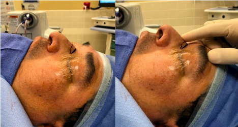 Figure 1: Preoperative age-related nasal tip ptosis (Left). Skin and soft tissue envelope pulled cephalically over the sellion causing cephalic tip rotation (Right). View Figure 1
Our impression was consistent with a patient who would benefit from a rhinolift, bilateral turbinate reduction and septoplasty. We discussed the procedure and risks at length. In addition, the patient was made aware of the expected scar, but he was comfortable with the cosmetic defect.
Redundancy of the SSTE was palpated at the sellion. The soft tissue was pulled cephalically over the sellion and resulted in cephalic tip rotation (Figure 1). Once the desired tip rotation was achieved, the redundant soft tissue enveloped was marked at its caudal and cephalic edge. The cephalic marking was placed in a horizontal rhytid, as is commonly seen in the aging face from overactivity of the procerus muscle. The SSTE was released and a symmetric elliptical incision was marked.
The nose was prepped and draped in sterile fashion and 1% lidocaine with epinephrine was injected along markings. Incision was made along the markings and carried down to the musculature overlaying the nasal bones. The soft tissue was then excised (Figure 2).
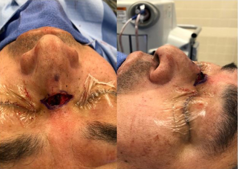 Figure 2: Soft tissue excision over the sellion. View Figure 2
Using an interrupted 4-0 monocryl stitch, the caudal subcutaneous tissue of the nasal envelope was approximated to the cephalic subcutaneous tissue (Figure 3). The cutaneous layer was then closed, tension-free, with a simple running 5-0 fast absorbable surgical gut suture.
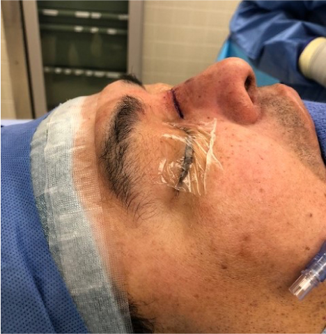 Figure 3: Caudal subcutaneous tissue of the skin and soft tissue envelope re-approximated to the cephalic subcutaneous tissue. View Figure 3
The patient was re-evaluated in clinic on postoperative day 6 (Figure 4). The patient noted significant improvement in his nasal breathing. However he noted tenderness, ecchymosis and swelling along his nasal dorsum. Examination revealed appropriate nasal contour, shape, and symmetry with resolution of his tip ptosis. There was mild swelling and ecchymosis along his sellion. The incision was clean, dry, intact and healing appropriately. One month follow up revealed resolution of the ecchymosis and edema over the sellion with no recurrence of his tip ptosis. The incision was well healed with minimal scarring and was contiguous with a pre-existing rhytid (Figure 5). The patient again noted improved nasal breathing. Exam at 3 months postoperatively again revealed no nasal tip ptosis (Figure 4). As compared to his preoperative baseline, the patient's nasal breathing was again noted to be subjectively improved.
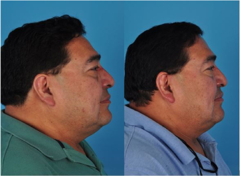 Figure 4: Postoperative day 6 (Left); 3 month postoperative visit (Right). View Figure 4
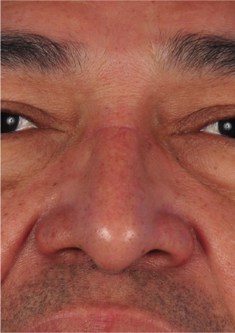 Figure 5: Modified Rhinolift 3 month postoperative scar. View Figure 5
Nasal obstruction is a quality of life impairment that affects many people [9]. Oftentimes, nasal obstruction is multifactorial, but in the aging patient, tip ptosis can play a prominent role [10]. In these patients, a modified rhinolift should be considered. It can be performed under local anesthesia in an office-based setting, mitigating the risks of general anesthesia [3]. The ideal candidate for our modified rhinolift technique is an elderly patient with an age-related tip ptosis. Exam should demonstrate a thickened SSTE and cephalic rotation of the tip with cephalic traction of the SSTE over the sellion.
The traditional rhinolift approach offers access to the scroll region through a direct supratrip incision [1,3]. This allows for potential reapproximation of the scroll region [3]. However, the added benefit of addressing the scroll may not outweigh the risks of an unsightly scar along the supratip. Our modified technique orients the scar within a natural crease in the sellion region, leading to superior cosmesis. Furthermore, we have achieved durable results without addressing the scroll region (Figure 6). As consistent with all operations, patients may require a second operation if desired results are not achieved in first operation.
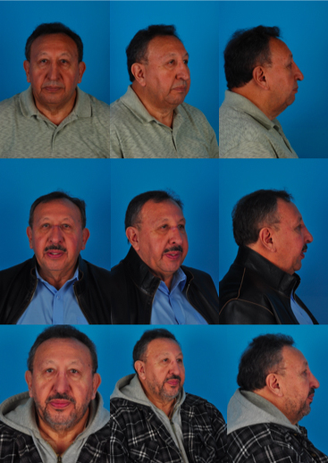 Figure 6: Preoperative nasal tip ptosis (top); 7 months postoperative (middle); 1.5 years postoperative (bottom). View Figure 6
The modified rhinolift is an effective intervention for nasal obstruction secondary to age-related nasal tip ptosis. The operation may be performed under local anesthesia, mitigating the risks of general anesthesia in patients with multiple comorbid conditions. Our technique offers durable results, while offering superior cosmesis over the traditional approach.
None.
There are no conflicts of interest for any of the authors mentioned above.