Silent sinus syndrome is a rare clinical entity affecting the maxillary sinus, causing progressive enophthalmos and hypoglobus resulting from retraction of maxillary sinus walls with increase in the orbital volume. We report a case of a 29-year-old male who presented with painless enophthalmos, without any disturbances in vision or ocular movements, with no history of sinusitis in the past. Computed tomography of paranasal sinus showed a relatively small and opacified maxillary sinus with inward retraction of roof, medial and anterolateral walls of maxillary sinus with hypoglobus. He underwent endoscopic middle meatal antrostomy following which he improved symptomatically without any progression of enophthalmos and hypoglobus.
Imploding antrum, Silent sinus syndrome
Silent sinus syndrome is a rare cause of enophthalmos and facial asymmetry. It poses a diagnostic challenge, as patients usually do not give any history of trauma or sinusitis in the past. The first two cases were reported in 1964 by Montgomery [1]. But the term "Silent Sinus Syndrome" was coined 30 years later by Soparker, et al. [2] Since then, many cases have been reported in both ophthalmology and otolaryngology literature. The etiopathogenesis is still not well understood, though the final pathway is due to obstruction of the maxillary sinus ostium leading to a chronic hypoventilated sinus [3]. Clinical diagnosis needs to be confirmed radiologically. Computed tomography of the paranasal sinuses, displays a completely developed maxillary sinus with thinned out and inward retraction of sinus walls, with associated loss of sinus volume. Infundibulum of maxillary sinus is seen to be occluded [3]. Management of silent sinus syndrome is surgical, by performing an endoscopic maxillary antrostomy [4]. Most of the patients may remain asymptomatic with well ventilated maxillary sinus, while a few may require orbital floor reconstruction as a second stage procedure [5]. We report a case of a 29-year-old male, presenting with painless enophthalmos and facial asymmetry. Computed tomography (CT) of paranasal sinuses (PNS) showed features of silent sinus syndrome. He underwent an endoscopic maxillary antrostomy which resulted in arrest of disease progression.
A 29-year-old male presented with a 4 month history of sunken appearance of his left eye, which was insidious in onset and gradually progressive, associated with facial asymmetry. He did not have disturbances in vision or ocular movements. There was no history of facial trauma, surgery of the face, nasal congestion, nasal discharge or any previous history suggestive of chronic sinusitis.
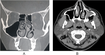
His general physical examination and systemic examination was normal. Ophthalmology evaluation was done, vision and ocular movements were found to be normal. He had 2 mm of enophthalmos and 4 mm of hypoglobus. Anterior rhinoscopy and diagnostic nasal endoscopy revealed a septal spur on the left side with normal pink nasal mucosa. Non contrast CT PNS was done, which showed a relatively small left maxillary sinus with inward retraction of the walls. Floor of the orbit and anterolateral walls were seen to have thinned out with resulting increase in the orbital volume causing hypoglobus (Figure 1A and Figure 1B). There was lateralization of the left uncinate process with obstruction of left osteomeatal complex. Complete opacification of left maxillary sinus was seen. The rest of the paranasal sinuses were normal. A diagnosis of Silent Sinus Syndrome was made.
 Figure 1: A,B) CT PNS coronal section and axial section showing small maxillary sinus on the left side, with inward retraction of the walls and thinned out bone of the sinus with hypoglobus and opacification of the sinus. View Figure 1
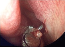
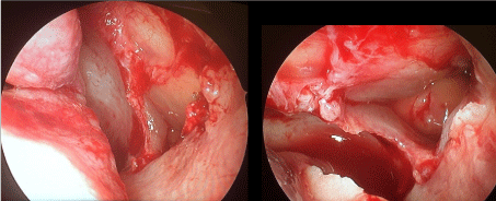
The patient underwent endoscopic middle meatal antrostomy on the left side under general anaesthesia. Careful dissection of the lateralized uncinate process was done to avoid injury to the globe. Thick mucoid discharge was suctioned out from the maxillary sinus (Figure 2). A wide antrostomy was done to ventilate the sinus. Prolapsed globe was seen through the antrostomy (Figure 3A and Figure 3B).
 Figure 2: Thick mucoid discharge seen from the maxillary sinus following uncinectomy. View Figure 2
 Figure 3: A,B) Prolapsed globe seen through the wide middle meatal antrostomy. View Figure 3
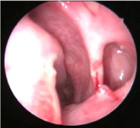
He was under regular follow up and after 4 months post-surgery, he had a well ventilated and normal maxillary sinus mucosa (Figure 4).
 Figure 4: Well ventilated and healthy maxillary sinus seen through the antrostomy, 4 months postoperatively. View Figure 4
Silent sinus syndrome, also termed as imploding antrum, is a rare disease with clinical features of spontaneous painless enophthalmos and hypoglobus, in the absence of significant symptomatic sinonasal disease. In both previously reported cases [2], and in the study done by Rose, et al. [6], the condition seems to present in third to fifth decades of life, similar to our case. There is no gender predilection [7]. The condition is non-progressive in the long term.
It was postulated that chronic hypoventilation of the maxillary sinus secondary to obstruction of the osteomeatal complex causes a negative pressure effect leading to inward retraction of the walls of maxillary sinus [8]. Bone resorption due to increased osteoclastic activity from a chronic inflammatory state, results in thinning of the orbital floor. With the weakening of orbital floor and the loss of sinus volume, the orbital content lacks its support and manifests as enophthalmos. There are proponents of a congenital theory, suggesting congenital hypoplasia of maxillary sinus with obstruction of the sinus ostium as the cause [2]. However there is enough evidence to prove that this is an acquired condition. Hourany, et al. reported a case of silent sinus syndrome in an adult male who had a normal maxillary sinus in a CT done ten years ago for some other reason [9]. Kruger, et al., in one of their case report of silent sinus syndrome seen in scuba divers, suggested barotrauma as a possible aetiology [10]. A similar aetiology was also seen in a 20-year-old male with silent sinus syndrome reported by Saldanha M, et al. [11]. The final pathway seems to be obstruction of maxillary sinus ventilation and mucous drainage. But surprisingly, incidence of silent sinus syndrome is rare compared to high prevalence of maxillary sinus obstruction [9]. Most of the reported cases of silent sinus syndrome involve the maxillary sinus. Silent sinus syndrome of frontal sinus, due to the obstruction of frontal recess, by a large type 3 Kuhn cell, was reported by Naik, et al. in 2013 [12].
Table 1 shows cases and series of Silent Sinus Syndrome reported in the literature [13-18].
Table 1: Review of literature. View Table 1
Diagnosis of silent sinus syndrome is confirmed by CT PNS, which show typical characteristic features of inward retraction of sinus walls causing reduction of sinus volume. The affected sinus almost always shows partial or complete obstruction of infundibulum, which is supposed to be the initiating factor. Illner, et al. in his case report noted lateralized uncinate process with opposition to the inferomedial orbital wall [3]. A similar finding was seen in our case as well. The downward displacement of orbital floor results in increase in the orbital volume causing enophthalmos [3].
Management of silent sinus syndrome is surgical, by performing a wide middle meatal antrostomy leading to restoring of sinus ventilation and drainage of mucous from the obstructed sinus. In the past, procedures like Caldwell-luc surgery and transconjunctival repair of orbital floor was done [4]. But at present, endoscopic middle meatal antrostomy is the preferred method. The surgeon needs to be very careful due to the lower than normal position of the globe. Risk of injury to the orbital contents is much higher than in usual patients undergoing endoscopic sinus surgery. After the surgery, sinus configuration may remain unchanged, improve slightly or revert to normal over time [3].
Repair of orbital floor can be done as a primary procedure or at a second stage later. Both alloplastic and autogenous materials are available. The more common alloplastic materials are titanium plates, nylon sheets, and Medpor. The most common autogenous materials are septal cartilage and split calvarial bone. The goal is to have it rest on a stable bone. Most commonly resting it on the orbital rim is possible, since this does not usually collapse with the floor. Usually, orbital rim and the floor is approached via transconjunctival or subcilliary incisions [5,13].
There is evidence to support correction of sinonasal and orbital repair as a two stage procedure [5]. There are reports which have shown spontaneous resolution of enophthalmos and hypoglobus following only aeration of maxillary sinus without repair of orbital floor [5,19,20].
As many patients recover from enophthalmos with only endoscopic middle meatal antrostomy, it is suggested to plan orbital floor repair as a second stage, if there is no improvement.
Silent sinus syndrome is a rare disease of the maxillary sinus with features of painless enophthalmos and hypoglobus resulting from collapse of sinus walls secondary to obstruction of the maxillary sinus ostium. Diagnosis is confirmed radiologically. CT PNS shows thinning and inward retraction of sinus walls, with obstruction of sinus ostium and opacification of maxillary sinus. Treatment is endoscopic middle meatal antrostomy, with utmost care, avoiding injury to the orbital contents due to the lateralized uncinate process and lower position of the globe. There is evidence that orbital repair may not be needed for many patients. Hence it is better to stage the procedure. Ophthalmologists and Oro maxillo facial surgeons also need to be aware of this rare disease as many patients first present to them.