Health indicators show a health-related characteristic that allows the study, evaluation and conclusion of a health process. Therefore, it allows us to influence public health policies to obtain an improvement in the health status of the population.
Iron deficiency is one of the most prevalent forms of malnutrition, without quality of life and can present in anaemia, with reduce of hemoglobin (it causes 50% of anaemia around the world) and in ferropenia, iron deficiency, without anaemia. The iron deficiency has a very heavy rates of mortality associated of it.
The iron element is a d-block transition element with an oxidation changeable ranging between -2 and 6+ which makes iron to be in different biological ways: ferrous (Fe2+), ferric (Fe3+), or ferryl (Fe4+) [1] states [1]. This capacity allows iron to bind to manifold ligands/proteins of the human organism, which are essential for all living cells; to participate in many metabolic and energetic pathways, like a production of energy in mitochondria, binding and transport of oxygen, regulation of cell growth and differentiation, synthesis and packaging of neurotransmitters and degradation of catecholamines [2], synthesis of the deoxyribonucleic acid or DNA [3] and others.
As free iron is toxic to cells, it is stored complexed to protein as ferritin. Thus, the serum ferritin level correlates with a total body iron, could discard ferropenia (iron deficiency without anaemia) and establish he quality of life.
Women, especially in their fertile stage, tend to have lower values because of menstrual losses, even during normal menses. In this small text we will try to reflect on how ferritin can be a parameter of evaluation of quality of life and normality of body functions.
Health indicator, Ferritin, Iron element, Iron deficiency, Erythropoiesis, Normal values, Quality of life
A health indicator is any health-related characteristic of a person or a population that demonstrates the magnitude of a health problem to reflect the change in the health status of a population throughout the time, to show differences in health among different populations and to assess to what extent the objectives of programmed have been achieved. These are variables depending on the countries, and the population in surveillance, according to the main health problems that this population faces [4]. The World Health Organization - WHO has more than 100 health indicators, classified by health status, health system, health services offer, and risk factors [5].
In other countries, the Healthy Life Years (HLY) are an indicator of health and contemplate the years in which people can live with diseases and health problems that cause them suffering and loss of quality of life, even if that do not produce the death immediately. This occurs in diseases and chronic symptomatology being often converted in limitations of leisure activities, daily life, work activities besides to be disabling [4].
Between 14% and 27% of primary care consultations are motivated by asthenia, which is 3 times more mentioned by women than by men and iron deficiency may be the cause [6]. The fatigue does not translate into death, but it does translate into poor quality of life, affecting the activities of daily living, as well as work activities, and productivity. It is at the bottom a public health problem.
Iron is a d-block transition element with an oxidation changeable ranging between -2 and 6+ which makes iron to be in different biological ways: ferrous (Fe2+), ferric (Fe3+), or ferryl (Fe4+) [2] states [1]. This capacity allows iron to bind to manifold ligands/proteins of the human organism, which are essential for all living cells; to participate in many metabolic and energetic pathways, since it can be reversibly oxidated (Fe3+) and reduced (Fe2+) -conferring iron with specific and needful properties in energy production, including electron transfer reaction on mitochondria, binding and transport of oxygen, regulation of cell growth and differentiation, synthesis and packaging of neurotransmitters and degradation of catecholamines [2], synthesis of the deoxyribonucleic acid or DNA [3] and others.
Iron-containing compounds in the human system are divided in 4 groups: heme-containing enzymes such as haemoglobin, myoglobin and cytochromes a,b,c (oxidases) and subtypes [7], MAOS enzymes like monoamine oxidases, tryptophan hydroxylase and tyrosine hydrolase [8], GABA transaminase: γ-aminobutyric acid and glutamate decarboxylase and t4-5'Deiodinase [9] and anothers; non-heme enzymes 4 such as metalloflavoproteins: 1-Xanthine-oxidase, NADH deshydrogenases, succinic deshydrogenases and α-glycerophosphate deshydrogenase, they are so important for the normal performance of krebs cycle and cellular replication and processes of mitosis 4. Iron element is participating as co-factor: ferredoxins [10], aconitase [11], proline and lysine hydroxylase [12], aldehyde oxidase 4 and another's; and in compounds of iron metabolism such as Iron Response Element (IRE), transferrin or ferritin in subunity L or H [13,14]. All of them are shown in the following schedule [3] (Table 1).
Table 1: Presence of iron element on the human body and its functions. View Table 1
Menstruation is one of the main causes of iron deficiency. In healthy women with a physiological normal menses the monthly loss varies between 40 ml and 495 ml of blood that means the equivalent to 16 mg and 200 mg of iron respectively, which means a loss of 7 mg of iron per day [15,16].
There are other aetiologies of iron deficiency such as a poor iron diets (due to malnutrition or the pressure of female beauty marketing against women), gastrointestinal diseases/symptoms, drugs administration, epistaxis, and renal diseases. H. pylori infection, diverticulitis, angioplasties intestinalis, autoimmune gastrointestinal diseases such as chronic disease and ulcerative colitis, or the bleeding haemorrhoids are the possible causes which must be discarded. Some of them are even very rare, very difficult to diagnose and there is a tendency to be misinterpreted and misdiagnosed as mental pathology or devalued.
Cancers are other causes, especially in the reduction of erythropoiesis in the lymphomas, iron loss such as gastrointestinal cancers. There are other causes such as hereditary factors such as sickle cell anaemia, or human habits such as alcohol consumption.
There are two types of iron available in the food. In its organic form, iron heme or Fe2+, ferrous state, which is what belongs to the haemoglobin of blood erythrocytes usually found in foods of animal origin such as red meat, pork, animal livers and chicken heart. Fe3+, iron non heme, or inorganic form, usually found foods of plant origin such as vegetables, some dry fruits (nuts) and small amounts in green vegetables such as spinach, chocolate and sugarcane.
Iron is absorbed from the diet or stores circulates in the plasma bound on the intestinal surface. Its absorption is favoured by the presence of acid and solubilizing agents such as sugars, so ferrous or divalent iron is more easily absorbed than Fe3+. This last one can be facilitated in the presence of vitamin C to the conversion of Fe3+ to Fe2+. Within the enterocyte Fe2+ can be stored as ferritin, or if there is a need for iron on the part of the organism it is transported via plasma through the transferrin protein. This last one is an 80 KDa glycoprotein synthesized and secreted by the liver, in addition to the retina, testicles and brain. By passing through the tissue, transferrin facilitates the release of iron to the cells. When this transport capacity is saturated, Transferrin Saturation, iron can circulate freely through the plasma, and can contribute to cellular damage in overload cases.
Transferrin and transferrin saturation as well as the iron element are parameters of ferric metabolism analysed in iron deficiency anaemia and iron deficiency as well as ferritin.
Free iron is toxic to cells, and the body needs to protect cells. For that the organism elaborated several protective mechanisms to bind iron in various tissue compartments. This way, the iron is stored complexed to protein as ferritin or hemoseriden. Ferritin is a complex globular protein, accumulates within cells of reticuloendothelial system, kidney and all cells, especially in the synthesis of composts with iron (eritroid precursors) and in the ferric reserve and metabolism (macrophags and hepatocits). Free form is apoferritin, when ferritin is not in connexion with iron element. Ferritin is a micromolecule with molecular weight 600000 dalton and it is made in the internal part by a polipeptidical fraction and until 4000 atoms of iron element and in normal situation this represents until 25% of total iron in a human body. Iron in the ferritin can be extracted for release by the RE system. So, ferritin serum level correlates with the total values of iron stores in organism stores and it is the most convenient laboratory test to estimate iron reserves. Its determination and value are needful to ensure a lot of iron activities in the human system.
The ferritin also could be used when the body needs of iron element and change to apoferritin. Ferritin is made on kidney, one of most important is reserve there and another is on the other cells. Then the remaining is available at the blood level and this is directly the representative part of the iron stored in the organism. It is therefore a ferric parameter very indispensable in the differential diagnosis of iron deficiency anaemia, ferropenia with or without anaemia, evaluation of ferric status. In some situations of inflammatory processes, ferritin may be increased, even in inflammatory anaemia, autoimmune diseases such as rheumatoid arthritis and others with hepatic injury [17].
Thus, its determination is very important to the efforts in the pathologies diagnosis and assessment of quality of life related to other basis pathologies or primary.
What if we focus on ferropenic anaemia and on ferropenia as result of absence of iron? What are the mínimum values to eliminate the anaemia and ferropenia?
Each erythrocyte has a billion atoms of iron and incorporates 2 × 10²⁰ atoms more every day. Iron is essential in erythropoiesis. To ensure enough iron values at marrow level, the values of serum ferritin must be higher than 50 ng/dL [18], otherwise the erythropoiesis on bone marrow does not take place even with values of ferritin above 70 µg/dL [19], and moreover, several studies refer that these values must be superior to 100-150 mg/L to exclude iron deficiency and iron deficiency anaemia [20]. Figure 1 [18] establishes the relationship between the iron levels and the increase of its body reserve. Above certain levels of the iron reserves there is no iron-deficiency anaemia (Level I); the plasmatic values of iron raise progressively and achieve a certain range, the iron deficiency stops (level II). Above this value, the iron rise results in the increase of reserves until they stabilize.
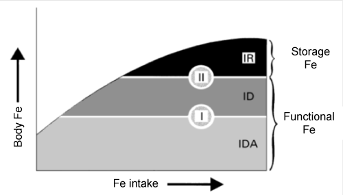 Figure 1: Relationship between iron reserves and iron in blood. In: Coenen JL, et al. [19]. View Figure 1
The iron deficiency anaemia [9] is characterized by an iron fall in the organism, and presents a progressive development with several stages, from a gradual reduction of the iron stores to a decrease of the erythrocyte size.
It concerns the following phases:
1. Deficiency Anemia pre-latent: characterized by the disappearance of the iron storage (bone marrow iron: serum ferritin lower than 40 ng/dL [21]), with a very decreased values of sideroblasts, below 5% (reference values of 30-40%). Iron in the blood can be normal and the plasmatic ferritin values can be decreased reflecting the absence of the iron of reserve.
2. Latent Iron Deficiency: decrement of the Transferrin Saturation Index (TSI), below 12% (reference values: 30-35%). Iron blood levels vary, although generally they are decreased as plasmatic ferritin does. It also goes with an increase of the Transferrin Saturation Capacity (CST), and a reasonable raise of % microcytes with a medium corpuscular volume below 60 fl.
3. Iron Deficiency Erythropoiesis: decay of the concentration of haemoglobin, macrocytosis and hypechromía (anaemia microcytic-hypochromic). It goes along with a reduction in all parameters of the iron metabolism: blood iron, ferritin and transferrin saturation index.
Because of its great action in the human system the iron deficiency is the reason of many hematologic and non-hematologic symptomsand signs: decreases in the amount and enzymatic activities in the Krebs cycle at mitochondrial level and others; brain alterations, fatigue, hypomielinization, increased absorption of lead and cadmium, memory decrement; alteration of neurocognitive development and behaviour in children [22], cardiac palpitations; liver, gastrointestinal tract alterations such as glossitis, angular stomatitis, less capacity to tolerate cold, hands and feet cold [4,6], lower immune response [1], thermoregulation mechanism, immune system, alterations of DNA synthesis and collagen formation that can make a strong contribution to the keloid scars [3].
Concerning to Attention Deficit Hyperactivity Disorder (ADHD), an increasingly frequent disorder in children and adolescents, characterized by neurological disorders, attention and memory deficit, nervousness, distraction, poor school performance, it is essential to rule out iron deficiency, since it is a differential aetiology [23].
Iron element protected against depression, speciality the maternal depression during the breastfeeding time.
Seems to a mental function on the protection a depressive nerves state, and mainly on the maternal depression during pregnancy. A retrospective study conducted in the Women's Health Concerns Clinic at St. Joseph's Healthcare Hamilton, analysed between 2009 and 2016, all women in the middle to late pregnancy (> 20 weeks gestation) were categorized as either iron deficient or not. The issues suggest that iron deficient pregnant women scored significantly higher on the EPDS (10.14 ± 5.69 vs. 7.87 ± 5.75; P = 0.03) and were more likely to develop antenatal depression (45% vs. 25%; P = 0.02) compared with women who were not iron deficient. Even, the odds of developing antenatal depression were two and one half times higher among iron deficient women (adjusted OR 2.51; 95% CI 1.14-5.52). The authors conclude that iron deficiency may be an important risk factor to maternal depression during pregnancy.
Other studies support that adjuvant therapy with iron in some situations can improve ferritin levels. A recent updated systematic review and meta-analysis about the treatment of Helicobacter pylori infection recommends adding iron therapy on the H. pylori eradication therapy when the ferritin and haemoglobin levels increase, and the way to contribute to the quality of life in a chronic disease [24].
The WHO recommends the iron supplementation in situations such as the menses period [25]. During this physiologic process women can take ferrous iron supplements, especially before lunch or dinner (coincides with the time of greater production of gastric acid, reducing the digestive intolerance to this), preferably accompanied with foods with vitamin C such as orange, lemon, tomato, peppers juices among others. The vitamin C promotes iron absorption at intestinal levels. The iron administered in its ferrous form presents better absorption than ferric iron.
The iron supplementation, as an adjuvant treatment, is to many other pathologies, very helpful in recovery and quality of life.
Recently another study realized in women with metrorrhagias (n = 236) during the menopause stage, divided in 3 groups, according to non-anaemics, iron-deficient anaemics (n = 63) and ferropenic anaemic (n = 140) evaluated the quality of life at 6 and 12 months after the primary treatment using the RAND-36, EUROQol 5 Dimensional and mental well-being questionnaires. At 12 months the 3 RAND-36 domain scores increased more in women who initially had anaemia than those who did not have (energy, p = 0.002; physical functionality, p = 0.04; social function, p = 0.05) and presented low levels in scores of anxiety (p = 0.02) and depression (p = 0.002). In women with ferropenies it was necessary 5 years to achieve normal ferritin levels. Such results allowed the authors to recommend the supplementation with iron in such situations and monitoring at 4 months to assure the quality of life [26].
Two studies in 2003 [27] and 2012 [28], point out the importance of therapy with iron in non-anaemic women, who had ferritin levels below 50 µg/dL and in whom the unexplained chronic fatigue has decreased significantly. Other medical causes of unexplained chronic fatigue were discarded and other symptoms such as anxiety and depression were considered and in the last study, quality of life and biochemical parameters of iron metabolism were also evaluated.
With a sampling of 136 and 111 cases respectively, studies concerning cases and control subjects were performed; the groups treated with iron during the period of 1 and 3 months respectively, presented recovery in the decrease of the fatigue levels from 1.82 vs. 0.85 of placebo group, with a difference of 0.97 (Confidence Interval, CI of 95%, standard deviation (SD): 0.32-1.62, p = 0.004) for the firststudy, with a scale for unexplained chronic fatigue - visual analogue scale, classified from 0 to 10 (maximum levels of fatigue) and a questionnaire of fatigue (from 0 to 40 points) (Table 2); for the second, using several scales of fatigue assessment, a decrease of 47.7% on the iron-treated group versus a 28.8% on the control group with a difference of 18.9% (CI: 95%, SD: -34.5 to -3.2) was verified. In this last study significant effects of increase in haemoglobin, ferritin, mean corpuscular volume and saturation of transferrin were found within a one and a half month of treatment.
Table 2: Change in level of fatigue after one month in woman receiving iron or placebo for unexplained fatigue in absence of anaemia. Values are means (standard deviations) unless stated otherwise. View Table 2
Another transversal non-published study were conducted in several hospitals in Barcelona [29] with 144 women (between 14 and 50-years-old, all presenting normal menstrual cycle) divided in two groups: the control group with ferritin levels higher than 50 ng/dL and the iron-treated group with lower ferritin levels. Other diseases were discarded.
45 women were included in the control group and 99 were included in the group with levels of ferritin lower than 50 ng/dL. Symptoms and signs were analysed and a Hamilton test which evidences quality of life was carried out. The objective was analysing the presence and grade of symptoms/signs and the personal evaluation of the quality of life in both groups, also to verify whether ferritin levels under 50 are related to a lower quality of life.
The control group presents a significant reduction of generalized symptoms related to an absence of iron; meanwhile, the group with ferritin lower than 50 ng/dL presented the following results (Figure 2).
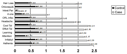 Figure 2: Symptoms related with low ferritin level. Graffic originally published. In: Ruiz-Cantero MT, et al. [30] View Figure 2
Asthenia, palpitations (p < 0.005), shakiness (< 0.034), ecchymosis at mild knocks (p < 0.01), hair loss (p < 0.036), brittle nails (p < 0.037), general muscle pain (p < 0.01), cold limbs, frontal headache, irritability, attention and memory diminishing and sleepiness (p < 0.037).
The Hamilton test and the Nottingham Health Profile also show that the control group presents lower levels of emotional state changes, sleepiness, nervousness, fatigue, sadness, difficulty in their relationship with other people, difficulty keeping control in stressful situations, difficulty solving problems, sexual life problems and crying easily than the group of women with iron deficiency.
The difference of symptoms between the control and case groups was significant in more than 50% of the elements of analysis, highlighting the importance of ferritin levels higher than 50 ng/dL.
There is some difference between a normal value or a reference values?
Conscious of the importance of ferritin in symptoms related to quality of life and to improve many other pathologies there are a vary of recommendations related to ferritin minimum value considered normal. The Department of Health of Catalonia, Spain, recommends the measurement of ferritin in all women of child-bearing age, pregnant or not, also the assessment of that when higher than 50 ng/ml as the lower limit, within an evidence-based medicine [6].
There are several studies referring to the importance of values of ferritin above 50 ng/dL to ensure that vital functions of the organism as erythropoiesis and ensure the quality of life.
Thus, why in many health centres the values of ferritin considered as normal are below 50 ng/dL? [30] Why do many those centres have different values of ferritin (and other biochemical parameters in the hemogram) for men and women (Table 3)? And why in these differences lower values for women are considered normal comparing with the values for men?
Table 3: Reference values of ferritin in public and private health centres in Spain. View Table 3
Is there any difference between the normal and reference values?
According to the Spanish Royal Academy [31], the reference value from the Latin word "refĕrens, -entis, referent, "it refers to something in relationship to something else depending or similar to another thing/value".
That is, the reference values indicate limits of evaluation in certain parameters on the population attended in centers and laboratories. It refers to the values of frequency in a sample and this definition can be confused with that of the normal values, which are the values for a population with a good quality of life and correct vital functions. They are the values of normality, of the natural state. Normal, derived from Latin normālis means something in its physiological natural state [31].
So, is it normal that women have values of ferritin lower than men when women lose up to 475 ml of blood monthly during a great part of their life? Or is it something frequent?
Yes, it is frequent that most women in child-bearing age have a negative balance of iron, but it is not normal, as the iron lack involves important alterations in vital functions of the organism and contributes to a poor quality of life of women. Biologically there is no evidence that this difference between men and women is normal/natural. Mammalian females present in many cases higher haemoglobin levels than males of their species.
In many cultures where iron kitchenware is used, women present elevated levels of haemoglobin (19.4 g/dL in Ethiopian women) [32], such as in our grandmothers' time because they cooked with iron kitchenware and they had iron diet. Later it was proved the decreased haemoglobin values are not "normal" for the women. The iron element does the same functions on the human body independently of whether it a women or men. It is even independent of the chronological age the woman has.
Recently a study performed in perimenopausal women between 40 and 55-years-old (age range with iron deficiency risk associate because there is possibility to the myomas such as one of the most important causes for the metrorrhagias) portrays how the iron deficiency remains a social and economic burden in European countries and others [33]. The authors conclude also that, underdiagnosed and undertreated, this problem has a strong negative impact on women's quality of life and put them to greater exposure to other pathologies than without iron deficiencies.
Ferritin is a parameter that allows to know the reserves of iron in the human organism. Iron is an essential element in the incorporation of hemoglobin, a protein that carries throughout the body oxygen allowing the normality of cellular functions to obtain energy. Iron also participates in multiple metabolic and energetic ways, as a coenzyme to allow them to work in their normality. Thus, by all the actions of iron in the human body, and by the structures in which this element incorporates, make relevant its normality values for a better optimization of cellular activities. Therefore, ferritin, being a parameter of evaluation of the iron reserves in the human organism, not only allows to agree with the presented clinical of iron deficiency but also as a predictive element of that clinic to avoid and to prevent them. Women, independently of the age range, may be the most benefited from this blood parameter, now suggested as a health indicator, since in a common but not normal way, they have iron deficiency. Thereby, ferritin as a health indicator would not only serve to detect early low iron values and relate them to the clinical and symptoms presented but also with the supplementation, if necessary. With ferritin levels above 50 ng/dl, according to what has been already mentioned, allow us to evaluate human health, to prevent it, and to maximize its optimization by ensuring that erythropoiesis is found in its normal productivity and other functions where iron collaborate are being developed in the most physiological and normal way possible, also collaborating directly on the quality of life. The quality of life render into a well being of the human person and thus contributes to the effective performance of work activities and personal fulfillment. This way, the ferritin value can play an indispensable to evaluation the health and quality of life by the today's societies.
Using the ferritin as health indicator is a good option to the prevention of many diseases and contribute to the well-being of each people.
CAPS: Centre d'Analisis I Programmes Sanitàris, Barcelona, Spain.
We acknowledge the contributions of Doctor Lucia Guilhermino and Doctor Willian dos Santos for all help in this revision.