These case reports evaluate oral supplementation with a resveratrol based red wine supplement, containing vitamin D3 and labile iron binding inositol hexaphosphate. We appraised function Visual Acuity (VA) and Structure Domain Optical Coherence Tomography (SDOCT) in 3 geriatric patients with atrophic AMD, without clinical alternatives. One female and two males (ages 96, 84, and 85 respectively) were treated under compassionate care guidelines and prescribed Longevinex® (a resveratrol based oral supplement), on a case-by-case-basis. Snellen VA was measured at every visit and photos/SDOCT were also taken to document and track retinal changes. Continual improved VA and retinal integrity were observed. Persistent enhancement is noted on follow up for all patients, continuing for several years for the 96-year-old, thus far. Low dose, low molecular weight nutraceutical molecules can render benefits in the aged with atrophic AMD. Improvement of subjective visual function and retinal integrity is consistent with our previous published reports, as well as reports across several medical disciplines, on the effectiveness of supplementation with a resveratrol-based nutraceutical. This account supports restoration of photoreceptor/RPE function, when other measures have been exhausted by a retinal specialist.
Age related Macular Degeneration (AMD), Retinal Pigment Epithelium (RPE), Resveratrol (RV), Epigenetics
Age Related Macular Degeneration (AMD) is the leading cause of vision loss in aging western societies, accounting for half of patients attending blind rehabilitation facilities. AMD eventually results in both loss of reading and driving vision, with resultant loss of geriatric independence [1]. It is most commonly characterized by degeneration of the retina with accumulation of waste called retinal drusen, thickening of Bruch's membrane as well as retinal and Choriocapillaris circulatory atrophy [2].
Resveratrol (RV), the medicinal component found in red wine, was discovered in the 1940s. It was later found to have broad-spectrum anti-cancer, anti-cardiovascular disease, and anti-inflammatory properties [3]. This weak antioxidant is concentrated in red wine via fermentation, from grapes grown under stressful conditions of northern latitude, high altitude, and cold temperature. RV works through a process known as hormesis - the upregulation of endogenous antioxidant protective systems [4].
RV is a small molecular weight, polyphenol phytoalexin, able to transit all cells, similar to allicin found in garlic. RV sparked interest in the world of medicine because of the French Paradox, where consumption of red wine showed positive health benefits of decreasing cardiac mortality in a smoking population consuming fatty foods, as shown by the Melbourne Collaborative Research Study [5,6]. With its comprehensive suite of biological actions, we have also observed healing effects in the human retina, in short and long-time periods [7]. Quite recently, we demonstrated stabilization/improvement of the dark adaptation curve, in AMD patients, employing a highly sensitive, specific and validated measure of photoreceptor/RPE health affected up to 3 years before the appearance of retinal drusen. (Accepted ARVO 2017 abstract, Baltimore, Maryland, manuscript in review) Even though dark adaptation testing was not performed for these reports, and the stage of disease was more advanced than the mentioned above study, it is worthy of exploring resveratrol's healing effects.
RV's multiple actions against systemic disease contribute to its broad biological actions. RV manifests germicidal, anti-inflammatory, vascular, brain, metabolic, and anti-carcinogenic properties. The latter include prevention of all 3 cancer stages: Initiation, promotion, and metastasis [3]. As an anti-inflammatory modulator, RV decreases COX2, CRP, and TNF. RV is considered to have superior anti-inflammatory action when compared to NSAIDs. In the vascular system, RV has anti-cholesterol, anti-hypertensive, anti-platelet, and anti-plaque effects, as well as mitigation of early atherosclerotic markers in a randomized human clinical trial [8]. RV has similar anti-clotting and analgesic qualities as aspirin without gastric complications, such as hemorrhagic risk. Neurologically, RV acts as an antidepressant by inhibiting Monoamine Oxidase (MOA), and reducing beta amyloid found in brain plaque and retinal drusen. Metabolically, RV reduces blood sugar and rescues pancreatic beta cells.
One female (age 96) and two males (age 85, 84), were prescribed Longevinex® under the compassionate care medical center guidelines of the Captain James A Lovell Federal Health Care Center (North Chicago, IL). The nutraceutical prescribed (Longevinex®) contains 100 mg trans resveratrol, red wine polyphenols, vitamin D3, and rice bran IP6. The recommended dosage is once daily by mouth. Longevinex® is an RV matrix of small molecular weight epigenetically active molecules having the ability to cross the blood aqueous and blood-retina barrier. The combination of a low dose Trans RV (100 mg), red wine polyphenols, 25 mg quercetin, fisitin as well as 1200 IU vitamin D3, and 17 mg rice bran IP6, has been shown to be synergistic [7]. Additionally, this brand of RV was chosen due to its published safety profile [7]. Patients returned for periodic eye examinations where Snellen visual acuity retinal macula spectral domain optical coherence tomography (SDOCT) (Cirrus Model 4000, Carl Zeiss Meditec, (Dublin, CA USA) and OptoVue RTVue (Fremont, CA) were used to document retinal health change. Color fundus photographs were taken for Case 1 and Case 2 using an OPTOS Wide field camera (Sydney, Australia).
A 96-year-old Caucasian female veteran presented to our clinic with her son with complaints of progressively decreasing vision at both distance and near OS > OD for many years secondary to both exudative and non-exudative AMD. She was diagnosed with atrophic AMD and moderately severe nuclear - cortical cataract and 20/150 Snellen visual acuity in her right eye (OD). She was diagnosed with exudative AMD status post 10+ intravitreal anti vascular endothelial growth factor pharmaceutical (anti-VEGF) injections. She also has a prosthetic intraocular lens in her left eye and light perception only (OS).
Systemic history includes sciatica, osteoporosis, osteoarthritis, Chronic Obstructive Lung Disease (COPD), depression, and history of pulmonary embolism. The patient is currently taking Warfarin 2 mg once daily by mouth and Calcium carbonate with 1000 IU vitamin D3 once daily by mouth. The patient denies smoking, alcohol consumption, and illicit drug usage. She is ambulatory and responsive. The patient was monitored every six months for thirty months (Baseline and 5 visits). Her son accompanied her at each visit and confirmed changes in his mother's ‘activities of daily living'.
An 85-year-old Caucasian male veteran presents to our clinic for driver's license renewal. He complains of seeing "holes" in his vision, at distance while driving, and at near while reading. The patient was diagnosed with primary open angle glaucoma, non-exudative AMD OS > OD and is pseudophakic (implant, post cataract surgery) both eyes. Entrance best corrected visual acuity was 20/50-2 OD, with the patient consistently missed the 2nd and 3rd letter of the 5 letter Snellen VA line, and 20/50-2 OS, with the patient consistently missing the 3rd and 4th letter. The resulting bilateral defect manifested as a disabling binocular central foveal visual field defect. A retinal photo was taken to document the appearance of the retina at time of evaluation (Figure 1a).
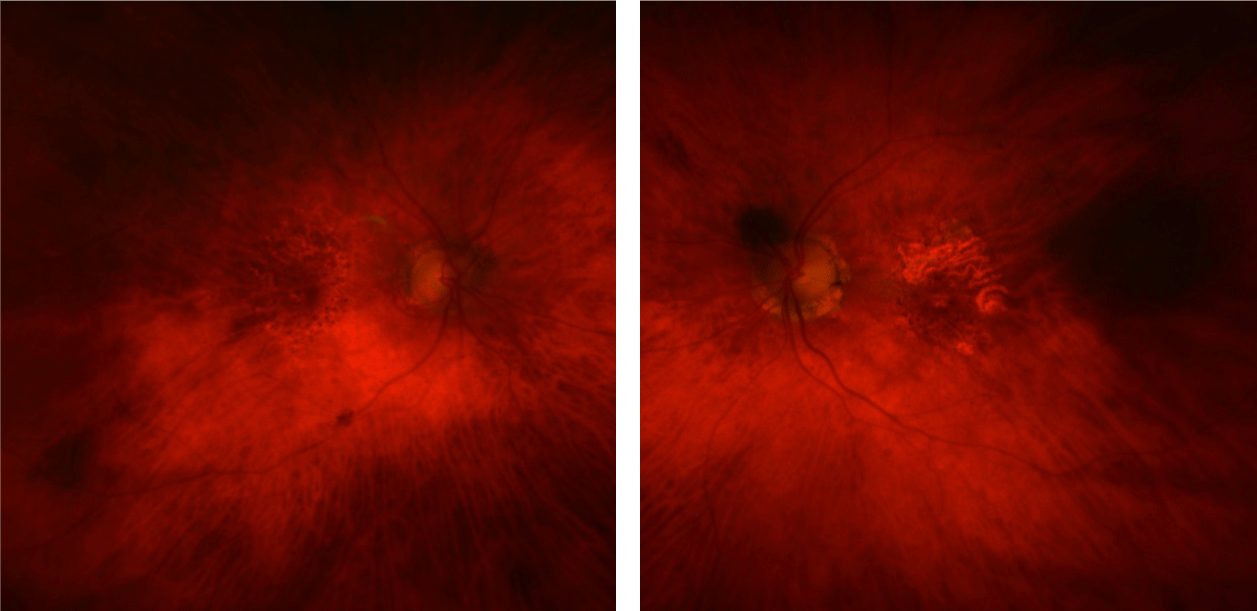 Figure 1A: 86-year-old male with atrophic AMD, decreased vision, and presence of bilateral central scotomas resulting in an absolute central binocular defect impeding his driving vision.
View Figure 1A
Figure 1A: 86-year-old male with atrophic AMD, decreased vision, and presence of bilateral central scotomas resulting in an absolute central binocular defect impeding his driving vision.
View Figure 1A
Systemic history includes hypertension, hyperlipidemia, thrombocytopenia, gout, chronic kidney disease and hypothyroidism. Current medications: Latanoprost ophthalmic drops daily, allopurinol 500 mg daily, Lisinopril 40 mg daily, pravastatin 80 mg one-half tablet by mouth daily with evening meal and terazosin 5 mg capsules once nightly by mouth.
An 84-year-old Caucasian male veteran presented initially with complaints of blurry vision distance and near OU. He had difficulties driving as well as reading standard newspaper text. He has a nuclear sclerotic cataract OD and prosthetic intraocular lens OS without secondary posterior subcapsular post-surgical opacification. The patient also has a mild epiretinal membrane (thickening of the internal limiting membrane of the retina) and is a bilateral glaucoma suspect based upon suspicious optic nerve cupping and increased intraocular pressure. Systemic history includes hypothyroidism. He is currently using latanoprost 0.005% ophthalmic drops daily, levothyroxine 0.088 mg two tablets daily by mouth, vitamin B complex capsules every morning.
Gradual continued improvement of vision was noted at each visit both objectively and subjectively in the better seeing eye for all 3 cases. In Case 1, initial VA was 20/150 OD, Light Perception OS eventually improving to 20/70 OD (3 lines improved) and 20/150 OS (now form vision), 2.5 years after RV-based nutraceutical treatment was prescribed (Figure 1b). However, there were no changes in the Optomap® color photographs (Figure 1c). S DOCT comparing initial visit with final visit OD shows reformation to a normal foveal contour and a decrease in signal scatter, suggesting improved RPE integrity (Figure 1d).
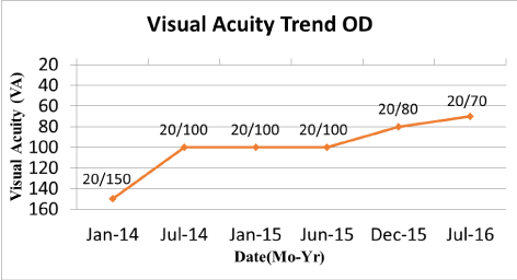 Figure 1B: 96-year-old Caucasian female patient demonstrates drastic Snellen visual acuity improvement, right eye, from 20/150 to 20/70 from Jan-2014 to July-2016 using Longevinex Advantage® for thirty months.
Supplementation was initiated after the first visit, but reduced to 50% dose by using Longevinex® Advantage formulation containing only 50 mg RV (1 capsule Longevinex Advantage®), as patient reported being "over energized".
View Figure 1B
 Figure 1C: 96-year-old female Optomap widefield retinal photos (OD retina (left images) and OS eye (right images) of the posterior pole comparing the patient's initial visit with the final visit 30 months later. No gross retinal changes are noted in the fundus photography even though the patient had continuous improved visual acuity and exhibited retinal remodeling on spectral domain optical coherence retinal imaging (SDOCT) (see Figure 1d). View Figure 1C
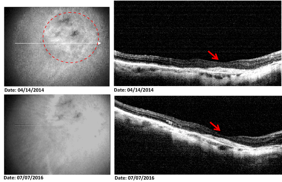 Figure 1D: 96-year-old female patient shows significant improvement in OD visual acuity, along with rebuilding of the fovea to a normal concave contour during the course of 2 1/2 years while she was using Longevinex® Advantage at 50% daily dose. More SDOCT retinal uniformity is noted in the foveal region corresponding to the markedly improved visual acuity of the patient.
View Figure 1D
Figure 1D: 96-year-old female patient shows significant improvement in OD visual acuity, along with rebuilding of the fovea to a normal concave contour during the course of 2 1/2 years while she was using Longevinex® Advantage at 50% daily dose. More SDOCT retinal uniformity is noted in the foveal region corresponding to the markedly improved visual acuity of the patient.
View Figure 1D
OCT showed decreased signal scatter in the foveal area (Figure 2a), indicative of RPE thickening. The initial VA was 20/50 - 2 OD, OS. Final VA was 20/25 + 2 OD, 20/25 + 1 OS (3 lines better) (Figure 2b). The patient was able to maintain his driver's license.
 Figure 2A: OCT 86-year-old SD OCT shows decreased signal scatter OD (the better seeing eye) in the foveal area, after Longevinex® supplementation which is indicative of RPE thickening. No changes were evident in his OS eye.
View Figure 2A
Figure 2A: OCT 86-year-old SD OCT shows decreased signal scatter OD (the better seeing eye) in the foveal area, after Longevinex® supplementation which is indicative of RPE thickening. No changes were evident in his OS eye.
View Figure 2A
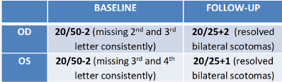 Figure 2B: Visual Acuity - Baseline versus follow up visual acuity in a 86-year-old male with atrophic AMD. Prominent improvement resulted after supplementation with a resveratrol-based nutraceutical supplement. View Figure 2B
Resolution of RPE disruption, more uniformity and thickening of the RPE in the foveal area was noted on OCT (and confirmed by our retinal specialist) (Figure 3a). Entrance VA was 20/60-1 OD, 20/50-2 OS. 1 lined of VA improvement (20/40) was noted in the better seeing eye at 1 month (Figure 3b). The patient now has the ability to read the newspaper clearly and has sufficient a sufficient VA to pass his driver's license test without a night restriction.
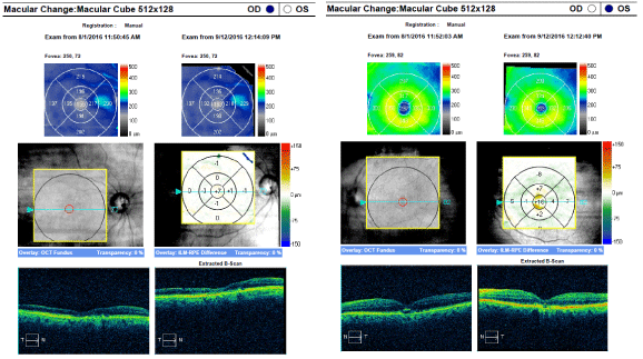 Figure 3A: OCT - 84-year-old male patient with RPE disruption is noted on SD OCT retinal image of the OS eye. More uniformity and thickening of the retinal pigment epithelium is noted in the OS foveal area, which was confirmed by a retinal specialist. No changes were observed in his OD eye. View Figure 3A
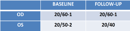 Figure 3B: Visual Acuity - Comparison between baseline and follow-up visual acuity in the better seeing eye (OS) post supplementation with Longevinex®. No changes were noted OD. View Figure 3B
The red wine polyphenols in the chosen combination are phytonutrients of the polyphenolic class and associated with disease prevention, due to their ability to epigenetically orchestrate helpful and harmful genes. Vitamin D3 is a potent anti-inflammatory, decalcifying agent, as well as a prohormone, for which several studies have shown its synergism with RV as well as the known association of Vitamin D deficiency with AMD status [7]. Lastly, IP6 is a labile iron and copper binding divalent mineral chelator derived from rice bran, and also aids in DNA repair and decalcification of the Bruch's membrane [7]. In the cases presented thickening of the RPE was observed, even though the tissue thins with age. AMD furthermore exacerbates RPE thinning [9,10]. Taken together, these cases are suggestive of improvement in retinal thickness and organization by resveratrol supplementation in helpless cases in patients when treatment options have been exhausted.
Nutraceutical intervention administered in low doses has the potential to render benefits to patients with advanced atrophic AMD. Improvement of visual acuity and retinal integrity is consistent with previous reports on the effectiveness of RV-based supplementation even in older AMD patients with advanced stages of the disease, for which few option remain. The oral supplement typically affects both eyes, with the less advanced retina receiving the best enhancement in structure and function. These cases suggest that even octogenarians with severe non-exudative AMD can have vision recovery after all medical resources have been exhausted by a retinal specialist. Yet today, we are left only with the limited set of nutrients from research conducted by the National Eye Institute, National Institute of Health USA [11]. Longevinex® has formally petitioned and been denied Fast Track evaluation by the US FDA [12]. Further research is warranted in order to prove the potential benefit of this nutraceutical supplement on advanced stages of macular degeneration in hopeless cases.
This article and the medical cases reported within are original clinical work supported by the Optometry and Ophthalmology Departments of the Captain James Lovell FHCC (Federal Health Care Center), North Chicago, IL.
Longevinex® was prescribed under compassionate care guidelines and informed consent was obtained in writing from the patient for the use of their health information.