Purpose: To present a case of long-term kissing choroidals in a patient following a glaucoma shunt procedure and to review related literature.
Materials and Methods: An 82-year-old patient developed hemorrhagic kissing choroidals after undergoing a glaucoma procedure. Due to underlying cardiac issues, he was unable to undergo choroidal drainage within the typical 2-3-week window, as he required continued anticoagulant therapy-likely a contributing factor to the complication. Instead, the patient was monitored for six months without intervention, during which moderate proliferative vitreoretinopathy (PVR) developed. Once operable, the patient underwent PVR repair with silicone oil tamponade. A review of relevant medical literature was conducted.
Results: The long-term choroidal detachment resolved, and the resultant PVR was successfully repaired with a favorable anatomic outcome. However, visual acuity remained unchanged due to the glaucomatous optic atrophy. Literature review indicates that standard management recommends drainage of kissing choroidals within 2-4 weeks to prevent adhesion of opposing retinal surfaces. However, cases of spontaneous resolution with notable visual recovery have also been reported.
Conclusion: While surgical drainage within 2-4 weeks is recommended to reduce the risk of PVR formation, long-standing kissing choroidals may still achieve significant anatomic and/or visual recovery, with or without intervention. The optimal timing for surgical management should be determined on a case-by-case basis, considering the patient's systemic condition.
Kissing choroidals, Proliferative vitreoretinopathy, Delayed treatment
"Kissing" choroidals are a rare but serious postoperative complication characterized by large choroidal detachments with central apposition, which can lead to profound vision loss and intraocular pressure derangement if not promptly managed (Hussain et al., 2018)[1]. Choroidal detachment most commonly occurs after glaucoma surgery, with reported postoperative incidences of 0.6–1.4% following trabeculectomy and 1.2–2.7% after tube shunt placement (Doniparthi et al., 2024)[2].These detachments result from either serous fluid accumulation or hemorrhage, with the latter being the mechanism in this case (Roa et al., 2019)[3].
This case presents a patient who underwent trabeculectomy then experienced a hemorrhagic choroidal detachment with central apposition. Management was complicated by anticoagulation with Eliquis for atrial fibrillation (A-fib) and coronary artery disease (CAD), delaying surgical drainage. During the waiting period, the patient developed proliferative vitreoretinopathy (PVR). Six months after the initial diagnosis, the patient underwent pars plana vitrectomy (PPV) with membrane peel, endolaser, and oil tamponade, leading to an anatomically successful outcome. This case highlights the complications associated with delayed intervention in hemorrhagic choroidal detachment and demonstrates the potential for a favorable anatomical and/or visual outcome with or without surgical intervention.
An 82-year-old male with a complex ocular history—including a failed corneal transplant following Descemet membrane endothelial keratoplasty (DMEK) in the left eye (OS), severe primary open-angle glaucoma (POAG), bilateral cataract extraction with intraocular lens implantation, and bilateral YAG capsulotomy—presented with worsening visual loss in his left eye. His medical history was significant for A-fib and CAD, for which he was on anticoagulation therapy with Eliquis.
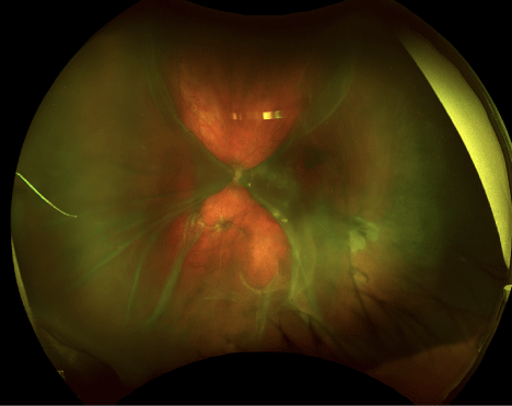
Initially evaluated by the glaucoma service, he subsequently underwent a trabeculectomy in the left eye for severe glaucoma. Postoperatively, he was prescribed mydriatic, steroidal, and antibiotic agents. Four weeks after surgery, he was referred to our retinal clinic, where a delayed massive suprachoroidal hemorrhage (MSCH) and choroidal detachment (CD) with central apposition were identified on B-scan and fundus photography (Figure 1). At this visit, his best corrected visual acuity (BCVA) was reduced to light perception (LP) OS, with an intraocular pressure (IOP) of 10 mmHg.
 Figure 1: Preoperative Fundus Photography of the Left Eye. Preoperative fundus photography reveals choroidal detachment with central apposition obscuring the visual axis. Proliferative vitreoretinopathy is visualized with irregular stellate folding of the posterior retina.
View Figure 1
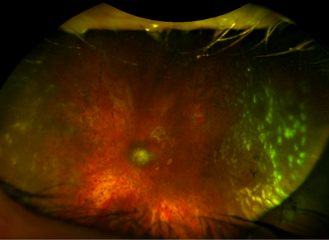
Drainage of the choroidal hemorrhage was discussed but ultimately deferred due to his anticoagulated status. He was monitored closely for six months, during which moderate PVR formation occurred. Once his cardiac condition stabilized, he underwent total PPV, retinal membrane peel, 360-degree endophotocoagulation, and oil tamponade without postoperative complications. Postoperative fundus photography revealed resolution of the choroidal detachment with a favorable anatomical outcome and 360-degree laser scarring (Figure 2). At his postoperative visit, his BCVA improved to 20/400. Given his limited visual potential and cardiac condition, oil removal was deferred.
 Figure 2: Post-Operative Fundus Photography of the Left Eye. Fundus photography reveals a flat macula with scarring from 360-degree endophotocoagulation. Post-operative imaging shows resolution of the "kissing" choroidals and vitreoretinopathy following proliferative vitreoretinopathy repair with oil tamponade.
View Figure 2
"Kissing" choroidals refer to large choroidal detachments with central apposition, creating a "kissing configuration." Choroidal detachment commonly occurs after glaucoma surgery—specifically, tube shunt and trabeculectomy procedures—as seen in this patient (Doniparthi et al., 2024)[2]. A major risk factor for hemorrhagic choroidal detachment is anticoagulant therapy, which exacerbates ciliary artery bleeding in the presence of postsurgical inflammation (Hussain et al., 2018; Schrieber & Liu, 2015)[1, 4]. In this patient, anticoagulation was mandatory due to his cardiac conditions and likely contributed to his hemorrhagic CD. Furthermore, his anticoagulated status and cardiac condition were the primary reasons for delaying surgical intervention, which led to the development of PVR.
Choroidal detachments are managed medically or surgically. Pharmacologic treatment typically involves steroidal and mydriatic agents, which this patient received postoperatively (Doniparthi et al., 2024) [2]. Surgical options include drainage via peritomy and sclerotomy (Ali et al., 2018)[5]. In cases of CDs with retinal involvement, sources report good outcomes with PPV (Yang, 1997)[6]. For hemorrhagic CDs, surgery is generally performed two weeks after onset to allow for clot liquefaction and lysis, optimizing outcomes (Ali et al., 2018)[5]. Additionally, prompt surgical management prevents adherence of opposing choroidal detachments and limits PVR formation. Despite pharmacologic treatment, this patient developed delayed hemorrhagic CD, necessitating eventual surgical correction. The timing of surgical intervention was dictated by his systemic conditions to minimize intraoperative and postoperative complications, underscoring the importance of case-by-case management.
Choroidal detachments are often associated with poor visual prognosis—particularly in hemorrhagic cases (Roa et al., 2019; Lee et al., 2015)[3,7]. However, multiple sources have identified favorable outcomes in MSCH with and without treatment. Chu et al. (1991)[8] reports the favorable outcome in eight eyes with choroidal detachments following glaucoma surgery. This source concludes that preoperative visual acuity was achieved in the majority ofeyes following spontaneous regression of choroidal detachment without treatment (Chu et al., 1991)[8]. Hussain et al. (2018)[1] presents the case of a 39-year-old female who underwent trabeculectomy with subsequent formation of choroidal detachment. The presented patient underwent a timely choroidal drainage and oil tamponade with a favorable return of visual acuity. Lastly, Lee et al. (2015)[7] presents the case of an 80-year-old female who underwent cataract surgery with immediate suprachoroidal hemorrhage formation with a kissing-configuration of the choroidal detachment. Without intervention, the choroidal detachments regressed over a period of one year with an improvement in best-corrected visual acuity (BCVA) from hand motion to 20/50. This case supports the notion that central apposition is not an absolute indication for surgical management and that favorable outcomes occur with and without treatment (Lee et al., 2015)[7] Furthermore, it supports that improvement of vision may occur even in long-term choroidal detachment.
Our patient underwent surgery six months after the initial diagnosis of hemorrhagic choroidal detachment and subsequent PVR formation. Despite the significant delay, a favorable anatomical outcome was achieved, as seen on fundus photography (Figure 2). His long-term BCVA remained limited, largely due to pre-existing optic atrophy from glaucoma and a failed corneal transplant. Nevertheless, his vision improved from light perception to 20/400, highlighting the potential for recovery even in complex cases with retinal involvement. This case reinforces that long-standing "kissing" choroidals may have favorable outcomes with or without treatment and that the timing of surgical intervention is influenced by multiple factors, including systemic disease.
Acknowledgements: The authors have no acknowledgements.
Consent to participate and publish: Informed verbal consent was received from the patient for participation in this report and for publication. Our institution does not require ethical approval for reporting individual cases or case series.
Funding Statement: The author(s) received no financial support for the research, authorship, and/or publication of this article.
Declaration of Conflicting Interests: The authors declared no potential conflicts of interest with respect to the research, authorship, and/or publication of this article.
Status of Manuscript: New.