Ota's nevus is a congenital benign oculodermal melanocytosis as a macular hyperpigmentation on the face. The color of lesion is mostly blotchy gray to blue or blue. Ota's nevus can cause emotional and psychological distress because the face can be a disfigurement, so appropriate treatment is necessary. However treating the Ota's nevus without side effects such as purpura, crust, postinflammatory hyperpigmentation and scarring is extremely difficult. Therefore, the authors introduce a new treatment using Dr. Hoon Hur's Golden Parameter with a high fluence 1064 nm Q-switched Nd: YAG laser that can effectively treat Ota's nevus without side effects and recurrences.
Ota's nevus, Dr. Hoon Hur's golden parameter therapy, A high fluence, Q-switched 1064-nm Nd: YAG laser
Ota's nevus is located unilaterally on the face and occurs at birth or around one year after birth. It is gray to blue or blue in color and is frequently accompanied by macules on the ocular and mucosal membranes [1-3]. The treatment of Ota's nevus is necessary because of cosmetic concerns. But the treatment of Ota's nevus without side effects such as purpura, crust, Postinflammatory Hyperpigmentation (PIH), scarring and recurrences is very difficult [4-6]. In this study, we report the new treatment of Ota's nevus using Dr. Hoon Hur's Golden Parameter with a high fluence 1064 nm Q-switched Nd: YAG laser without side effects and recurrences.
This study was performed on 12 Korean patients (age range: 15-42 years old, mean age: 22.6 years old) who were clinically diagnosed with Ota's nevus (Figure 1 and Figure 2). No significant medical or familial history was found in the patients. After obtaining written informed consent, all of the 12 patients were received 30 treatment sessions of a 1064 nm Q-switched Nd: YAG laser (Spectra Laser, Lutronic, South Korea) at a one-week interval with a spot size of 7 mm and a fluence of 2.4 J/cm2. The laser energy was irradiated three passes slowly using a sliding-stacking technique at a pulse rate of 10 Hz to the lesion of the Ota's nevus. After the treatment, the entire face was cooled with ice packs, and the patients applied a broad-spectrum sunscreen to the entire face daily throughout the treatment period. But the use of emollients was not performed after the treatment. The patient was photographed on the day of treatment and 4 weeks after the final treatment. The patients were evaluated with standardized digital photographs pictured by a Canon Camera G11 (Japan). The patients were asked to notify immediately if any pain, discomfort, or side effects occurred during treatment. All of the 12 patients with Ota's nevus were achieved complete clearance of the pigmented lesions and there were no significant side effects including purpura, crust, PIH, and scarring except slight pain during laser treatment (Figure 3, Figure 4, Figure 5 and Figure 6).
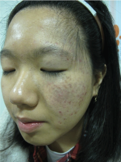 Figure 1: Unilateral blue-gray hyperpigmented macules and patches scattered along the first and second divisions of trigeminal nerve on the face (before treatment). View Figure 1
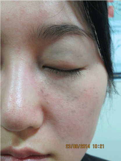 Figure 2: Unilateral blue-gray hyperpigmented macules and patches on the left periorbital area and left maxillary area (before treatment). View Figure 2
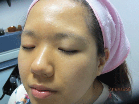 Figure 3: A complete clearance of Ota's nevus (after treatment with Golden Parameter: 5/30/2015). View Figure 3
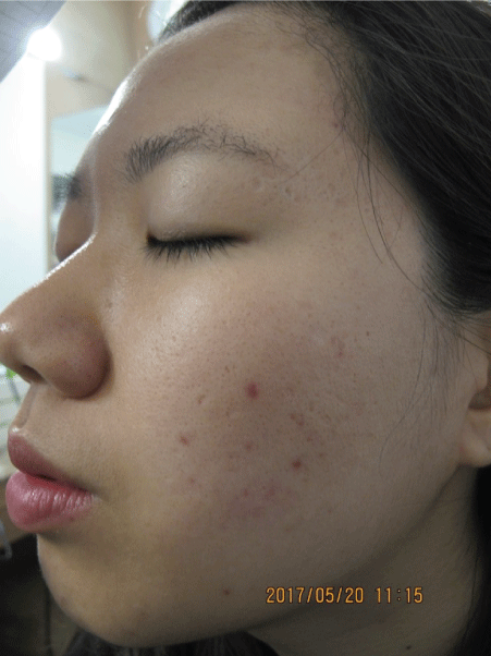 Figure 4: There is no recurrence at 24 months follow-up (5/20/2017). View Figure 4
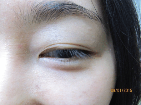 Figure 5: A complete clearance of Ota's nevus (after treatment with Golden Parameter: 1/9/2015). View Figure 5
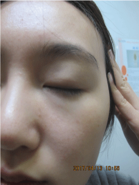 Figure 6: There is no recurrence at 24 months follow-up (4/13/2017). View Figure 6
Ota's nevus is a congenital benign oculodermal melanocytosis. Clinically, Ota's nevus occurs as a gray to bluish patch on the face from birth to about one year after birth and is distributed at ophthalmic and maxillary branches of the trigeminal nerve [1-3]. Ota's nevus may be almost unilateral or bilateral, and may involve ocular or oral mucosa in addition to skin. Ota's nevus is most commonly found in Asian populations; it occurs in 0.2%-0.6% of Japanese people and appears more frequently in females [1-3]. Ocular melanoma associated with Ota's nevus has been reported in the choroid, orbit, iris, ciliary body, optic nerve and brain [1-3]. Histopathologically, Ota's nevus shows highly dendritic, deeply pigmented melanocytes and melanophages dissecting bundles of dermal collagen in the superficial layer of the dermis or deep layer of the dermis or throughout the dermis [3]. Differential diagnosis should include agminated blue nevus and melanoma. Histopathologically, agminated blue nevus reveals brownish pigmented fusiform and oval-shaped melanocytic cells in the upper and deeper dermis, and not forming nests but arranged in bundles [7]. But melanoma shows marked atypical melanocytes and pagetoid spread of melanocytes in the epidermis [8]. The etiology and pathogenesis of Ota's nevus is idiopathic. Although not confirmed, Ota's nevus may indicate melanocytes that have not completely migrated from the neural crest to the epidermis at the embryonic stage [9]. Also specific mutations have been detected in dermal melanocytes, most commonly GNAQ or GNA11 and they may be associated with hormones that play an important role in the developmental process [10,11]. And because ultraviolet radiation cannot reach the deep dermal melanocytes, the role of ultraviolet radiation may be limited. Conventional laser treatments had been used widely for many years. However, some side effects of conventional laser treatments might occur, such as purpura, crust, PIH, scarring and recurrences. It is difficult to treat Ota's nevus without inducing PIH [4-6]. Although the exact cause of PIH is still unclear, some reasons are thought to be the possible cause of PIH occurrence when using conventional laser therapy in Ota's nevus. The 515-755 nm of intense pulsed light, 694 nm of ruby laser, 532 nm of Q-Switched Nd: YAG laser and 755 nm of alexandrite laser are generally absorbed by much more melanin than 1064 nm of Q-Switched Nd: YAG laser. This greater absorption leads to the destruction of the epidermal melanocytes and damages the surrounding keratinocytes. These damaged keratinocytes secrete Interleukin-1 (IL-1), which stimulates keratinocytes to secrete some keratinocyte injury-induced cytokines, which are endothelin-1, α-Melanocyte Stimulating Hormone (MSH), Adrenocorticotropic Hormone (ACTH) and Prostaglandin (PGE2, PGF2α). These cytokines are responsible for the activation of melanocytes and the increase of melanin synthesis, which located in the melanosomes, therefore provoking PIH [12-15]. The single-chain Urokinase Type Plasminogen Activator (sc-uPA) is also secreted by the damaged keratinocytes. Plasminogen is converted to plasmin by the sc-uPA. The plasmin then stimulates the keratinocytes to secrete basic Fibroblast Growth Factor (bFGF). The melanocytes then get activated by this bFGF, increasing the melanin synthesis in the melanosomes, which cause PIH [12-15]. Treatment with conventional laser therapy can provoke purpura and crusts, which can be accompanied by damage of fibroblasts, mast cells, lymphocytes, macrophages and vascular endotheliums due to laser energy. Then, damaged fibroblast-derived Stem Cell Growth Factor (SCF) and Hepatocyte Growth Factor (HGF) activate melanocytes and increase melanin synthesis in the melanosomes, eventually leading to PIH [12-15]. Finally, the damaged keratinocytes produce reactive oxygen species such as nitric oxide, free radical oxygen and peroxide, which activate melanocytes and increase melanin synthesis in the melanosomes, and eventually induce PIH [12-15]. To avoid side effects such as crust, purpura, PIH and scarring caused by conventional laser therapy, the authors devised Dr. Hoon Hur's Golden Parameter Therapy (GPT) using a high fluence 1064 nm Q-switched Nd: YAG laser without side effects or recurrence [15-17]. Dr. Hoon Hur's Golden Parameter Therapy using a high fluence 1064 nm Q-switched Nd: YAG laser may destroy dermal melanocytes without keratinocyte damage, and the end products of damaged melanocytes will be removed through transepidermal elimination [15-17]. Also the end products, including the dispersed melanosomes and melanins of damaged dermal melanocytes are phagocytized by the macrophages and are removed through the lymphatic system [15-17]. By using Dr. Hoon Hur's Golden Parameter Therapy with a high 1064 nm Q-switched Nd: YAG laser, the authors believe that destroying epidermal melanocytes or dermal melanocytes can be done with minimal epidermal damage and accelerating apoptotic melanocytic cell death program, and improving various skin diseases such as café au lait spot, partial unilateral lentiginosis, Becker's nevus and congenital melanocytic nevus without side effects such as PIH and scarring are also achievable [15-17]. The authors think that the wavelength of 1064 nm used in Dr. Hoon Hur's Golden Parameter Therapy is less absorbed by the epidermal melanin. This mechanism is able to destroy the epidermal melanocytes or dermal melanocytes while minimizing the epidermal damage, therefore not causing purpura and crusts. Performed weekly, this Dr. Hoon Hur's Golden Parameter Therapy is able to destroy melanocytes completely and accelerates apoptotic melanocyte cell death. The dispersed melanosomes and melanins, which are the end products of damaged melanocytes, are either removed by the transepidermal elimination or are removed by dermal melanophages via the lymphatic system [15-17]. In the end, it is possible to achieve complete clearance of Ota's nevus without any side effects or recurrences. In our study, patients with Ota's nevus were treated with a high fluence 1064 nm Q-switched Nd: YAG laser (Spectra Laser, Lutronic, South Korea) in 30 treatment sessions with a one-week interval. We used a spot size of 7 mm, a fluence of 2.4 J/cm2 and a pulse rate of 10 Hz. The dermal melanocytes were destroyed with minimal epidermal damage using the slowly three passes of this parameter by a sliding-stacking technique to Ota's nevus. Due to the less absorption by epidermal melanin in Dr. Hoon Hur's Golden Parameter Therapy, it is possible to deliver sufficient energy to destroy dermal melanocytes and in the same time salvaging normal background tissue, preventing PIH and scarring from being triggered, and minimizing epidermal damage without inducing purpura and crusts. However, this Dr. Hoon Hur's Golden Parameter Therapy requires the continuous 30 treatment sessions for 8 months. In our study, 12 patients with Ota's nevus (Figure 1 and Figure 2) were treated with Dr. Hoon Hur's Golden Parameter Therapy using a high fluence 1064 nm Q-switched Nd: YAG laser. All of the 12 patients with Ota's nevus were achieved complete clearance of the pigmented lesions and PIH and scarring were not found (Figure 3 and Figure 5). No recurrences have been detected after a follow-up of 24 months (Figure 4 and Figure 6). All patients were satisfied with the results of Dr. Hoon Hur's Golden Parameter Therapy without any side effects, including PIH and scarring.
In this study, Dr. Hoon Hur's Golden Parameter Therapy using a high fluence 1064 nm Q-switched Nd: YAG laser achieved complete clearance of Ota's nevus without side effects and recurrences. We suggest Dr. Hoon Hur's Golden Parameter Therapy using a high fluence 1064 nm Q-switched Nd: YAG laser will be a new and good option for treating Ota's nevus.