Central Retinal Artery Occlusion (CRAO) is the partial or complete blockage of the central retinal artery presenting as acute painless monocular vision loss with increased risk in those with cardiovascular disease. Diagnosis of CRAO requires a dilated fundoscopic exam performed by an Ophthalmologist. In resource limited hospitals, access to these expertise and resources may be difficult. Early recognition of CRAO in the emergency department (ED) can lead to reduced negative consequences and reduced cost to healthcare. Here we present a case using Point-of-Care Ultrasound (POCUS) to identify CRAO in a patient.
Case report, Central retinal artery occlusion, Point-of-care ultrasound, Early diagnosis, Vision loss
Acute vision loss at presentation in the ED can progress to blindness or severe visual impairment. CRAO is the partial or complete blockage of the central retinal artery that presents as painless monocular vision loss [1]. Risk factors of CRAO are similar to those of cardiovascular or stroke risk factors related to thromboembolic disease, including hypertension, cardiac valvular disease, and diabetes [2]. The most common cause of CRAO is by an embolus from the aortic arch, ipsilateral carotid artery, or heart [3]. This can result in vision loss, rapid cell damage, and retinal hypoperfusion [4]. Dilated fundoscopic exam is the standard assessment for diagnosing diseases related to painless vision loss. However, diagnostic tools of that nature are limited by availability of specialists needed to complete the exam at a community hospital POCUS can be used as a cost-effective, noninvasive diagnostic tool to accurately diagnose CRAO in patients presenting to the ED.
A 65-year-old woman presented to the ED with moderate right eye peripheral vision loss with onset 3 days prior to arrival. At admission, the patient had normal ocular motility. She has a prominent past medical history of hypertension, hypercholesterolemia, and diabetes mellitus. The patient was sent in by ophthalmology to be evaluated for CRAO. She was originally evaluated for posterior circulation infarct due to the loss of vision in her eye. The non-contrast computed tomography (CT) of the head showed no evidence of intracranial hemorrhage or infarct. An electrocardiogram was performed showing mild bradycardia. Neurology and cardiology consultants were performed in the ED and the patient was given aspirin 325 mg. The patient was admitted to the hospital with the diagnosis of CRAO and sent to the Intensive care unit for closer observation.
One day after presenting to the ED the patient received a magnetic resonance imaging of the brain showing no evidence of any acute infarct or intracranial pathology. After 2 days in the ICU, the ICU Attending performed a POCUS on the patient's eye using a high frequency linear ray probe. There were no findings of vitreous hemorrhage or detachment and no findings of retinal detachment. The POCUS showed an irregular optic nerve sheath measuring larger than 5 millimeters (Figure 1). The findings were consistent with CRAO.
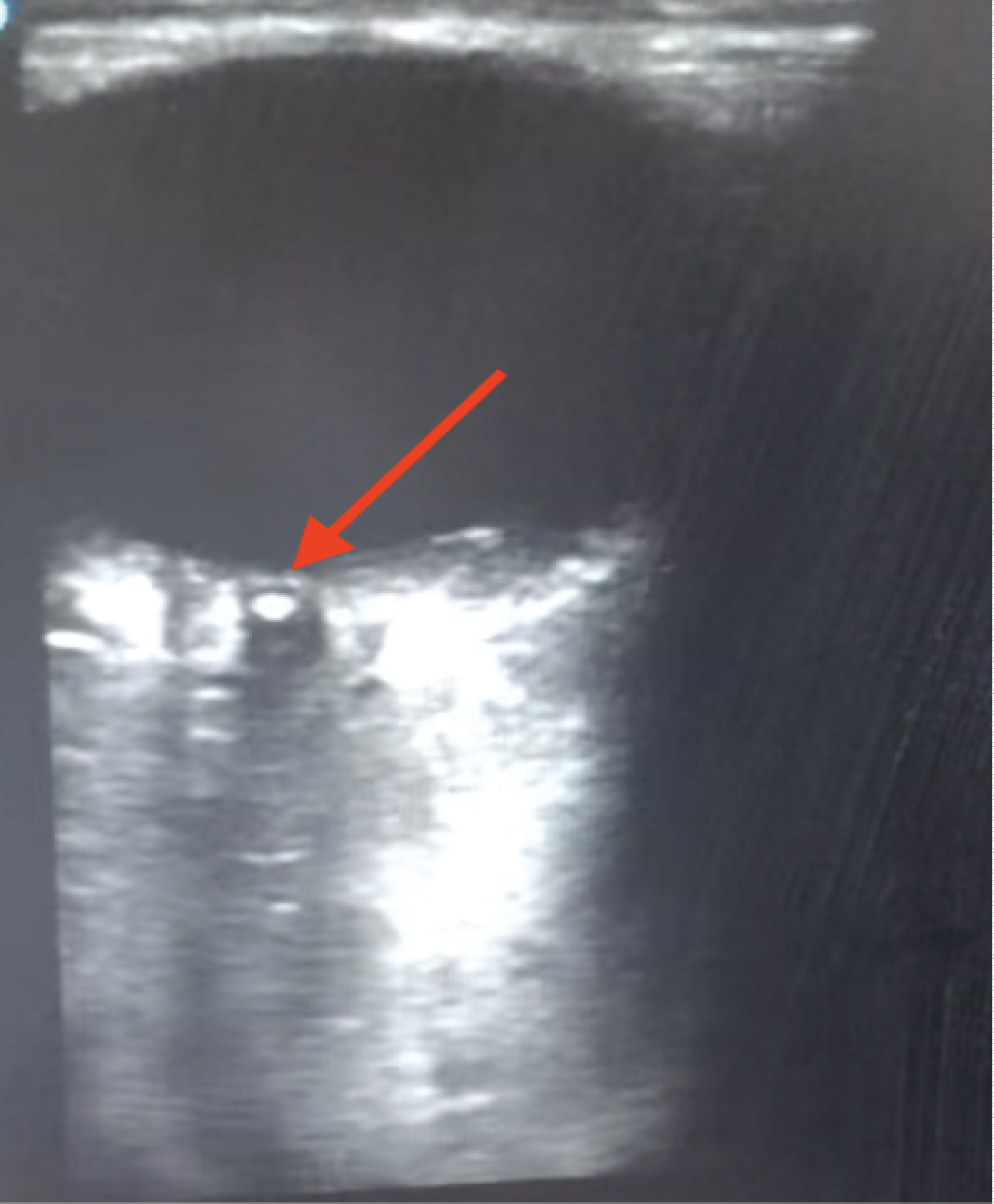 Figure 1: POCUS image of the right eye using a high frequency linear ray probe. Displaying a clot (red arrow).
View Figure 1
Figure 1: POCUS image of the right eye using a high frequency linear ray probe. Displaying a clot (red arrow).
View Figure 1
Patient was diagnosed with bilateral severe carotid stenosis based on the ultrasound findings. The carotid stenosis was worse on the right side with no evidence of a cardiac source of embolism. The patient underwent a carotid endarterectomy with patch right intraoperative shunt and was not expected to have improvement in right eye vision. Patient was sent home with new medications including aspirin 325 mg once a day and apixaban 5 mg two times a day.
CRAO is a medical emergency, presenting as painless vision loss, which can lead to permanent vision loss or impairment. POCUS is an effective, cost-efficient tool to accurately evaluate ocular pathology in the ER without the need for costly diagnostic tests. One meta-analysis reported that practitioners can utilize POCUS as a helpful and sensitive diagnostic tool for ocular emergencies [5]. This tool can help differentiate immediate ophthalmologic emergencies from those that can be managed as outpatients [6].
The efficacy of POCUS to diagnose various ocular emergencies has been described in several cases involving retinal detachment, vitreous hemorrhage, and vitreous detachment with high sensitivity (96.9%) and specificity (88.1%) [7]. POCUS can aid in the diagnosis of patients with CRAO through the detection of a hyperechoic object known as a "spot sign" [8]. A prospective study found that of 12 patients diagnosed with embolic CRAO, 10 had a visible "spot sign" using sonographic assessment [9]. Direct fundoscopy has previously been used as the primary diagnostic tool for CRAO [4]. Many physicians cannot reliably visualize ocular abnormalities leading to a decrease in confidence for the ability to diagnose CRAO using only a direct fundoscopy [10]. One retrospective study shows that the thrombus was directly visualized using a dilated fundoscopic exam only 11% of the time in CRAO patients [11]. Thus, highlighting the lack of reliability of fundoscopic exams in diagnosing CRAO. A review of the diagnostic value of POCUS for CRAO found a high interobserver agreement (Chohen's kappa 0.98) and albeit small, studies determined a strong specificity (100%) and sensitivity (83%) for POCUS reliability [4,9-12].
Prompt recognition of CRAO is critical for positive outcomes. Previous case studies show that rapid POCUS can be used as an accurate reliable tool to diagnose CRAO [11-13]. Once diagnosed, specialists in ophthalmology and neurology can help determine the best course of treatment such as intravenous tissue plasminogen activator (tPA) [14]. Intravenous tPA, a therapy used to treat acute ischemic strokes, requires onset of symptoms within a 4.5-hour window leading to limited patient enrollment. However, it has been shown to improve long-term outcomes [15]. One meta-analysis of observational studies found that in patients diagnosed with acute CRAO, thrombolysis drug therapy resulted in a 50% recovery in patients who qualified for treatment [16].
In our patient case, she was referred to the ER by the ophthalmologist for CRAO. A fluorescein angiography was not performed nor was an ESR or CRP level obtained for the patient because of the presumed diagnosis of CRAO prior to coming into the ED. Intravenous tPA was not administered because the patient failed to meet inclusion criteria due to the onset of symptoms being 3 days prior [17]. POCUS was not performed in the ED due to availability and familiarity. The evidence found in the POCUS indicates that the patients can be accurately diagnosed with CRAO in the ED allowing quicker intervention in the absence of ophthalmology service.
In small hospitals with limited resources, POCUS is a reliable, cost-effective alternative tool to diagnose patients with CRAO. Therapies that can improve long-term outcomes for patients require an early diagnosis. Our case report highlights the ability to utilize POCUS as an alternative diagnostic method to decrease time to definitive therapy in the absence of ophthalmology specialists and proper resources.
The authors have not declared a specific grant for this research from any funding agency in the public, commercial, or not-for-profit sectors.
We would like to thank the patient who entrusted us to be part of his care and permitted us to share his case.
The authors declare that they have no relevant financial interests.
Our institution does not require ethical approval for reporting individual cases or case series.
Written consent has been obtained from the patient for publication of the case report and accompanying images.