The prognosis of cancer is highly dependent on its stage at diagnosis, with early diagnosis strongly correlating with better treatment outcomes. The incidence of discovering cancer during pregnancy has steadily increased over the years, with 1/2000 cases reported in the 1960s to 1/1000 cases currently reported.1 However, these diagnoses are primarily driven by patients experiencing symptoms that prompt their healthcare team to investigate the possibility of cancer. Incidental findings of cancer during a Cesarean section (C-section) are extremely rare. In this case report, a 25-year-old G1P0 patient underwent a C-section at 41.1 weeks due to prolonged rupture of membranes and failure to progress. During the C-section, multiple nodules were found incidentally on the patient’s uterus, bladder, abdominal wall, and omentum. The ovaries and fallopian tubes were also examined but no gross abnormalities were reported. A uterine nodule biopsy was sent to pathology and, combined with the results of a CT of the abdomen and pelvis, resulted in a diagnosis of low grade serous ovarian carcinoma. The patient’s care was transferred to a gynecologic oncologist, where she completed six cycles of depo-Lupron and anastrozole chemotherapy in addition to an omentectomy and a total abdominal hysterectomy with bilateral salpingo-oopherectomy. While rare, it is important to be aware of the occurrence of incidental cancers found at the time of cesarean section. OB/GYN’s must biopsy any abnormal intraoperative findings during a Xesarean section, as the results may lead to a life-saving diagnosis.
The incidence of cancer during pregnancy has increased from 1/2000 cases in the 1960’s to 1/1000 cases in the present day [1]. This increased incidence could be due to higher rates of advanced maternal age, as well as increased detection via recent advances in interpretation of noninvasive prenatal testing (NIPT) [2]. The most common malignancies identified during pregnancy are breast cancer (41%), lymphomas (12%), cervical cancer (10%), leukemias (8%), and ovarian cancer (7%) [3].
Identifying cancer in its early stages strongly correlates with better treatment outcomes. However, diagnosing cancer during pregnancy is a complex process as many physiologic changes that occur during pregnancy resemble symptoms of malignancy. Fatigue, bloating, nausea, vomiting, pelvic/abdominal pain, breast changes, hyperpigmentation, nevi changes, and anemia are all common changes in pregnancy. These mirror classic symptoms of malignancy. Physical exam findings can be unreliable due to expected changes to the maternal body during pregnancy, such as a gravid uterus and enlarged tender breasts. Additionally, physiological changes in pregnancy and concerns for fetal safety make it challenging to use standard diagnostic imaging and routine lab work to diagnose cancer in pregnant patients [1,4].
While measuring tumor markers may be useful in diagnosing cancer in the general population, many well-known tumor markers have varying levels during pregnancy. Tumor markers altered for at least one trimester in pregnancy include CA 15-3, CA 125, SCC, AFP, and inhibin B1,4. Altered serum levels in pregnant patients compared to nonpregnant patients make these tumor markers unreliable for use in diagnosing cancer in pregnancy.
Serous ovarian carcinoma, a subtype of ovarian cancer, can be further divided into low-grade serous ovarian carcinoma (LGSC) and high-grade serous ovarian carcinoma (HGSC). LGSC, the malignancy featured in this case, is a rare cancer that comprises 2% of all epithelial ovarian cancers. LGSC is typically diagnosed in women on average in their mid-40's, with a poor prognostic factor being discovery in women younger than 35 [5]. Most ovarian carcinomas are asymptomatic in the early stages and typically present with vague abdominal and pelvic symptomology at later stages; therefore, early detection and diagnosis is difficult. The most common abnormality detected that prompts further investigation to rule out ovarian cancer is the presence of a complex ovarian cyst, discovered by transvaginal ultrasound [6]. The 5-year survival rate for patients with LGSC is dependent on their stage at diagnosis, with stage I at 89%, Stage II at 87%, stage III at 49% and stage IV at 25% [7]. This case study reflects on the rare nature of a nulliparous woman with labor dystocia who underwent an unplanned C-section which resulted in the incidental finding of an asymptomatic LGSC. This provided the opportunity for diagnosis and treatment of a potentially fatal malignancy which would have otherwise gone unnoticed had the patient completed her desired spontaneous vaginal delivery. Despite C-section being one of the most common surgeries in the world, cancerous findings during this surgery are extremely rare. There are no statistics currently available concerning the discovery of malignancy during a C-section - a fact which further highlights the truly anomalous nature of this patient’s case.
The patient is a 25-year-old white primigravid female at 41.1 weeks gestation. The pregnancy was complicated by failure of 1 hour glucose tolerance test (GTT) with refusal of 3-hour GTT. Ultrasonography at 35 and 6/7 weeks indicated the fetus was large for gestational age (LGA) but did not otherwise indicate any abnormalities, including the presence of abnormal uterine, adnexal, or pelvic organ abnormalities. She presented to labor and delivery from a birthing center for prolonged labor and prolonged rupture of membranes. Despite Pitocin augmentation, uterine contractions were inadequate and transition to cesarean delivery was recommended for failure to progress. A healthy male infant was delivered via C-section. Uterine atony was noted. Quantitative blood loss was 1326 mL, conferring a diagnosis of postpartum hemorrhage. This was managed successfully with intraoperative methergine administration. Abnormal intraoperative findings included uterine, bladder, abdominal wall, and omental nodules. A uterine nodule biopsy was obtained intraoperatively and sent to pathology, where it was initially identified as a low-grade serous neoplasm. The tumor immunohistochemistry is shown in table 1. The patient and her baby progressed through the acute postpartum period without further sequelae and were discharged home on postpartum day 2. On postpartum day 18, the patient underwent a computed tomography (CT) scan which revealed multiple partially calcified masses seen primarily in the parametrial fat which were suggestive of peritoneal based disease. Imaging studies are shown in figures 1a-1c. The patient’s care was transferred to a gynecologic oncologist, where she completed six cycles of Lupron Depot and anastrozole chemotherapy in addition to an omentectomy and a total abdominal hysterectomy with bilateral salpingo-oopherectomy. At the time of this case report, the patient is receiving combination therapy which includes traditional chemotherapy and “integrated holistic therapy” coordinated by an independent clinic in California. The information described in this case summary was collected via direct involvement in the case and thorough chart review.
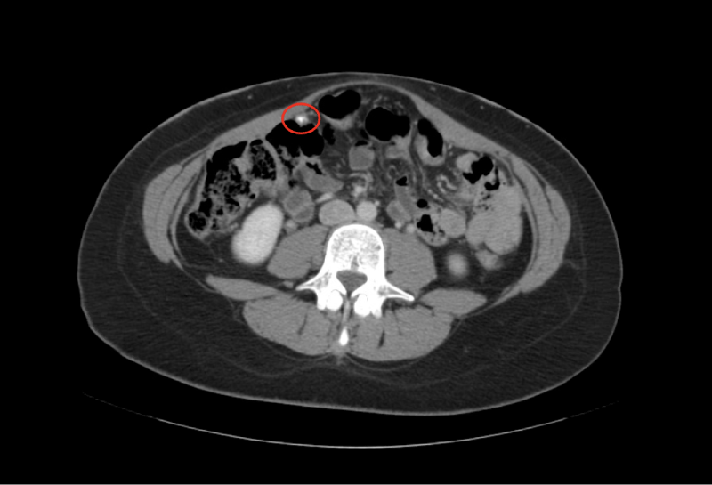 Figure 1a: Computed Tomography demonstrating partially calcified peritoneal mass at the lateral margin of the right colon proximal to the hepatic flexure.
View Figure 1a
Figure 1a: Computed Tomography demonstrating partially calcified peritoneal mass at the lateral margin of the right colon proximal to the hepatic flexure.
View Figure 1a
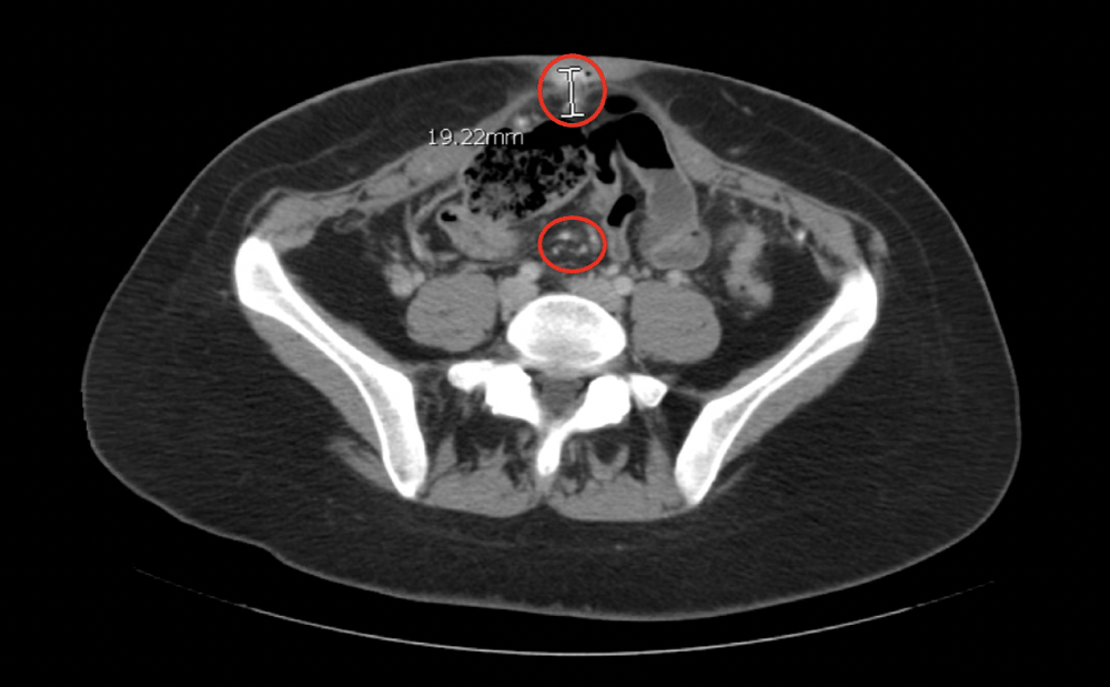 Figure 1b: Computed Tomography demonstrating small partially calcified peritoneal masses and a 1.9 cm partially calcified mass in the region of the umbilicus.
View Figure 1b
Figure 1b: Computed Tomography demonstrating small partially calcified peritoneal masses and a 1.9 cm partially calcified mass in the region of the umbilicus.
View Figure 1b
 Figure 1c: Computed Tomography demonstrating 2.3 cm mass adjacent to the anterior aspect of the uterine fundus within the parametrial fat.
View Figure 1c
Figure 1c: Computed Tomography demonstrating 2.3 cm mass adjacent to the anterior aspect of the uterine fundus within the parametrial fat.
View Figure 1c
Table 1: Tumor Immunohistochemistry. A uterine nodule biopsy was sent to pathology, which demonstrated a low-grade papillary neoplasm of unknown origin. Immunoprofiling of the uterine nodule biopsy showed that the cells were PAX8+, ER+, PR+, claudin 4+, Ber-EP4+, WT1-, D240-, calretinin focally+, and had WT p53 expression, which suggests serous differentiation. Positive tumor markers are displayed in bold.View Table 1
Ovarian masses are detected during pregnancy at a rate of 1-2%, with the majority of those being incidental findings on routine prenatal ultrasonography [24]. Of those, most abnormalities are benign findings which spontaneously resolve as the pregnancy progresses. In exceedingly rare cases, 2-3% of ovarian and adnexal masses discovered during pregnancy are found to be malignancies [24]. Ovarian cancer in the general population is frequently diagnosed at later stages due to the absence of specific symptoms [25]. Nonspecific symptoms, along with physiologic confounding of tumor markers, pose significant challenges to diagnosis of ovarian masses in the pregnant patient. Diagnosis in pregnancy thus becomes heavily reliant on skilled ultrasonographic imaging in the prenatal period. In this case report, the patient's ultrasounds showed no evidence of ovarian masses throughout her pregnancy.
The patient in this case received prenatal care at an outside facility and her prenatal course did not rouse suspicion for malignancy. Despite previously expressing a desire for spontaneous vaginal delivery, conversion to C-section resulted in appreciation of grossly abnormal nodules on the uterus, bladder, abdominal wall, and omentum. Biopsy at the time of discovery resulted in expedient diagnosis of this patient’s primary ovarian carcinoma, thus drastically increasing her odds of survival compared to if the malignancy had continued undetected [7].
This case report highlights the importance of maintaining a high index of suspicion during surgical procedures, even in seemingly routine cases. The incidental discovery of asymptomatic primary ovarian carcinoma in a nulliparous woman undergoing an unplanned C-section underscores the rarity but significance of such findings. Recognition of abnormal tissue and promptly initiating further evaluation can lead to timely diagnosis and treatment, ultimately improving patient prognosis. This case serves as a reminder of the importance of a comprehensive understanding of normal anatomy and the vigilant examination of surgical specimens.
All data within this case report is included with the submission.
The authors declare no conflicts of interest.
No funding was received for this research.
We would like to thank the patient for her verbal consent to proceed with this case report and the care teams that have worked tirelessly for the improvement of the patient’s outcome.