Encephalopathy, Corpus callosum, Splenium, Transient ischemic attack, Diffusion, MRI
A 21-year-old man was admitted to emergency department due to slurred speech, weakness and loss of consciousness. The previous history is unremarkable except a slight cold 2 weeks ago. The first magnetic resonance imaging (MRI) revealed a distinct lesion involving in the splenium of the corpus callosum. This lesion showed restricted diffusion with hyperintense signal on diffusion-weighted imaging (DWI) and hypointense signal on apparent diffusion coefficient (ADC) sequence (Figure 1). A follow-up brain MRI scan was performed 9 days after onset of disease. Reduced lesions with decreased signals were discovered in DWI and ADC.
Mild encephalitis/encephalopathy with reversible splenial lesion (MERS) is a newly minted syndrome that was first described by Tada, et al. in 2004 as a rare clinico-radiological syndrome in 2004 [1]. Patients with MERS presented with mild central nervous system symptoms such as consciousness disturbance, seizures and headache and recovered completely within a month [2]. Many child-onset MERS cases have been reported in the litarature. However, adult onset MERS is rare [3]. The pathogenesis of MERS is still unknown. Several hypotheses have been proposed for pathogenesis of the specific lesion, such as intramyelinic edema, hyponatremia, axonal damage, and oxidative stress [1,3]. High signal intensity on DWI and decreased ADC values of splenium of the corpus callosum have been observed in MERS. ADC may return to normal within a week if the intramyelinic edema or inflammatory infiltrate resolves quickly [2].
None.
None.
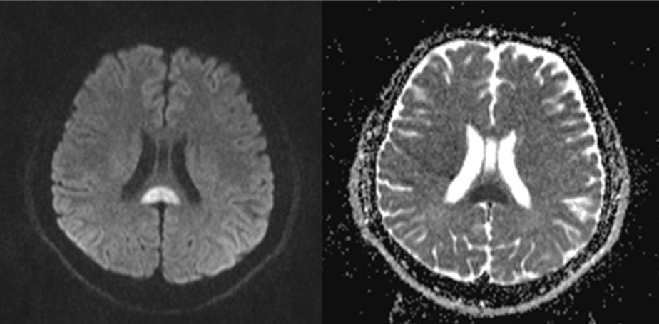
Figure 1: A lesion in the splenium part of the corpus callosum shows restricted diffusion with hyperintense signal on diffusion-weighted imaging (DWI) and hypointense signal on apparent diffusion coefficient (ADC) sequence.