The uterine lipomatous tumors are extremely uncommon benign neoplasms. Lipoleiomyoma is a benign tumor that is a leiomyoma variation. Lipoleiomyoma consists of variable proportions of mature lipocytes and smooth muscle cells. Unlike conventional leiomyomas in which the size somewhat declines after menopause, uterine lipoleiomyomas are characterized by progressive enlargement even after menopause [1]. We report a case of uterine lipoleiomyoma because of its rarity. Their reported incidence varies from 0.03 to 0.2% [2].
Here we present a case of a 61 years old female, menopaused over 10 years, the patient initially had her first episode of postmenopausal bleeding 5 years ago however subsequently defaulted her follow up. She presented this time with hypovolemic shock from massive postmenopausal bleeding requiring resuscitation. During the hypovolemic event, her uterus was equivalent to a 30 weeks size gravid uterus, non-tender, firm, mobile, and regular margin, with no other organomegaly. Pelvic examination was unremarkable except for some fullness at the Pouch of Douglas.
Bedside ultrasonography showed a grossly enlarged uterus with well demarcated heterogenous hyperechoic mass occupying the endometrial cavity. There were no adnexal masses or free fluid. Endometrial sampling was attempted however came back as non-representative sampling. A CT scan imaging which was done showed large lobulated mass extending from the pelvis to the upper abdomen, measuring 14.0 cm (AP) x 20.6 cm (W) x 20.0 cm (CC), which had predominantly fat content with ill-defined enhancing components within. Coarse calcifications were seen within this lesion and it was well-encapsulated within a thick enhancing wall (Figure 1). An incidental finding of pulmonary embolism was also diagnosed. The patient was then subjected to diagnostic ultrasound guided biopsy of the mass which only reported fat- containing lesion.
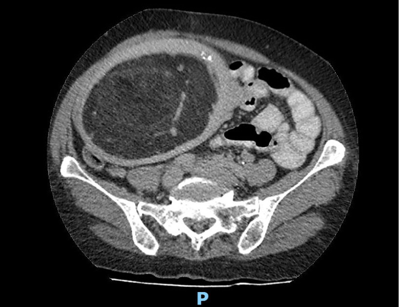 Figure 1: CT scan demonstrated a large lobulated mass, which had predominantly fat content with ill-defined enhancing components within.
View Figure 1
Figure 1: CT scan demonstrated a large lobulated mass, which had predominantly fat content with ill-defined enhancing components within.
View Figure 1
She underwent total abdominal hysterectomy bilateral salphingoophorectomy after a course of anticoagulant. Intraoperatively, the uterus was equivalent to 24 weeks size of a gravid uterus. The dissected specimen revealed a huge well - circumscribed posterior intramural firm fibro-fatty mass measuring 18 x 23 x 16 cm pushing the endometrial cavity anteriorly. It is 1mm away from the nearest serosa. It had patchy areas of fibrotic tissues. There was no obvious area of haemorrhagic necrosis, or calcification. The endometrium was grossly normal and the uninvolved endomyometrium measured 15-20 mm in thickness (Figure 2).
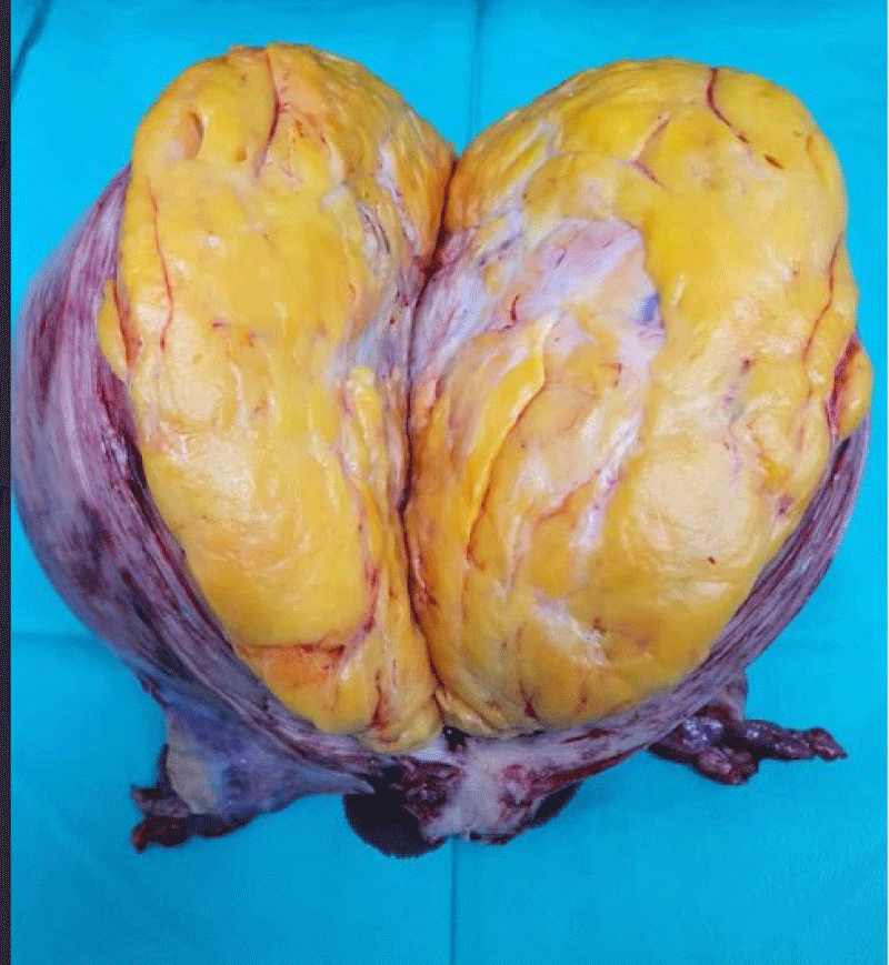 Figure 2: Cut section of intramural firm fibro-fatty mass.
View Figure 2
Figure 2: Cut section of intramural firm fibro-fatty mass.
View Figure 2
Histopathology examination revealed uterine lipoleiomyoma. The intramural mass is composed of mainly mature fat cells without nuclear atypia mixed with bland, spindle - shaped smooth muscle cells in a whorled pattern. The rest of the study was normal.
The patient recovered well post-surgery and was discharged from the gynaecology clinic after review.
Rare lipomatous uterine tumors can be difficult to diagnose. The common presenting complaint of uterine lipoleiomyoma would be progressive uterine enlargement with postmenopausal bleeding mimicking presentation of leiomyosarcoma. This patient of mine however presented with massive postmenopausal bleeding resulting in shock and pulmonary embolism. The inconsistent abdominal finding of the initial presentation and final specimen may suggest intrauterine blood collection during the presentation. Management dilemma was made worse with the patient having pulmonary embolism which would subject the patient to high risk immediate surgery and the anti-coagulant started could have worsened her uterine bleeding.
The patient’s final diagnosis of uterine lipoleiomyoma, though rare, is actually the most common variant of leiomyomas in post-menopausal women (85.7%) [3]. Based on the World Health Organisation publication, variant forms of leiomyomas accounted for about 10% of total leiomyomas [4]. Generally accepted pathogenesis of lipoleiomyoma could be either from lipomatous metamorphosis of a pre-existing leiomyoma [5] or lipomatous tissue arise from direct transformation of smooth muscle cells or transformation of totipotent mesenchymal cells within the uterine myometrium [1]. In both conditions, lipoleiomyomas formation was attributed to conditions of low oxygen / hypoxia, or serum starvation [1]. Given that the majority of patients are postmenopausal women, it is hypothesized that abnormal intracellular storage of lipids may be promoted by a variety of lipid metabolic disorders or other related problems that are linked to estrogen insufficiency could contribute to lipomatous metaplasia [6]. Microscopically, the tumor is composed of mainly mature fat cells without nuclear atypia mixed with bland, spindle-shaped smooth muscle cells in a whorled pattern like in this patient (Figure 3).
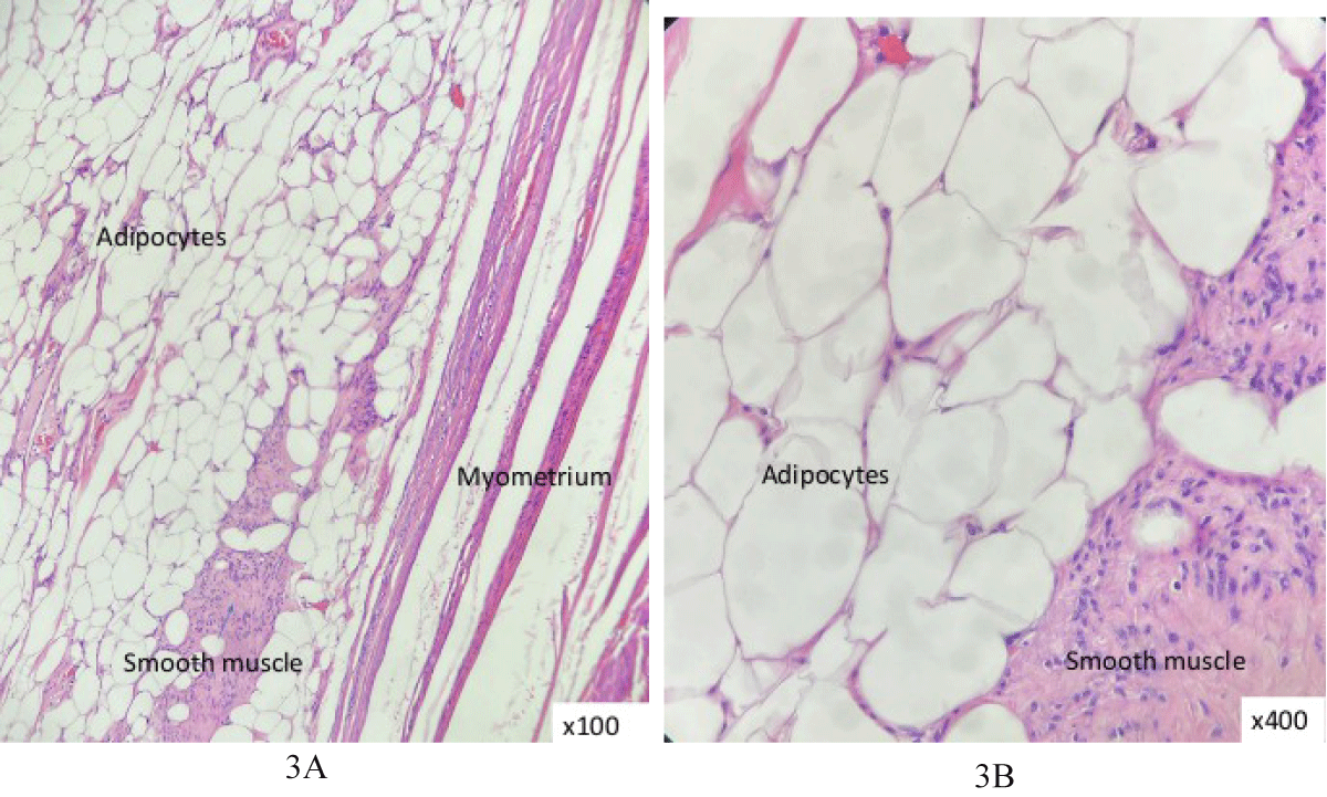 Figure 3: (A): Well-circumscribed intramural mass composed of mainly adipocytes admixed with spindle-shaped smooth muscle cells in a whorled pattern; (B): The mass is composed of mainly mature fat cells without nuclear atypia admixed with smooth muscles cells. No lipoblasts seen. The smooth muscle cells show uniform nuclei with finely dispersed chromatin and small nucleoli.
View Figure 3
Figure 3: (A): Well-circumscribed intramural mass composed of mainly adipocytes admixed with spindle-shaped smooth muscle cells in a whorled pattern; (B): The mass is composed of mainly mature fat cells without nuclear atypia admixed with smooth muscles cells. No lipoblasts seen. The smooth muscle cells show uniform nuclei with finely dispersed chromatin and small nucleoli.
View Figure 3
Even though lipoleiomyomas have low malignant potential, patients may present with severe symptoms and the rapid growing tumour size would make clinician suspicious of malignant uterine tumours. Hysterectomy would be commonly offered to post-menopausal women with such symptoms and final histopathological examination would confirm the diagnosis of this benign condition. Optimally resected lipoleiomyoma has shown no documented disease recurrence [1]. Rarely, small, asymptomatic lipoleiomyomas do not require treatment and can be managed conservatively similar to leiomyomas [7].
We would like to express our sincere gratitude to the Department of Radiology and Department of Pathology (Histopathology Unit) from Hospital Raja Permaisuri Bainun, Perak, Malaysia.
The authors declare that there is no conflict of interest.