Bursitis, Ultrasonography, Chondrocalcinosis, Calcium pyrophosphate dihydrate
With respect to the article "An Unusual Association: Iliopsoas Bursitis Related to Calcium Pyrophosphate Crystal Arthritis" published in Case Reports in Rheumatology by Di Carlo, et al. [1] describing the unusual association of iliopsoas bursitis and calcium pyrophosphate crystal (CPPD) arthritis as first clinical manifestation of chondrocalcinosis, we would present a similar case in order to increase the awareness of this potential manifestation of chondrocalcinosis.
We describe a 78-year-old daily hiker woman with no previous significant clinical records presented with a history of acute hip pain, limping, with no previous trauma. Clinical examination showed inability to extend lower left limb, intense pain at hip mobilization with intense limitation of hip range of motion. X-Ray showed severe coxofemoral osteoarthritis with typical CPPD calcification (Figure 1B) and radiocarpal joint calcification (Figure 1C), Ultrasonography revealed both a large hypo-echoic image located before coxofemoral joint and moderate joint synovial hypertrophy (Figure 1A); Magnetic Resonance Imaging confirmed the presence of severe iliopsoas bursitis, severe coxofemoral osteoarthritis with mild joint effusion (Figure 1D and Figure 1E). An iliopsoas bursa aspiration was performed, obtaining 15 cc of a non-inflammatory featured synovial fluid with the presence of rhomboid crystals with a weak positive birefringence at microscopy polarized light. Patient rapidly showed major improvement after ultrasonography-guided triamcinolone injection of the bursa.
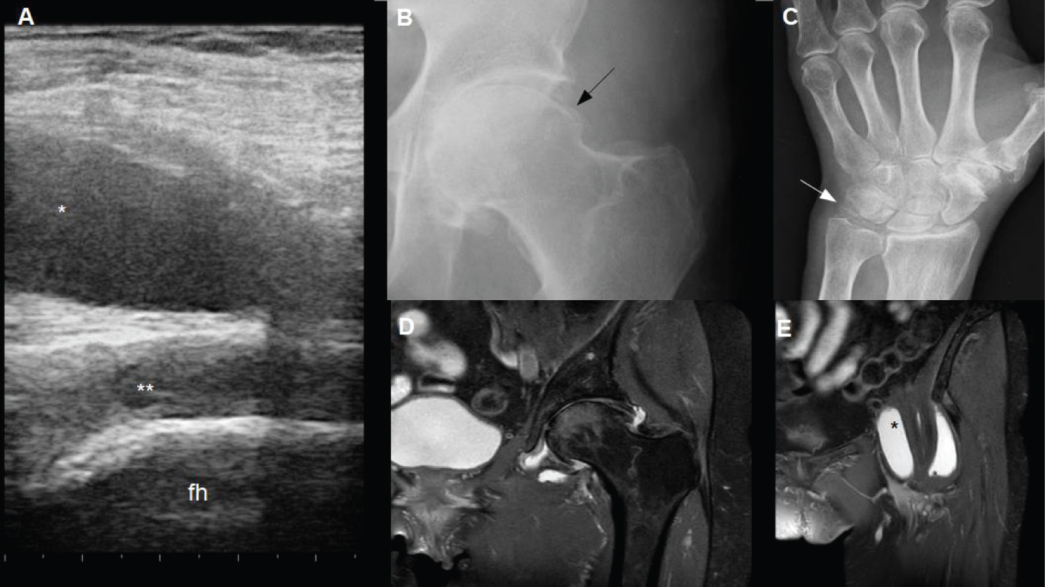 Figure 1: Anterior Hip Longitudinal Ultrasonography scan (A) showing fluid effusion (*) anterior to left coxo-femoral joint (femoral head, fh) and coxo-femoral synovial hypertrophy (**); X-ray of (B) both left coxo-femoral and (C) radiocarpal joints showing calcifications of the soft tissues near the left hip (black arrow) and radiocarpal (white arrow) joints suggestive of calcium pyrophosphate crystal deposition; Magnetic Resonance Imaging: STIR sequenced coronal image of coxo-femoral joints and pelvic area evidencing osteoarthritis and mild joint effusion (D), and STIR sequenced coronal image of the iliopsoas bursa (E).
View Figure 1
Figure 1: Anterior Hip Longitudinal Ultrasonography scan (A) showing fluid effusion (*) anterior to left coxo-femoral joint (femoral head, fh) and coxo-femoral synovial hypertrophy (**); X-ray of (B) both left coxo-femoral and (C) radiocarpal joints showing calcifications of the soft tissues near the left hip (black arrow) and radiocarpal (white arrow) joints suggestive of calcium pyrophosphate crystal deposition; Magnetic Resonance Imaging: STIR sequenced coronal image of coxo-femoral joints and pelvic area evidencing osteoarthritis and mild joint effusion (D), and STIR sequenced coronal image of the iliopsoas bursa (E).
View Figure 1
Chondrocalcinosis is a microcrystalline disease characterized by multiple foci of calcification in hyaline and fibrocartilage of the joints. Hip is a complex joint and surrounding iliopsoas bursa is one of the largest articular recesses of the human body. Several diseases may lead to an iliopsoas bursitis: Rheumatoid arthritis, osteoarthritis, osteonecrosis, synovial chondromatosis, pigmented villonodular synovitis, septic arthritis, chondrocalcinosis, and complications of total hip arthroplasty [1]. To our knowledge, this is the second reported case of this unusual first presentation of CPPD-chondrocalcinosis, although other pseudotumoral presentations have been also published [1-3].
In our opinion, patients suffering PPCD deposition and hip pain, irrespective of the presence of previous clinical symptoms, should be evaluated in order to detect iliopsoas bursitis, an uncommon presentation.
The authors do not have conflict of interest for this study.
The patient signed an informed consent to give permission for publication.
The manuscript has been approved by CSAPG-Ethics Committee.