This study aimed to examine acute effects of scapular mobilisation on upper limb neurodynamic test 1 (ULNT1) in asymptomatic adults.
This study was a crossover randomised controlled trial. 12 young healthy individuals (10 men and two women, age 21.1 ± 0.3 years, body mass index 20.4 ± 1.9) were recruited. At two separate sessions, participants received randomly assigned interventions; scapular mobilisation or placebo intervention. Range of motion in elbow extension and pain during ULNT1 were assessed before and after each intervention.
There was a statistically significant improvement in ULNT1 only after scapular mobilisation (p < 0.05). No significant change in pain level was identified in the two groups. The scapular mobilisation group displayed large or moderate effect sizes to improve ULNT1 and pain, whereas effect sizes of placebo intervention were small.
Large-amplitude end-range scapular mobilisation significantly improved ULNT1 in asymptomatic participants. Scapular mobilisation might be able to affect mechanosensitivity of the nervous system. Further research is required to test its effects among symptomatic patients with nerve-related neck and arm pain.
Upper limb neurodynamic test 1, Scapular mobilisation, Crossover randomised controlled trial
Cervical and/or upper limb pain due to peripheral neuropathy is a common clinical condition in general population [1-3]. Although the exact prevalence or incidence is uncertain, cervical radiculopathy is estimated to have a yearly incidence of 0.8-3.5 cases per 1,000 people, with a peak at 50 to 54 years of age in the US [4,5]. Carpal tunnel syndrome, another common peripheral neuropathy has been reported to have a mean annual incidence of 3.3 cases per 1,000 person-years [6]. Other common forms of peripheral neuropathy which can occur in cervical or upper limb region include thoracic outlet syndrome, cubital tunnel syndrome and traumatic brachial plexus injuries [7-9]. Considering the high prevalence of cervical and/or upper limb pain due to peripheral neuropathy, its global economic burden is thought to be substantial [10].
The term 'peripheral neuropathic pain' is often used to describe clinical symptoms where pain develops after injuries or diseases affecting somatosensory nervous system [11,12]. Typical symptoms of peripheral neuropathy include radiating pain, nocturnal pain, spontaneous pain, paresthesia, dysesthesia, hypoesthesia, anesthesia, muscle spasm and muscle weakness [13,14]. In addition to these physical complaints, patients with peripheral neuropathic pain often suffer from sleep disturbance and elevated levels of mental distress, including depression and anxiety [15]. Due to pain, physical disabilities and psychological stress, patients' activities of daily living and health-related quality of life can be variably compromised [10,16].
A variety of physical examinations have been suggested to diagnose and assess cervical or upper-limb peripheral neuropathy. These examinations include the following; palpation, manual muscle testing, superficial sensory testing, tendon reflex and upper limb neurodynamic tests (ULNTs) [13,14]. ULNTs have been thought to test the sensitivity of the nervous system by using multi joint movements of upper limbs, physically challenging the neural structures and surrounding interface [13,14,17,18]. Although four ULNTs have been proposed, ULNT1 is most frequently used to assess patients with cervical radiculopathy or median nerve neuropathy [14]. In this particular test, patients' upper-limb joints are passively mobilised or stabilized in the following order: Stabilization of the shoulder girdle, shoulder abduction, wrist dorsiflexion and thumb/finger extension, forearm supination, shoulder external lateral rotation and elbow extension [14]. The reliability and the validity of ULNT1 have been reported to be acceptable for clinical use [19-21].
Numerous manual interventions have been proposed to treat patients with nerve-related neck and arm pain [14,17,22]. Cervical lateral glide mobilisation was found to improve the results of ULNT1 immediately among both symptomatic and asymptomatic adults [23,24]. Since this specific technique is a direct passive intervention towards cervical spine, however, it can provoke pain for some patients with cervical radiculopathy. In author's clinical experiences, passive scapular mobilisation can be also effective to improve the results of ULNT1 for patients with cervical radiculopathy, without provoking their pain during the intervention. Although using scapular mobilisation technique for patients with neuropathic cervicobrachial pain was originally described by Robert Elvey in 1986, its individual effects have not been tested and the evidence is still anecdotal [25]. The author hypothesised that scapular mobilisation might be also effective to decrease the mechanosensitivity of the nervous system and improve the results of ULNT1.
The purpose of this study was to investigate the acute effects of scapular mobilisation on ULNT1 of asymptomatic adults.
This research was approved by an ethical committee at Tokyo University of Technology before the commencement of experiments (registration number: E17HS-024). Prior to data collection, all participants were provided with information regarding the risks and benefits of taking part in the study. Each participant volitionally signed an informed consent document. All participants were able to withdraw at any stage of the experiments. The protocol of this trial was registered in University Hospital Medical Information Clinical Trials Registry (registration number: UMIN000031877) in advance.
A randomised, placebo-controlled crossover study involving two intervention groups was designed to test the author's hypothesis. This design was adopted in order to acquire as large of a sample size as possible. The study included two intervention groups; scapular mobilisation and placebo intervention groups. Participants received two interventions in two separate occasions in a random order. Range of motion (ROM) in elbow extension and perceived pain during ULNT1 were chosen as primary outcome measures.
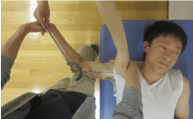
12 healthy asymptomatic young collegiate students (10 men and two women, age 21.1 ± 0.3 years, body weight 58.5 ± 6.2 kg, height 169.6 ± 5.2 cm, body mass index 20.4 ± 1.9) with limited elbow extension in screening ULNT1 were recruited for this study. Limited elbow extension in ULNT1 was defined as larger than 0-degree flexion in this study. In the recruitment process, ULNT1 as a screening test was performed to identify participants with limited ULNT1 in their dominant arms. Eligibility criteria were as follows: (1) No history of radiating pain in their arms, (2) No obvious neurological symptoms, including sensory loss and motor impairments, (3) No musculoskeletal pain in neck or dominant arm, which limits the execution of ULNT1, and (4) The presence of limitation in ULNT1. The presence of neurological symptoms was verbally asked among potential participants. In this study, ULNT1 was modified to increase the sensitivity of the test and enable efficient recruitment and measurements as follows; 30-degree contralateral cervical lateral flexion, 90-degree shoulder abduction, maximal wrist dorsiflexion and thumb/finger extension, maximal forearm supination, 90-degree shoulder lateral rotation, and elbow extension to the limit of maximum resistance (refer to Figure 1).
 Figure 1: ULNT1. View Figure 1
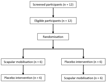
A sampling process is shown in Figure 2. At first, 12 healthy collegiate students volunteered to participate in this research. Following screening assessments, all students were found to meet the eligibility criteria, and were included in this study. Most eligible participants were right-handed (n = 11), except for one person who was left-handed (n = 1). Subjects were instructed to refrain from any vigorous overhead exercises or stretching which involves neck and arms for 24 hours before experimental sessions in order to minimise potential confounding bias. Although it was not possible to blind participants due to the nature of the interventions, the author did not explain details and the supposed mechanism of the interventions to minimise placebo or nocebo effects.
 Figure 2: Flowchart of the study. View Figure 2
After the recruitment of 12 participants, each subject participated in two experiments in a random order, enabling the allocation concealment (Figure 2). Two testing sessions were separated by a minimum of 24 hours, in order to minimise potential carry-over effects of the interventions. In first experimental sessions, anthropometric measurements were taken and body mass index was calculated accordingly. In both intervention sessions, the baseline data for ULNT1 on the dominant side was collected in the same manner as the screening test. All screening tests, and pre- and post-intervention measurements were performed by the same two assessors. Whilst one physiotherapist (KM) performed ULNT1, elbow extension ROM was measured by one 4th-year undergraduate physiotherapy student, using an analogue goniometer (OG wellness, Japan) (see Figure 1). Elbow extension ROM was measured by identifying three bony landmarks; the centre of the humeral head, medial epicondyle of the humerus and the ulnar head. ULNT1 was performed by a physiotherapist (KM) who had completed Master degree in musculoskeletal and sports physiotherapy. Maximum ROM in elbow extension was defined as when the primary assessor experienced resistance which limited further passive elbow extension. The level of pain was verbally asked using 11-point numerical scale (NRS). 11-point NRS consists of 11 numbers from 0 through 10; 0 representing 'no pain' and 10 representing 'worst imaginable pain'. This scale has been reported to be valid and reliable to assess the level of pain [26]. ULNT1 was performed for each participant lying in supine on the same treatment table (MC Healthcare, Japan) without pillow (Figure 1).
In the first testing sessions, following baseline measurements, one assessor who was responsible for measuring ROM with the goniometer left the room, and the physiotherapist (KM) performed a randomly chosen intervention (either scapular mobilisation or placebo intervention). Excel 2016 (Microsoft, USA) was used for randomisation after the baseline measurements. Scapular mobilisation was performed on the dominant sides and directed towards elevation and depression alternately into maximal resistance in side-lying (Figure 3). An oscillatory manoeuvre was repeated once per four seconds towards each direction respectively for 40 seconds in total. The dosage of the mobilisation technique corresponded with grade lll++ at 0.25 Hz [27]. This large-amplitude end-range mobilisation was repeated three times with 10-second intervals, which means that it took 2 minutes and 20 seconds to complete the whole mobilisation intervention. In placebo interventions, the therapist placed his hands on participants scapula as in scapular mobilisation, but stayed still in the same position for 2 minutes and 20 seconds (Figure 3). This placebo technique was designed to imitate positioning of the scapular mobilisation. During both interventions, two soft pillows (Sanmoto, Japan) were placed under participants' heads and arms to ensure the neutral positions of cervical spine and upper limbs (Figure 3). After each intervention was completed, the other tester entered the room for post-intervention measurements. Post-intervention measurements of ULNT1 were performed for the dominant side in the same manner as the baseline evaluations.
 Figure 3: Scapular mobilisation (left; elevation, centre; depression) and placebo intervention (right). View Figure 3
For the second experimental sessions, participants were again assessed in the same manner as the first session. In this session, however, the other intervention was performed (scapular mobilisation or placebo intervention). Thus, each participant received two different interventions with at least 24 hours between sessions to enable comparisons between the two interventions. The information regarding the order of stretching methods were not given to one assessor who was in charge of measuring ROM, until the completion of post-intervention assessments in second sessions, which guaranteed partial blinding for assessors.
All testing sessions were conducted in the same room at the same room temperature of 26 degrees Celsius. Participants were instructed to wear the same short-sleeve T-shirts in the experiments to minimise the impact of resistance from cloths during ULNT1. This also maintained patient modesty for female participants, maximising the recruitment. Efforts and cares were taken to ensure that all participants received the same verbal instructions and visual cues to minimise potential bias between the two groups.
The results are presented as mean ± standard deviation (SD) values. The baseline data in ULNT1 and pain levels from two testing sessions were utilised to calculate intraclass correlation (ICC) and determine the reliability of the two outcome measures [28,29]. ICC was evaluated accordingly; > 0.75 as excellent, 0.40-0.75 as fair to good and < 0.40 as poor [30]. A repeated measures one-way analysis of variance and Friedman test were used to examine significant differences in ULNT1 and pain respectively. A Bonferroni post-hoc test was also performed to compare pre- and post-intervention data. Statistical tests were conducted with SPSS (IBM, USA). Differences were considered statistically significant at p < 0.05. Hedges' g and 95% confidence intervals (CI) were calculated to determine within-group effect sizes [31]. Effect size was categorised as large (> 0.8), moderate (> 0.5) or small (> 0.2) [32].
All participants (n = 12) completed both testing sessions and there was no dropout (Figure 2). ICC of ULNT1 and NRS for pain level were 0.87 and 0.93 respectively. Thus, the two outcome measures used in this study had an excellent reliability. Descriptive statistics for the results are summarised in Table 1. There was no significant difference between each baseline data set in the two groups (p = 1.00), which confirmed the baseline comparability.
Table 1: Results of ULNT1 and pain assessments. View Table 1
Statistical tests revealed significant improvements in ULNT1 after scapular mobilisation (p < 0.05), however not after placebo intervention (p = 1.00). There was no statistically significant change in pain level after the two interventions (p = 0.07 and 0.12 respectively). The scapular mobilisation group displayed large within-group effect size (Hedges' g = 0.99, 95% CI -2.09 to 4.07) to improve ULNT1, whilst placebo intervention had a small effect size (Hedges' g = 0.19, 95% CI -3.30 to 3.67). The effect size for pain reduction was moderate in scapular mobilisation group (Hedges' g = 0.66, 95% CI 0.18 to 1.15), whereas the effect size was small in placebo intervention group (Hedges' g = 0.37, 95% CI -0.32 to 1.06).
This is the first study which investigated the immediate effects of scapular mobilisation technique on ULNT1 in asymptomatic participants. Generally, neural mobilisation techniques for nerve-related neck and arm pain involve cervical lateral glide, and slider and tensioner techniques adapting ULNTs [22]. Although a similar scapular mobilisation technique in supine position was proposed by Robert Elvey in 1986, this technique has not been examined in formal research [25]. In one RCT, another similar scapular mobilisation technique in prone position was incorporated into management, along with various other interventions, such as cervical lateral glide mobilisation, thoracic mobilisation, glenohumeral mobilisation, muscle re-education and home exercises [33]. Although they reported significant improvements in pain and physical disability among patients with cervicobrachial pain, the individual effects of scapular mobilisation were not clear. Before the initiation of this study, the author hypothesised that there would be significant improvements in ULNT1 after scapular mobilisation. The results of this study were in line with the hypothesis and suggested that scapular mobilisation might be effective to immediately improve ULNT1 in asymptomatic participants.
The findings in this study indicate that clinicians may be able to affect the mechanosensitivity of the nervous system by the scapular moblisation technique. One potential mechanism of this positive effect might be related to centrally-mediated hypoalgesic effects of manual therapy. A growing evidence suggests that passive manual interventions can produce neurophysiological hypoalgesic effects mediated by the periaqueductal gray and the dorsal horn of the spinal cord [34]. Furthermore, slow large-amplitude end-range mobilisation adopted in this research might have reduced participants' fears of passive upper-limb movements during the test, resulting in non-specific analgesic effects and increased mobility [35].
In addition to the potential involvement of the central nervous system, scapular mobilisation might have physically improved the mobility of peripheral neuromusculoskeletal tissues, leading to improved ULNT1. Numerous studies have reported that peripheral nerves can slide through surrounding musculoskeletal tissues longitudinally, depending of the movements of limbs and spine [36,37]. In the last phase of ULNT1, cervical nerve roots, brachial plexus and peripheral nerves in the upper arm are thought to move distally due to elbow extension. This distal sliding movement might have improved by the use of end-range scapular mobilisation towards depression, which encouraged cervical nerve roots and brachial plexus to move distally and pass through musculoskeletal interfaces, such as cervical intervertebral foramen and scalene muscles. Increased mobility of the peripheral tissues might have led to less mechanical nociceptive inputs during ULNT1, enhancing the elbow extension ROM.
Several methodological limitations in this research must be acknowledged. Since this study recruited only asymptomatic participants, the findings might not be applicable to patients with peripheral neuropathic pain. Cervical lateral glide has been reported to be effective for patients with cervical radiculopathy to improve ULNT1 in the short term [24]. Further research is required to compare effects of scapular mobilisation with other conventional neurodynamic techniques, including cervical lateral glide, slider and tensioner in the positions of ULNTs [22]. A potential advantage of scapular mobilisation over cervical lateral glide is that scapular mobilisation is less likely to provoke pain for patients with cervical radiculopathy, for it does not involve cervical movements. Failure to assess the credibility of the placebo intervention is another limitation in this study. The lack of fully blinded assessors might have introduced measurement bias. However, no statistically difference in pain levels in pre- and post-intervention assessments in both groups suggests that ULNT1 may have been performed up to maximum ROM regardless of interventions, which implicitly supports the soundness of ULNT1 measurements in this study. It is necessary to consider these potential limitations carefully when we interpret the findings of the study and apply them to clinical practice.
Large-amplitude end-range scapular mobilisation in the side-lying position significantly increased elbow extension ROM during ULNT1 in asymptomatic adults. The findings imply that this mobilisation technique might be able to affect mechanosensitivity of the nervous system and improve ULNT in the short term. Further research is required to test the effects of scapular mobilisation in symptomatic patients with peripheral neuropathic pain.
The author would like to thank Tokyo University of Technology for funding this research project.
This work was supported by Tokyo University of Technology.
None declared.