Background: Kidney transplantation is the treatment of choice for patients with end-stage chronic kidney disease. In the first six months post-transplantation, surgical complications represent a significant cause of graft loss. Situs Inversus Totalis (SIT) is a rare anatomical anomaly that poses technical challenges in organ procurement and transplantation. Evidence regarding the viability of organs from donors with SIT is scarce.
Methods: We report the successful procurement of both kidneys from a cadaveric donor with SIT, a finding that was detected intraoperatively. The right kidney showed a longer renal artery and vein, while the left kidney showed three converging arteries and one vein. Both kidneys were transplanted into different recipients. The first recipient evolved favorably, while the second recipient with multiple arteries in the graft presented acute graft dysfunction secondary to renal infarction, massive hemorrhage, and death.
Results: SIT is not an absolute contraindication for organ donation. However, anatomical variants such as the presence of multiple arteries increase surgical complexity and the risk of postoperative complications, highlighting the importance of an adequate anatomical evaluation and surgical planning before transplantation. Kidney grafts from donors with SIT can be used successfully, provided that an adequate selection and individualized management of the recipients is performed.
Conclusion: This report emphasizes the need for more rigorous imaging protocols and consideration of graft vascular anatomy in perioperative decision making.
Kidney transplant, Situs inversus totalis, Cadaveric donor, Surgical complications, Multiple arteries, Thrombosis
Kidney transplantation is the treatment of choice for patients with end-stage chronic kidney disease, as it presents superior benefits to dialysis in terms of patient survival and quality of life [1]. During the initial 6 months after kidney transplantation, surgical complications present a higher risk of graft loss compared to immunologic allograft rejection [2], so preoperative anatomic evaluation of living donors is crucial to minimize surgical complications and optimize outcomes in recipients [3].
SIT is a very rare genetic anomaly, affecting 0.01% of the population. SIT represents a total vertical transposition of the thoracic and abdominal organs that are inverted in a mirror image of the normal position [4]. For many years, donors with this pathology were discouraged for organ donation in particular for liver and heart donation, considering the associated anomalies of the blood vessels that appear in more than 40% of cases [5]. In the medical literature, case reports describing renal donors with situs inversus are limited, and even fewer those evaluating the postoperative evolution of the recipients in the short term.
In this report, we present the case of a cadaveric donor with SIT, from whom both kidneys were procured for transplantation, detailing the anatomical characteristics found, the surgical approach used and the short-term clinical evolution of the recipients.
The patient was a 32-year-old female, who presented a cranioencephalic trauma and brain death confirmed by cerebral angiotomography. Her blood type was O with Rh positive and non-reactive viral panel, being compatible for the recipient. Physical examination revealed no abnormalities, it was not until a chest X-ray was performed as part of the pre-surgical protocol, where dextrocardia was not clearly identified due to poor technique, it was decided that based on the rest of the normal initial approach, she was a good candidate for donor.
During the procurement, it was performed according to our standard, the approach was by exploratory laparotomy + thoracotomy. During surgery, reverse transposition of all abdominal organs was evidenced, the liver and gallbladder in the left hypochondrium, the spleen in the right hypochondrium (Figure 1), the stomach with the major curvature to the right of the spine and the minor curvature to the right of the spine, gut malrotation with left appendix (Figure 2), transposition of the inferior vein cava and aorta (Figure 3), and in the thorax, dextrocardia (Figure 3 and 4), deciding to remove both kidneys, of which, the right kidney had 1 artery and the left kidney 3 arteries originating from the abdominal aorta, which reached the renal hilum, both had 1 vein, exited the renal hilum and converge in the inferior vena cava, the right vein being longer. The ureter and veins were evidenced without any anatomical variation (Figure 5).
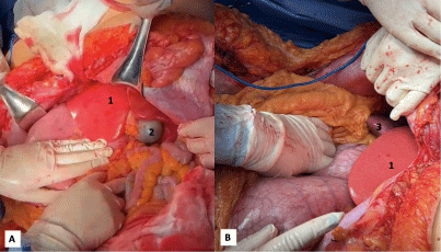 Figure 1: (a) Liver (1), gallbladder (2); and (b) spleen (3) shown in inverted position (upper left quadrant).
View Figure 1
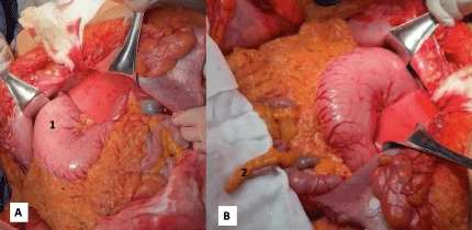 Figure 2: (A) (1) The stomach is shown with the greater curvature to the right of the spine and the lesser curvature to the left; (b) (2) Cecal appendix in left iliac fossa.
View Figure 2
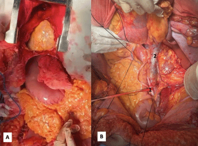 Figure 3: (a) Dextrocardia is shown, with the tip of the heart to the right; (b) The inferior vena cava (2), which was to the left of the vertebral column and the common iliac artery (1) to the right of the vertebral column.
View Figure 3
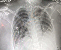 Figure 4: A chest X-ray is presented, with the cardiac silhouette with a tendency of cardiac apex to the right, without well delimiting the hepatic silhouette and the gastric bubble due to poor technique.
View Figure 4
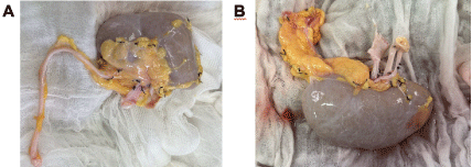 Figure 5: (a) The right kidney of the cadaveric donor is observed with 1 vein and 1 renal artery and 1 ureter without macroscopic alterations; (b) The left kidney of the cadaveric donor is observed with its 3 renal arteries joined with a carrel patch and a vein.
View Figure 5
In the case of the recipient, a 29-year-old female, with advanced chronic kidney disease and arterial hypertension, on hemodialysis treatment for 12 years, the right kidney was placed in the right iliac fossa via Rutherford Morison incision, and the terminolateral anastomosis of the renal artery to the external iliac artery and the extravesical ureteroneocystostomy (Lich-Gregoir) were performed, with 100% perfusion at reperfusion (Figure 6), without eventualities, with adequate evolution, she was discharged on her 7 th post-transplant day, with adequate graft function so far, with adequate evolution, he was discharged on his 7 th post-transplant day, with adequate graft function until now.
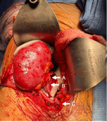 Figure 6: (a) The right kidney with 100% perfusion, the renal artery; (b) the renal vein; and (c) the Lich-Gregoir ureterovesical junction are observed during kidney transplantation. In figure 1 the liver and gallbladder are observed in the left hypochondrium.
View Figure 6
On the other hand, the second 23-year-old recipient with advanced chronic kidney disease on peritoneal dialysis for 3 years, with standard criteria, underwent heterotopic renal transplantation, via Rutherford Morison incision, the left kidney was placed in the right iliac fossa, a terminolateral anastomosis of the carrel patch (3 arteries) to the external iliac artery and an extravesical ureteroneocystostomy (Lich-Gregoir) was performed, with 100% perfusion without eventualities, until day 9 post-transplantation, showing acute graft dysfunction secondary to infection by E. Coli blee infection, with torpid evolution, with subsequent transfictive pain in the right flank associated with graft infarction, it was decided to reoperate, showing ischemia and multiple areas of graft necrosis, nephrectomy of the graft was performed, when performing arteriotomy of the external iliac artery, he presented incoercible bleeding with grade IV hypovolemic shock, cardiorespiratory arrest, with reperfusion at 22 minutes, with subsequent diagnosis of brain death. She died 28 days after transplantation.
SIT is a very rare anomaly characterized by a total vertical transposition of the thoracic and abdominal organs that are inverted in a mirror image of the normal position [4]. All internal organs are inverted 180° in patients with SIT, but the relative position between all organs is normal [6]. Its prevalence is estimated to be between 1:8,000 and 1:20,000 live births [7].
During the procurement, classic and previously described findings of SIT were observed. In the case of kidney grafts, the right kidney with 1 renal artery, with a vein longer than the left one which crossed the aorta anteriorly up to the right renal hilum, as it has been evidenced in other cases of SIT, where the visceral branches of the aorta (such as renal arteries, superior mesenteric, celiac) can be arranged in a specular manner [8], remembering the normal anatomy, the left vein is usually longer, which has been seen to allow a technically easier venous anastomosis, by reducing the tension on the suture and facilitating the manipulation of the graft in the iliac fossa [9]. However, in more current studies of nephrectomy in living donors, no differences have been seen in terms of morbidity and mortality [2]. Although there are not many studies that tell us about the follow-up and results in cadaveric donors with SIT; in most of those reported, both right and left have been used indistinctly, with good results in the short and long term [7].
In the case of the recipient of the left kidney, which presented 3 kidney arteries and 1 kidney vein with the previously mentioned characteristics, which were anastomosed with terminolateral technique to the external iliac artery, with carrel patch, with monofilament polypropylene suture 6-0, which had a torpid post-surgical evolution, with acute graft dysfunction due to probable infarction, which required reintervention, massive transfusion secondary to hypovolemic shock that progressed to cardiorespiratory arrest and brain death; these events could be due to multiple causes described in literature, such as the increased risk of thrombosis when presenting multiple arteries, which refers to presenting two or more arteries that irrigate different segments of the graft with an incidence of 27% in the general population, with an even lower incidence of 2. 5% in the case of presenting 3 arteries [10]. The decision of whether or not to use these organs can be difficult, presenting a higher risk of post-transplant vascular complications, such as thrombosis or arterial stenosis, with poor prognosis [11], presenting greater surgical complexity and increasing surgical time and therefore the time of hot ischemia [10,12]. Although the anatomical condition of the donor is not directly related to this outcome, the technical difficulties derived from the complex vascular anatomy could have contributed to the unfavorable evolution. Therefore, this fact highlights the importance of surgical planning and adequate vascular evaluation and the establishment of criteria in current guidelines regarding donor anatomical variations at the time of transplantation.
The SIT was once a contraindication to organ transplantation due to concerns about visceral and vascular abnormalities [7]. Currently, none of the current renal transplantation guidelines clearly state whether this group of SIT constitutes a contraindication to organ transplantation. However, cadaveric donor transplantation with SIT has been reported since 2003, with good short- and long-term surgical outcomes [5].
SIT is a rare condition in which the internal organs of the body deviate from their normal position. Its cause is unknown. It is often an incidental finding as in our case. The present case demonstrates that SIT in a cadaveric donor is not an absolute contraindication for kidney transplantation, provided that proper anatomic evaluation and surgical planning is performed. Despite the associated visceral and vascular variants, the procurement was technically feasible and allowed successful implantation of both kidneys. Nevertheless, complex anatomical conditions, such as the presence of multiple arteries, may increase the risk of postoperative complications, as was evidenced in one of the recipients, whose clinical evolution was unfavorable. This report underscores the importance of establishing more rigorous preoperative imaging protocols and highlights the need for an individualized approach in the selection and management of recipients with risk factors.
All the authors fulfilled the following functions:
• Substantial contributions to the conception or design of the work.
• Acquisition, analysis, and interpretation of data for the work.
• Drafting the work, revising it critically for important intellectual content.
• Final approval of the version to be published.
• Agreed to be accountable for all aspects of the work in ensuring that questions related to the accuracy or integrity of any part of the work are appropriately investigated and resolved.
No funding was received.
The authors have no conflict of interest to declare.
This case report exempted from ethical approval.
We have a confirmation with familiar of patient for this case report.