Intracranial extension of a dermoid cyst in temporal region of scalp is extremely rare and we present one such case along with its management and follow-up. Dermoid cysts are benign tumors which result from aberrant closure of the neural tube. They have thick capsules which are lined by squamous epithelium and contain skin appendages. About 7 percent of all dermoids are craniofacial dermoid cysts with an incidence of 0.03 to 0.14 percent [1]. Although congenital dermoid cysts are present at birth, only 40% are noted then; 60% are diagnosed by the age of 5 years [2] and thus rarely present in adults. They usually have a good prognosis as no recurrence has been documented after complete en bloc resection.
A 52-year-old male patient came to out-patient department with complaints of swelling over right parietal scalp which was slowly growing since 8 years (Figure 1).
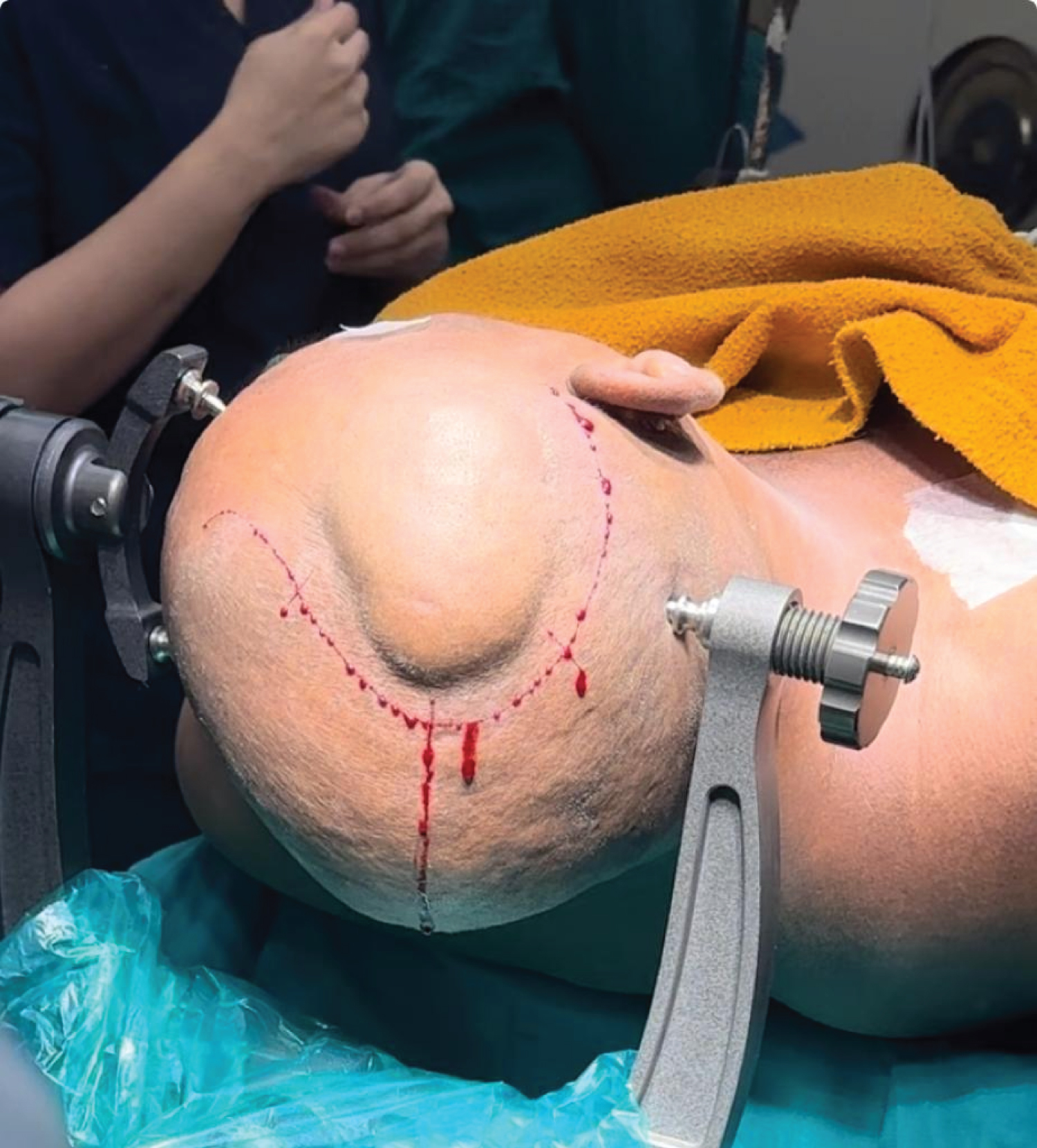 Figure 1: Right parietal scalp swelling.
View Figure 1
Figure 1: Right parietal scalp swelling.
View Figure 1
X-Ray of skull was done which showed a lytic lesion in the right parietal scalp (Figure 2). CT brain (plain plus contrast) was suggestive of 5 × 2 × 5 cm hypodense lesion in right parietal scalp (Figure 3). MRI brain was suggestive of a focal osteolytic lesion in right parietal bone showing internal blooming with a hyperintense collection seen in right subcutaneous plane (Figure 4).
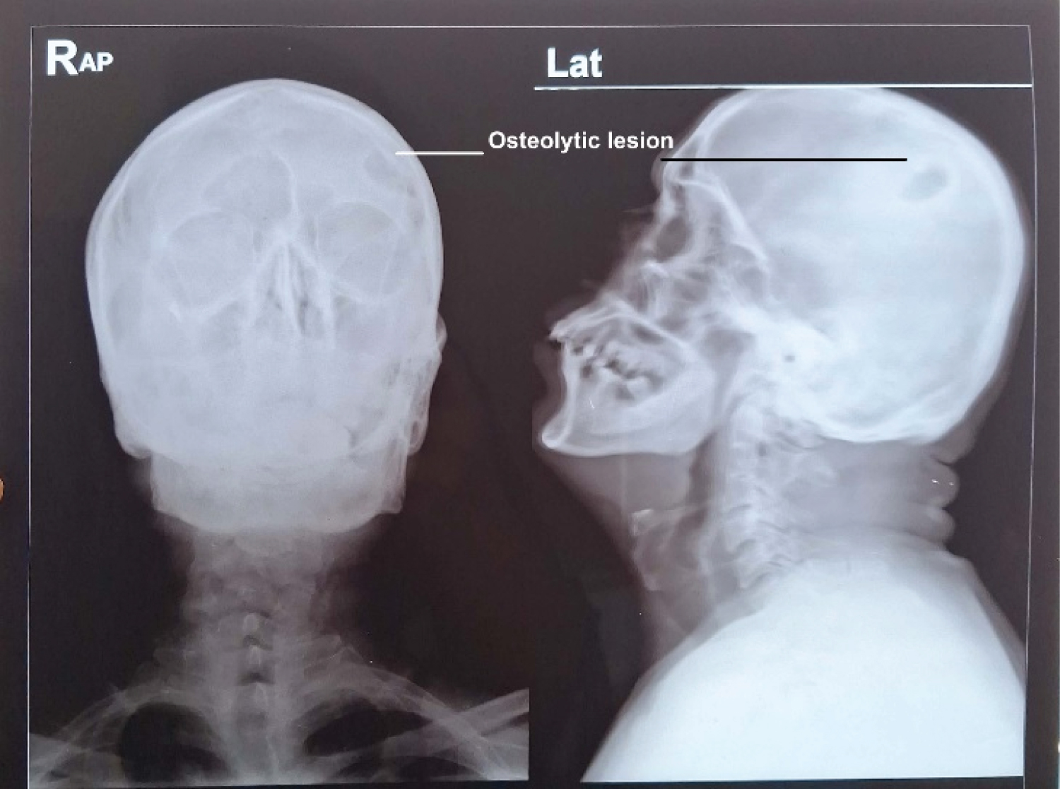 Figure 2: Osteolytic lesion on skull X-ray.
View Figure 2
Figure 2: Osteolytic lesion on skull X-ray.
View Figure 2
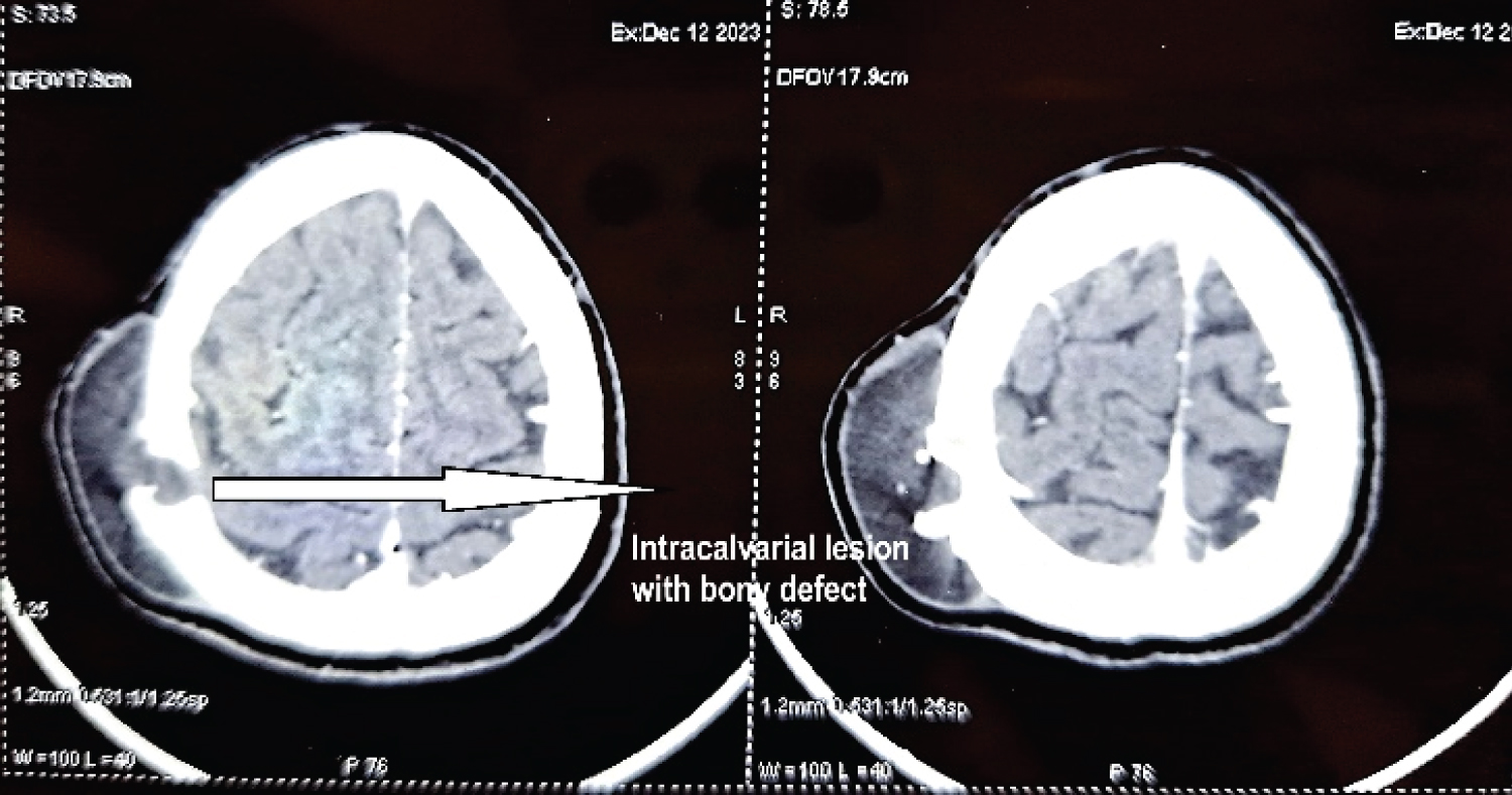 Figure 3: CT scan with calvarial defect and lesion.
View Figure 3
Figure 3: CT scan with calvarial defect and lesion.
View Figure 3
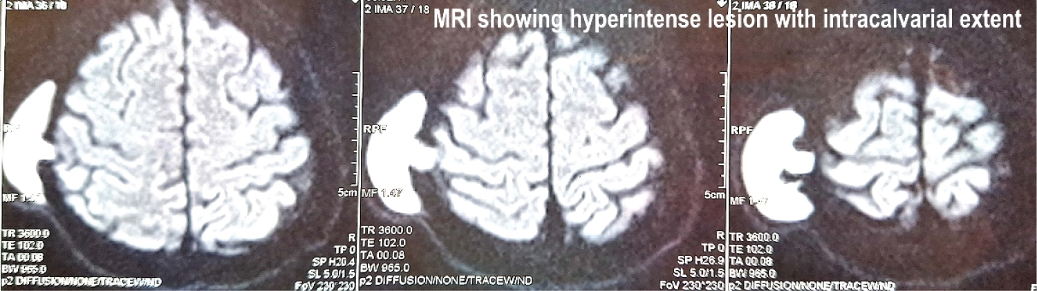 Figure 4: MRI brain showing intracalvarial extent of tumour.
View Figure 4
Figure 4: MRI brain showing intracalvarial extent of tumour.
View Figure 4
In supine position, under general anaesthesia, head was turned to left and fixed with three point fixation device. A C-shaped incision was taken around the lesion as shown in Figure 1. Skin flaps were raised. The tumor was found to have eroded the calvarium and was firmly attached to the periosteum. The tumor was dissected from all sides. The wall ruptured while removing, and approximately 30 cc thick, brown, paste-like material was removed (Figure 5). A small craniotomy was done to remove the wall completely from the bone. Dura was found to be intact (Figure 6). A thorough wash was given with saline and diluted betadine. The calvarial defect was closed with a titanium mesh and mini screws (Figure 7). Hemostasis was achieved and the flap was closed in 2 layers.
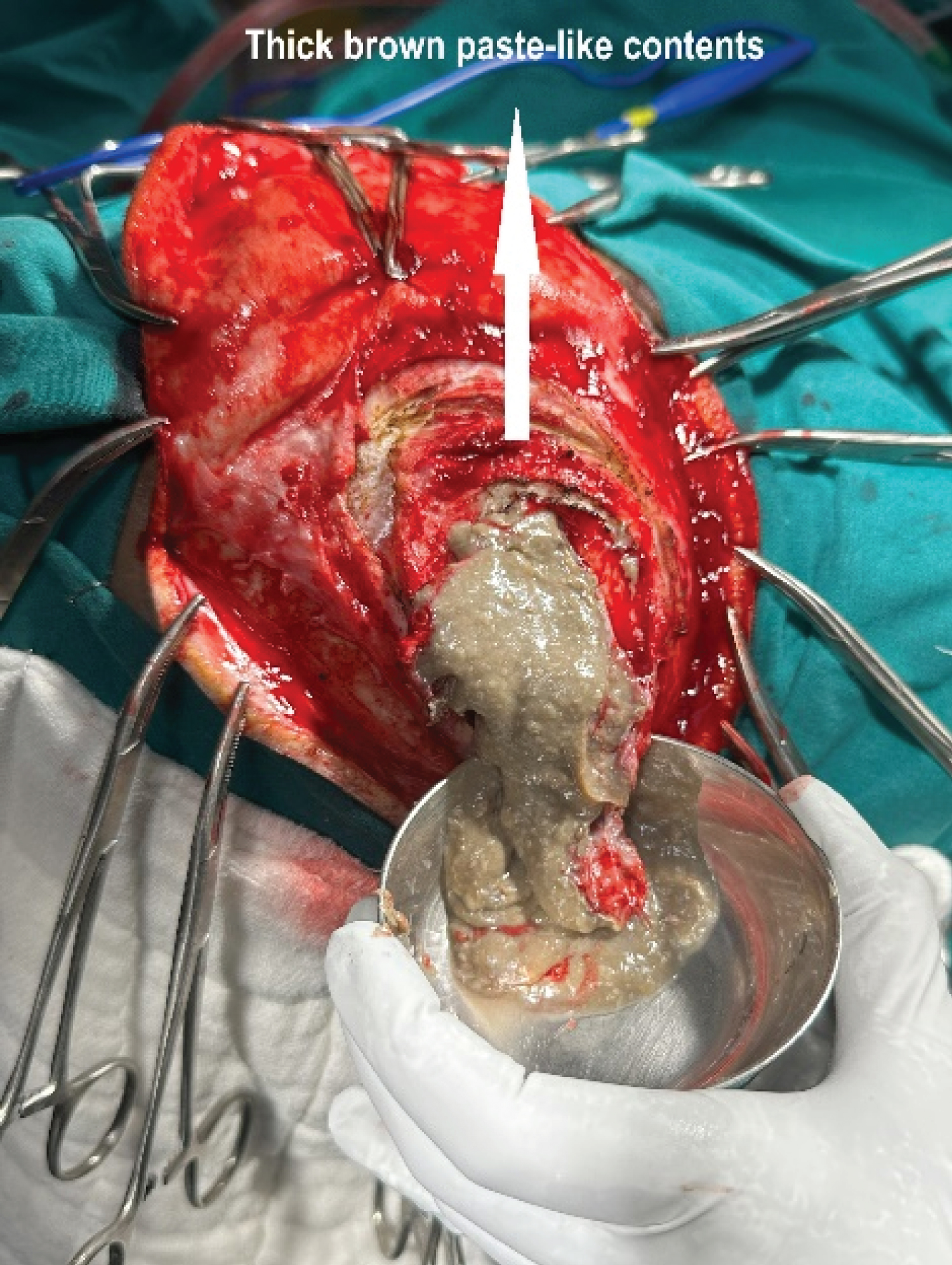 Figure 5: Thick brown paste-like content of cyst.
View Figure 5
Figure 5: Thick brown paste-like content of cyst.
View Figure 5
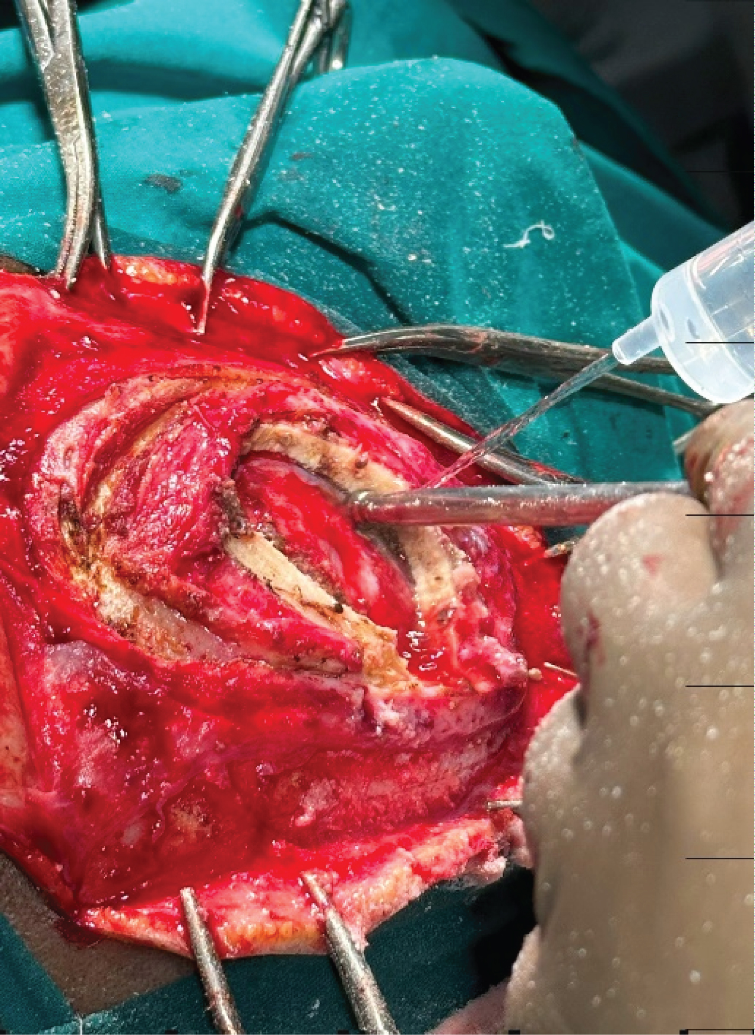 Figure 6: Craniotomy.
View Figure 6
Figure 6: Craniotomy.
View Figure 6
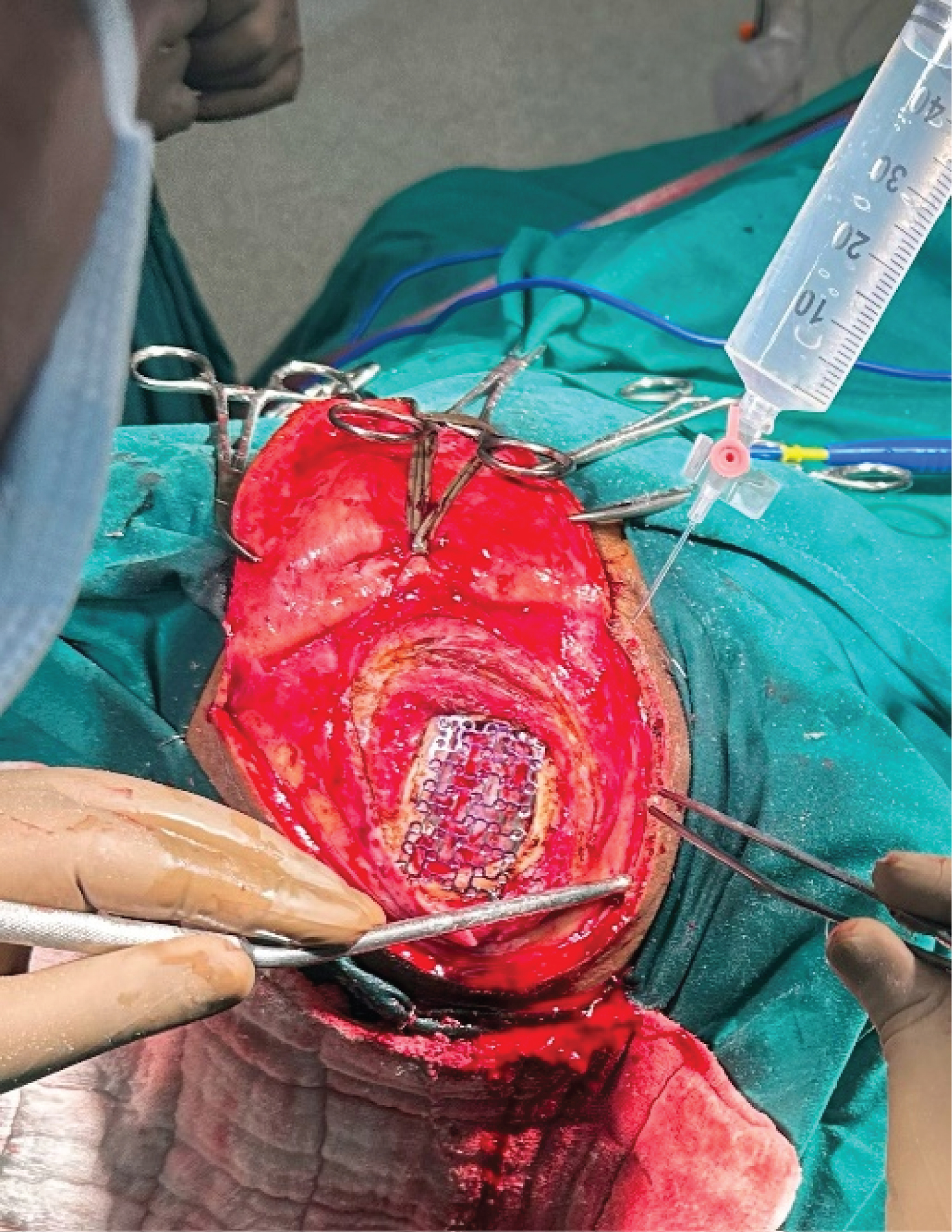 Figure 7: Titanium mesh.
View Figure 7
Figure 7: Titanium mesh.
View Figure 7
The postoperative period was uneventful and the patient was discharged on Day 3. The patient was followed up regularly, initially at 1 week intervals and then at 2 week intervals for 3 months. There were no signs of recurrence of swelling.
The exact cause of dermoid cysts is largely unclear, yet they are regarded as benign congenital developmental anomalies resulting from the entrapment of ectodermal cells along the embryonic closure lines. These cysts are true hamartoma, featuring a lining of stratified squamous epithelium. Their lumen is filled with keratin and hair; and mature skin appendages are found on their wall. Although they are considered to be congenital, only about 40% of dermoid cysts are diagnosed at birth, while about 60% of the dermoid cysts are diagnosed by five years of age [2]. Diagnosis of dermoid cyst in adults is rare. They are very slow-growing tumours and occur most commonly on the head and neck, with 84% of these cysts occurring in this region [3].
Intracranial dermoid cysts account for about 0.1%-0.7% of all intracranial tumors [4]. Intracalvarial dermoid cysts become symptomatic when they become infected, which may lead to cerebral abscess or recurrent meningitis while cyst rupture may result in chemical aseptic meningitis. They can also present with local compressive symptoms [1].
Hartley, et al. [5] described midline nasal lesions on the basis of depth, and Overland, et al. [6] modified the same system to describe any head and neck lesion. The lesions were divided into superficial (type 1), subpericranial/stalk to suture (type 2), intraosseous (type 3), intracranial extradural (type 4), and intracranial intradural (type 5). As per this criteria, our patient falls into type 4 lesion. Also according to this study [6], temporal dermoid was present in 5.6% of the patients (aged 2 years to 18 years). It was also concluded in this study that temporal dermoids have a high rate of intracranial extension, however no such report is available showing similar results in adults.
Ramnarayan, et al. [7] have described scalp dermoids in three adults, aged 18-27 years. The lesion in all three patients was limited to the periosteum, without breach in the outer table.
A recently published report by Shalmiyev R, et al. [8] is one of the rare references in literature citing intradural extension of dermoid cyst in an adult which was also repaired similarly by craniotomy, excision and closure by mesh placement.
Dermoid cysts of temporal bone in adults are rare. Intracalvarial extension of the dermoid cyst in an adult is extremely rare. We thus present a rare case of intracalvarial dermoid cyst in an adult which was excised successfully and the craniotomy defect was reconstructed using a titanium mesh.
Nil.
Nil.