This study investigated if resistance exercise performed at differing Arterial Occlusion Pressures (AOP) causes oxidative stress and muscle damage. Twelve males completed 4 sets of 10 repetitions of knee extension at 20% of 1RM, with 30 s rest intervals between sets, that varied only in the amount of restriction pressure applied: 1) CON (no pressure), 2) AOP-50 (50% of the total AOP), 3) AOP-75 (75% of the AOP), and 4) AOP-100 (100% of the AOP), with each condition separated by 7 days. Ratings of Delayed-Onset Muscle Soreness (DOMS), Maximal Isometric Strength (MIS), serum levels of Creatine Kinase (CK), Lactate Dehydrogenase (LDH), Lipid Peroxide (LP), and the Antioxidant Activity of Plasma (AAP) were assessed at rest, 1, 24, and 48 h post-exercise. DOMS, MSI, CK, LP, and AAP did not differ among the conditions (p > 0.05) across the different time points, however, plasma levels of LDH were significantly lower (p < 0.01) at 24 h post-exercise (265.83 ± 17.55 UI/L) in comparison to 48 h post-exercise (294.96 ± 17.51 UI/L) averaged across all groups. It was concluded that resistance exercise with differing occlusion pressures does not cause prolonged muscle damage or oxidative stress, regardless of the amount of AOP.
Blood flow restriction, KAATSU, Strength exercise, Metabolic stress, Occlusion training
Exercise-induced muscle damage has been shown to occur following acute bouts of high-intensity resistance exercise, mostly due to the eccentric component of the contractions performed [1-3]. Although some researchers have investigated the physiological mechanisms underlying the muscle damage phenomenon in both human and animal models, these responses remain unclear. Several factors contribute to muscle damage following resistance exercise, such as gender [1], exercise volume [4], rest periods between sets [5], and the type of the exercise performed [1].
Concerning exercise intensity, there is no evidence that low-intensity resistance exercise is capable of inducing significant muscle damage post-exercise [6]. However, low-intensity resistance exercise (20-50% 1RM) with Blood Flow Restriction (BFR) has gained some attention regarding the occurrence of muscle damage linked to this model of exercise [7] and it is speculated that it may cause minimal muscle damage [8,9].
Oxidative stress has been linked to muscle damage following resistance exercise [10]. In this sense, muscle damage related to BFR could be better explained by internal physiological factors rather than exercise workloads, since BFR exercise uses significantly lower loads (20%-50% 1RM) compared to traditional resistance training (70%-80% 1RM). Thus, muscle damage observed following BFR exercise may be triggered by the formation of hypoxia-induced oxygen species in the active muscles. During BFR resistance exercise, pneumatic cuffs are attached to the exercising limbs, inflated to a target pressure, and then deflated following completion of the exercise. The venous pooling of blood in the limbs and the resultant increased venous return from the muscle following cuff deflation promotes the creation of reactive oxygen species which could potentially damage muscle tissue [11]. However, it is still unclear whether low-intensity resistance exercise at distinct restrictive pressures (50%, 70%, or 100% of total occlusion) would be able to induce significant acute oxidative stress and muscle damage.
The majority of studies that have attempted to examine the muscle damage response after BFR exercise have a common limitation that needs to be addressed. Most of these studies have applied an arbitrary pressure [9,12] or have based the restrictive pressures on the subjects' brachial Systolic Blood Pressure (SBP) [8,13]. By applying the same amount of pressure to all subjects, it is very likely that subjects with different size limbs would experience differing amounts of BFR resulting in either an overestimation or underestimation of the blood flow going to the working muscle. Additionally, studies that have used SBP at the brachial artery (arm) to determine the restrictive pressure for the lower limbs may underestimate the restrictive effects of the pressured being used. Recent evidence suggests that the Arterial Occlusion Pressure (AOP) should be individually prescribed for each subject [14,15] and based on the amount of pressure required to totally occlude blood flow to the limbs [16]. Since occlusion pressure, as an independent factor, plays an important role in the muscle damage response to BFR resistance exercise due to the ischemia-reperfusion phenomena, different degrees of BFR should be evaluated.
To the best of our knowledge, no study has examined how oxidative stress and muscle damage responses are affected by BFR resistance exercise performed at different degrees of occlusion pressure. The fact that a wide range of occlusion pressures could be applied during BFR resistance exercise and that muscle performance is reduced by muscle damage, new information in this area should be helpful for physiotherapists, athletic trainers, and strength and conditioning trainers to improve BFR exercise training programs. Therefore, the purpose of the current study was to examine if BFR resistance exercises performed at different degrees of AOP cause oxidative stress and muscle damage in young trained men. Taking into consideration that muscle damage after BFR exercise can be triggered by the hypoxia-induced oxygen species formation in the active muscles [11], we hypothesized that resistance exercise with a higher AOP would result in a higher oxidative stress response and greater muscle damage, as well as a longer recovery period.
Twelve trained males (20.58 ± 2.39 yrs, 72.77 ± 8.42 kg, 1.79 ± 0.07 m, 22.71 ± 1.71 kg/m2) involved in resistance training exercise for at least 6 months prior to this study were included. Only healthy, non-smoking subjects (without any history of cardiovascular or osteomuscular disease, non-obese, with BMI values between 20.0 and 29.9 kg/m2, with an ankle-brachial index between 0.9 and 1.30, and without any known injuries) were included. Subjects were asked to avoid any resistance exercise throughout the study period and to refrain from alcohol and caffeine at least 24 h prior to each exercise session. The study protocols and procedures were explained to each subject, and written informed consent was obtained prior to participation. This study was approved by the Institutional Review Board of the Federal University of Paraiba.
Subjects were required to complete all four experimental conditions, which varied according to the amount of AOP applied to the lower-body. The four experimental conditions were performed using a contralateral counterbalanced design as follows: 1) Unilateral knee extension with no AOP (CON); 2) Unilateral knee extension at 50% of AOP (AOP-50) using the contralateral leg; 3) Unilateral knee extension at 75% of AOP (AOP-75); and 4) Unilateral knee extension at 100% of AOP (AOP-100) using the contra lateral leg. Each experimental condition was separated by 7 days. The percentages of blood flow restriction (0%, 50%, 75%, and 100%) were based on the pressure required to totally occlude the arterial blood flow to the lower extremities. The order of the experimental conditions were randomized by using a random numbers table.
Participants attended the laboratory on several different occasions. On the first visit, height, weight, resting arterial blood pressure, ABI, and AOP were measured. Subjects also completed a standardized maximal strength test (one Repetition Maximum-1RM) for both knee extensors (right and left legs), such that 20% of 1RM could be determined for each experimental condition. In subsequent visits, subjects performed the experimental conditions and the oxidative stress and muscle damage markers were measured before and at 1 h, 24 h and 48 h following each exercise protocol.
The pressure required to achieve total AOP for each leg was determined using a standardized protocol first proposed by Laurentino, et al. [16]. With subjects in a supine position, a vascular Doppler probe (Medpej, DV-2001, Brazil) was placed on the tibial artery to detect the auscultator pulse. Then, a standard 15 cm wide blood pressure cuff [17] located at the most proximal portion of the thigh was inflated until the pulse could no longer be detected and then slowly released until the moment in which the pulse could be detected again. This value was considered to be 100% of the AOP and was used to determine the different percentages of occlusion pressure used in the different exercise sessions (0%, 50%, 75%, and 100%).
The 1RM test was randomly performed for each leg separately as described by Baechle and Earle [18]. The initial warm-up period utilized a weight that could be easily lifted for 5-10 repetitions of knee extension. Then, following a 1 min rest period, the weight was increased by 10-20%, and 5 more repetitions were performed. The last warm-up set of 2-3 repetitions occurred after a 2 min rest, with another weight increase of 10-20%. Participants then began single attempts of knee extension with increased weight until the weight could not be lifted through a full range of motion. No more than 5 attempts were allowed to reach maximal strength, and 3 min rest periods separated each attempt. After a 10 min rest, the second leg performed the same test sequence. There was no significant difference between the maximum strength values measured from both legs (p > 0.05).
Oxidative stress was verified according to the Lipid Peroxide (LP) levels and the Antioxidant Activity of Plasma (AAP). The lipid peroxide levels were quantified using the reaction of thiobarbituric acid with the decomposition products of the hydroperoxides, as described by Ohkawa, et al. [19]. For this, 250 μL of serum was incubated in a water bath at 37 ℃ for 60 minutes. Samples were then precipitated with perchloric acid AA at 35% and centrifuged at 14,000 rpm for 20 minutes at a temperature of 4 ℃. The top layer of supernatant was transferred to new micro tubes, to which 400 μL of thiobarbituric acid at 0.6% was added and incubated at 100 ℃ for 60 minutes. After cooling, the samples were analyzed using an ultraviolet spectrophotometer (Bioespectro, model 22, Brazil) at a wavelength of 532 nm and room temperature.
The antioxidant activity of the plasma was determined in agreement with Brand-Williams, et al. [20]. In detail, an aliquot of 1.25 mg of 2,2-Diphenyl-1 Picrylhydrazyl (DPPH•) was diluted in 100 ml of ethanol (99.5% absolute ethanol), kept under refrigeration, and protected from the light (using aluminum foil or amber-colored glass). In appropriate centrifuge tubes, 3.9 ml of DPPH• solution was added, followed by 100 μL of plasma. The tubes were vortexed and left to stand for 30 minutes. Then, tubes were centrifuged for 15 minutes at a speed of 10,000 rpm and a temperature of 20 ℃. The supernatant was used to perform the analysis in a spectrophotometer at 515 nm. The results are expressed as percentages of the antioxidant activity, using the following equation:
AAP = 100 - [DPPH•R]t/[DPPH•R]B 100)
[DPPH•R]t and [DPPH•R]B correspond to the remaining concentration of DPPH• after 30 minutes, assessed on the sample (t) and on the white (B) and prepared with distilled water.
The maximal isometric strength of the exercising leg was measured using a portable digital dynamometer (Instrutherm Ltd., DD-300, Brazil) attached to the knee extension machine. Participants had their knee positioned at an angle of 60° (where 0° represented a full knee extension), and a belt was fastened across the hip to minimize extraneous body movements, which could affect the strength measurements. Subjects were then asked to perform three maximum voluntary contractions and to hold each for 3 seconds. One minute of rest was given between each attempt. The highest score was registered and used for statistical analysis. In addition, a familiarization session was performed for the first week, before the main testing.
Serum levels of Creatine Kinase (CK) and Lactate Dehydrogenase (LDH) were used as indirect biomarkers of muscle damage. Venous blood samples (5 ml for each point of measurement) were obtained from the antecubital vein by a nurse; the samples were placed in assay tubes and centrifuged at a speed of 3,000 rpm for 15 min. The serum was stored at -20 ℃ until analysis. An immunochemical automatic analyzer (Labtest, Labmax 240 premium, Brazil) was used to run the analysis using specific kits for CK and LDH (Labtest, Brazil). All the procedures were performed according to the manufacturer's specifications.
Delayed-Onset Muscle Soreness (DOMS) was measured through an analogic verbal scale proposed by Nieman, et al. [21]. This scale ranges from 0 to 10 as follows: 0 (no soreness), 2 (dull, vague ache), 4 (slight soreness), 5 (more than slight soreness), 7 (sore), 8 (very sore), and 10 (unbearably sore). Subjects were introduced to the scale and asked if they had any questions.
During each exercise session, subjects performed 4 sets of 10 repetitions of unilateral knee extensions, through a 90° range of motion, with a load of 20% of 1RM and a pace of 1.5 seconds for both the concentric and the eccentric portion of the contraction. A total of 30 seconds of rest were allowed between sets. BFR was used in all conditions, except for the CON, varying only in the amount of pressure applied. In this regard, the cuff was positioned on the inguinal portion of the exercising limb and then inflated to the target pressure just prior to the start of the first set; it was deflated at the end of the fourth set. The amount of AOP for each condition was 66.58 ± 9.72 mmHg (AOP-50), 99.25 ± 14.95 mmHg (AOP-75) and 129.50 ± 18.73 (AOP-100). There was a significant difference between the conditions.
Sample size was calculated using G*Power version 3.1.9.2 for OS X (Franz Faul, Universitat Kiel, Germany) and was set at 12 subjects, corresponding to an effect size of 0.60, a power of 0.80, α = 0.05, test family = F test and statistical test = ANOVA repeated measures, within-betwechanges in the levels en interactions [22]. The Shapiro-Wilk test was performed to confirm the data normality. A two-way repeated measures ANOVA (condition [CON, 50-AOP, 75-AOP and 100-AOP] x time [rest, 1 h, 24 h, and 48 h post-exercise]) was used to compare the main effects of time, condition, and the condition by time interaction. When a significant difference was found, a Bonferroni post hoc test was carried out to identify where the difference was located. Given that the delayed-onset muscle soreness data did not present a normal distribution, the Kruskal-Wallis test was used with the Mann Whitney U test to find mean differences according to Bonferroni corrections. Data are presented as the mean ± SE. The significance level was set as p < 0.05. Data were analyzed using SPSS version 22 for OS X (IBM, Chicago, IL).
As presented in Table 1 and Figure 1, no significant condition (F = 0.16, p = 0.92) or time (F = 0.70, p = 0.56) main effects and no significant condition by time interaction (F = 0.56, p = 0.83) were observed for LP. For the AAP, there was no significant time main effect (F = 1.55, p = 0.22) or significant condition by time interaction (F = 1.55, p = 0.18), but there was a significant condition main effect (F = 3.32, p = 0.03); however, follow up analyses reviewed that such difference does not exist (p > 0.05) (Table 2 and Figure 2).
Table 1: Comparison of lipid peroxide levels (nmol/L) at rest, 1 h, 24 h, and 48 h post-resistance exercise performed at different levels of AOP (n = 12). View Table 1
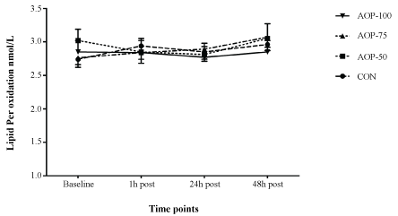 Figure 1: Comparison of lipid peroxide levels (nmol/L) at rest, 1 h, 24 h, and 48 h after resistance exercise performed at different levels of AOP (n = 12). CON: Control condition with no occlusion; AOP-50: Experimental condition at 50% of Arterial Occlusion Pressure; AOP-75: Experimental condition at 75% of Arterial Occlusion Pressure; AOP-100: Experimental condition at 100% of Arterial Occlusion Pressure. View Figure 1
Table 2: Comparison of the antioxidant activity of plasma (%) at rest, 1 h, 24 h, and 48 h post-resistance exercise performed at different levels of AOP (n = 12). View Table 2
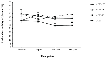 Figure 2: Comparison of the antioxidant activity of plasma (%) at rest, 1 h, 24 h, and 48 h after resistance exercise performed at different levels of AOP (n = 12). CON: Control condition with no occlusion; AOP-50: Experimental condition at 50% of Arterial Occlusion Pressure; AOP-75: Experimental condition at 75% of Arterial Occlusion Pressure; AOP-100: Experimental condition at 100% of Arterial Occlusion Pressure. View Figure 2
As presented in Figure 3, there was no significant condition main effect (F = 1.00, p = 0.40) or condition by time interaction (F = 1.21, p = 0.30) for changes in the levels of maximal isometric strength, but there was a significant time main effect (F = 3.30, p = 0.04); however, follow up analysis revealed that only the comparison between the time points at 1 h post-exercise (79.95 ± 3.79 UI/L) and at 24 h post-exercise (84.27 ± 3.70 UI/L) approached statistical significance (p = 0.08).
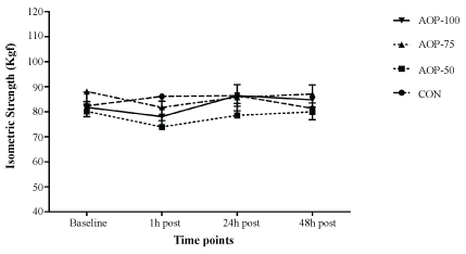 Figure 3: Comparison of isometric strength levels at rest, 1 h, 24 h, and 48 h after resistance exercise performed at different levels of AOP (n = 12). CON: Control condition with no occlusion; AOP-50: Experimental condition at 50% of Arterial Occlusion Pressure; AOP-75: Experimental condition at 75% of Arterial Occlusion Pressure; AOP-100: Experimental condition at 100% of Arterial Occlusion Pressure. View Figure 3
Figure 4 presents the serum levels of CK. There was no significant condition (F = 2.63, p = 0.07) or time (F = 1.25, p = 0.31) main effects and no significant condition by time interaction (F = 1.22, p = 0.29). For plasma levels of LDH (Figure 5), the main effect of condition was not significant (F = 1.10, p = 0.12) and there was no significant condition by time interaction (F = 0.98, p = 0.46). However, there was a significant time main effect (F = 3.29, p = 0.048), in which plasma levels of LDH were significant lower (p < 0.01) 24 h post-exercise (265.83 ± 17.55 UI/L) compared to 48 h post-exercise (294.96 ± 17.51 UI/L).
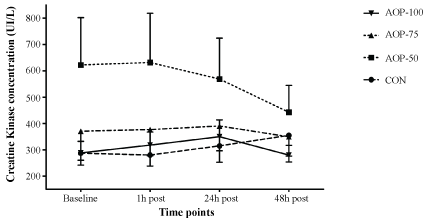 Figure 4: Comparison of creatine kinase concentrations at rest, 1 h, 24 h, and 48 h post-resistance exercise performed at different levels of AOP (n = 12). CON: Control condition with no occlusion; AOP-50: Experimental condition at 50% of Arterial Occlusion Pressure; AOP-75: Experimental condition at 75% of Arterial Occlusion Pressure; AOP-100: Experimental condition at 100% of Arterial Occlusion Pressure. View Figure 4
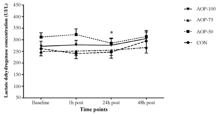 Figure 5: Comparison of the lactate dehydrogenase concentrations at rest, 1 h, 24 h, and 48 h post-resistance exercise performed at different levels of AOP (n = 12). CON: Control condition with no occlusion; AOP-50: Experimental condition at 50% of Arterial Occlusion Pressure; AOP-75: Experimental condition at 75% of Arterial Occlusion Pressure; AOP-100: Experimental condition at 100% of Arterial Occlusion Pressure. *Significantly lower than 48 h post-exercise (p < 0.05). View Figure 5
There was no significant difference for levels of delayed-onset muscle soreness following any of the post-exercise testing times (1 h, 24 h and 48 h: p > 0.05) (Table 1 and Table 3).
Table 3: Comparison of delayed-onset muscle soreness at rest, 1 h, 24 h and 48 h post-resistance exercise performed at different levels of AOP (n = 12). View Table 3
Even though several studies have investigated BFR exercise-induced oxidative stress and muscle damage [7,11,23-25], this was the first study to analyze oxidative stress and muscle damage responses to resistance exercise performed at different degrees of AOP. The findings of the present study do not support our previous hypothesis that applying high levels of AOP would result in higher oxidative stress responses and muscle damage, as well as longer recovery periods.
Our findings are consistent with those from other studies that investigated the relationship between BFR and exercise-induced muscle damage [14,26,27]. Using elastic bands, Wilson, et al. [27] observed a significant elevation in the determinants of hypertrophy without identifying significant elevations in the indicators of muscle damage (muscle soreness, muscle swelling and power) up to 24 h post-exercise. This supports the idea of using BFR resistance exercise as an effective alternative for promoting significant enhancements in the levels of strength and hypertrophy for those who are unable to train at high loads [28,29]. It also supports the relative safety of this method of training [30].
Prolonged reductions in muscle strength have been reported to be one of the most reliable indirect markers of muscle damage [6,31]. In the present study, the maximal isometric strength was measured at 1 h, 24 h, and up to 48 h post-exercise, however, there were no decreases in isometric strength levels at any time point, regardless of the AOP applied. Similar findings were reported by Loenneke, et al. [32] who did not observe any impact of BFR by itself or in combination with exercise (BFR pressure relative to participants' thigh circumference) on levels of isometric strength at 1 h and 24 h post-exercise.
Furthermore, there was no increase in the ratings for DOMS for any of the exercise conditions in the current study. Interestingly, different findings were reported by Umbel, et al. [8], who reported increased DOMS ratings above baseline levels at 24 h and 48 h post-exercise. However, it is important to note that the authors designed the study so that the experimental group (BFR at 130% of upper arm SBP applied to the upper thigh) completed the repetitions until muscle failure, however, the control group (not occluded) only completed the same number of repetitions as the experimental group without reaching muscle failure since they were non-occluded.
The serum levels of CK and LDH were also measured as indirect biomarkers of muscle damage. In this regard, Clarkson and Hubal [6] stated that CK levels are expected to rise over 100% following resistance exercise in comparison to their baseline levels and that it remains elevated for several days following resistance exercise. However, no significant increases from baseline values in the levels of CK and LDH were observed over time for any of the conditions tested. Although LDH levels significantly increased for all conditions 48 h post-exercise in comparison to 24 h post-exercise, this difference is likely due to normal daily variations in serum levels of LDH rather than the exercise performed, as no significant difference was observed neither between the conditions tested nor between 1 h and 48 h post-exercise. Other studies have shown, that even at high BFR pressures ( ≥ 200 mmHg), there were no significant changes in the levels of CK at 24 and 48 h post-exercise [13]. Takarada, et al. [33] applied a BFR pressure of 214 mmHg to young subjects who exercised at 20% of 1RM and also did not observe significant elevations in levels of CK at 24 h post-exercise. Coupled with the findings of the present study, this evidence suggests that BFR exercise does not result in muscle damage, even when the exercise is performed at very high pressures (over 200 mmHg).
The acute oxidative stress response was also measured in the present study. Oxidative stress is known for its deleterious effects on DNA and skeletal muscle due to the formation of reactive oxygen species, which can also lead to muscle damage [34,10]. Since BFR exercise is commonly performed at very low intensities (i.e. 20%-30% of 1RM), it is very unlikely that the mechanisms known for evoking muscle damage during traditional resistance training such as physical damage to muscle fibers due to high mechanical stress associated with high intensities and volumes [1,4], is applicable to this model of exercise. Therefore, perhaps a more reasonable explanation for the muscle damage linked to BFR exercise would be more metabolic in nature with increases in hydrogen ions concentrations, stimulation of the endocrine system, or an increase in reactive oxygen species. In this context, previous research has reported that an ischemia-reperfusion protocol has been shown to stimulate the formation of reactive species of oxygen in a rat model [10]. However, no changes in any of the oxidative stress markers (LP and AAP) were observed in the current study. Likewise, Takarada, et al. [33] did not observe alterations in the levels of LP up to 24 h following BFR exercise. Interestingly, the authors also did not observe any increases in the determinants of muscle damage. Additionally, Garten, et al. [11] observed that BFR combined with low-intensity resistance exercise (30% of 1RM) actually attenuated the oxidative stress response.
It is known that subsequent bouts of resistance exercise, known as the repeated bout training effect, may provide protective adaptations for muscle and help prevent possible damage [35]. However, it is important to note that the design employed in this study attempted to avoid the repeated-bout effect to tamper the muscle damage response. Even though the participants were submitted to a within individuals cross-over design, there was a large period of recovery time allowed between trials, similar to previous studies [36,37]. Additionally, it seems that protective adaptations from previous bouts of exercise are mainly related to high intensity exercise. In this regard, Nosaka and Newton [38] have shown that submaximal resistance exercise (at 50% of 1RM) does not exacerbate or offer any protection for additional subsequent bouts of exercise.
By analyzing collectively, all the variables assessed in the current study, no evidence was found to support the hypothesis that resistance exercise with either partial or total AOP were able to induce muscle damage up to 48 h post-exercise. Usually, muscle damage is observed following high intensity resistance exercise within 24 to 48 h post-exercise, and it is generally attributed to the eccentric portions of the contractions performed, in which fewer muscle fibers are recruited in comparison to the concentric contractions. However, Thiebaud, et al. [24] reported that eccentric contractions with BFR did not increase muscle activation or result in prolonged muscle damage up to four days following eccentric exercise. It has also been speculated that muscle damage may occur post BFR exercise due to the formation of reactive oxygen species, from the ischemia/reperfusion response that occurs after releasing blood flow [11]. In this regard, even though total AOP was applied to the subjects of this study, it is still very likely that there was some arterial blood flow into the muscle during the exercise, which might diminish the ischemia/reperfusion process.
It has also been suggested that exercise intensity and exercise volume may play a role in the exercise-induced muscle damage response [1]. Thus, it is worth noting many of the previous BFR studies performed BFR exercise at high volumes (75 reps) [39] or until failure [40]. This is a limitation of the present study, which applied an exercise volume of only 40 reps. It is important to highlight that this moderate exercise volume (4 sets of 10 reps) and it was used in order to standardize all the tested conditions, since the AOP-100 condition would not allow subjects to perform the exercise with a higher number of repetitions. Additionally, the low load traditionally used during BFR resistance exercise (< 50% of 1RM) may be responsible for the absence of muscle damage in this training method. Moreover, we did not measure the oxidative stress and muscle damage responses beyond 48 h post-exercise; and it may be possible that the measures of damage might occur after 48 h post-exercise. Finally, this study only included young male resistance trained participants, which makes it difficult to extend these findings to other populations, such as the elderly, women, and the untrained. It is very likely that untrained individuals may have responded differently to resistance exercise performed at different degrees of AOP.
In conclusion, low-intensity resistance exercise with BFR does not result in acute oxidative stress and muscle damage responses in young trained men, regardless of the level of AOP applied during the exercise. From a practical standpoint, BFR resistance training seems to have no detrimental effect on isometric muscle strength and soreness, and a similar recovery time after resistance training at different AOPs could be expected (50, 75, and 100%). Moreover, these findings confirm the relative safety of this method of training, however, we encourage future studies to investigate this response in other populations, such as the untrained, women, and the elderly subjects.
The authors would like to thank the participants and the nurses that dedicated their precious time in contribution to this study.