Pituitary adenoma is a benign tumor of the pituitary gland. It represents 8-10% of intracranial masses [1]. Pituitary adenomas are categorized based on primary cell origin and type of hormone secreted. If the adenoma does not secrete a sufficient level of hormones to be detectable in the blood or to result in clinical manifestations, it is considered nonfunctioning. Prolactinomas comprise 40% to 57% of all adenomas, followed by nonfunctioning adenomas (28% to 37%), growth hormone-secreting adenomas (11% to 13%), and Adrenocorticotropic Hormone (ACTH)-secreting adenomas (1% to 2%). Pituitary adenomas that secrete Follicle-Stimulating Hormone (FSH), Luteinizing Hormone (LH), or Thyroid-Stimulating Hormone (TSH) are rare [2]. Tumors are also categorized based on size. If the tumor is 10 mm or larger, it is considered a macroadenoma; if it is less than 10 mm, it is considered a microadenoma. Microadenomas are slightly more common than macroadenomas (57.4% vs. 42.6%) [3].
Pituitary adenomas present clinically in three ways: endocrine syndrome, neurological syndrome, and an acute intrasellar syndrome.
- Endocrine syndrome: It can be a syndrome of hormone hypersecretion, the most common of which are hyperprolactinemia, acromegaly, and Cushing disease, or a syndrome of hypopituitarism.
- Neurological syndrome is characterized by decrease of visual acuity, visual field deficit, and headaches.
- Acute intra-seller syndrome or pituitary apoplexy characterized by ophthalmoplegia, intracranial hypertension and a meningeal irritation.
We report a series of 3 cases of pituitary macro-adenoma with different clinical presentations.
A 32-years-old patient with a previous history of surgically operated anal fistula has consulted for a worsening progressive decrease of visual acuity associated with eye pain which lasted 5 years. The corrected visual acuity was 1/50 Parinaud 20 right and 1/20 Parinaud 4 left. The ophthalmologic examination was normal. The visual field noted a bitemporal hemianopia with central deficit on the right eye. Brain MRI revealed a large pituitary adenoma of 38 mm × 37 mm × 25 mm (Figure 1). The hormonal check-up revealed a hyperprolactinemia at 116.7 ng/l (normal range: 2.54-16.11 ng/ml).
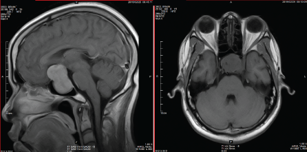 Figure 1: MRI images of the macroadenoma. View Figure 1
The prolactinoma diagnosis was done and an adenomectomy through endonasal approach was carried out with simple postoperative monitoring. The surgical treatment was decided based on the size of the macroadenoma and the severity of the visual impairment. An adjuvant therapy by cabergoline was instituted. A significant improvement of visual function was noted six weeks after the operation. The corrected visual acuity was 6/19 Parinaud 5 in right eye and 6/7.5 Parinaud 2 in the left. The visual field has improved with the persistence of a temporal hemianopia at the right, while the left eye was normal (Figure 2).
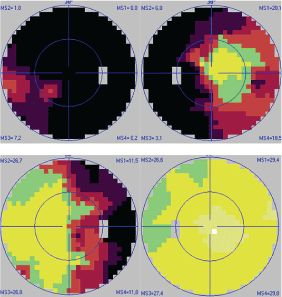 Figure 2: Visual fields, at the diagnosis (up) and postoperatively (down).
View Figure 2
Figure 2: Visual fields, at the diagnosis (up) and postoperatively (down).
View Figure 2
A 55-years-old patient, without special medical history was consulted for the prescription of glasses. She had low bilateral visual acuity for 6 months. The questioning did not found any headache or any endocrinal dysfunction sign. The corrected visual acuity was 2/10 Parinaud 2 at the right and 1/50 Parinaud 12 at the left. The ophthalmological examination was normal. The fluorescein retinal angiography was normal. The visual field revealed a bitemporal hemianopia with a marked central deficit on the left. Brain CT revealed a 33 mm × 24 mm × 23 mm, homogenous intra-seller mass (Figure 3). The hormonal check-ups were normal. The diagnosis of non-secreting pituitary macroadenoma was made and the patient underwent a trans-sphenoidal adenomectomy. After the operation, there was an improvement in visual acuity on the left from 1/50 Parinaud 12 to 2/10 Parinaud 2, the right visual acuity remains the same at 2/10 Parinaud 2. There was a gradual improvement in the consecutive visual fields with reduction of deficit. One year after the operation, the visual field has noticed an upper bilateral quadranopia with a central deficit more important on the right eye (Figure 4).
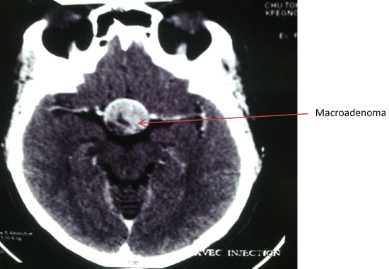 Figure 3: TDM image of the macroadenoma.
View Figure 3
Figure 3: TDM image of the macroadenoma.
View Figure 3
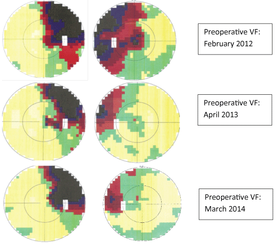 Figure 4: Visual field changes.
View Figure 4
Figure 4: Visual field changes.
View Figure 4
A 56-years-old patient with no particular medical history was consulted for headaches which started for two weeks associated with diplopia in the right eye. The corrected visual acuity was 5/10 Parinaud 5 on right and left eye. The ophthalmologic examination revealed right nerve VI palsy. Brain MRI revealed a 26 mm × 24 mm × 16 mm, enhanced, homogenous intra and supra-seller mass evoking a pituitary macro-adenoma (Figure 5). The hormonal tests were normal. The visual field was within normal limits on the right and left. The surgery was scheduled.
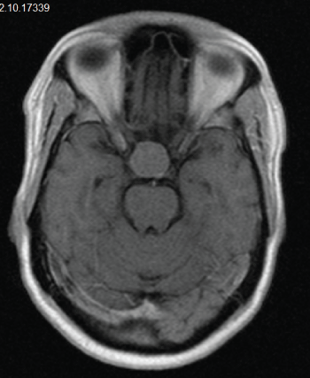 Figure 5: MRI image of the macroadenoma.
View Figure 5
Figure 5: MRI image of the macroadenoma.
View Figure 5
Our three observations have in common visual disorders ranging from decrease of visual acuity to diplopia associated or not with headache or eye pains. These neurological symptoms are the most common ones especially in case of nonfunctioning adenoma. Bitemporal hemianopia and the nerves II, III, IV and VI palsies can directly be linked to the size of the tumor [4]. If a pituitary adenoma is suspected, MRI is the best imaging, with a sensitivity of 61% to 72% and a specificity of 88% to 90% [5,6]. But if MRI is unavailable, CT with thin coronal sections helps to do the analysis of the seller lesions diagnosis [7].
The resection of the adenoma has enabled the reduction of the compression and a gradual improvement of the visual field in both cases. Prolactinoma can be managed with medically with dopamine agonist such as cabergoline or bromocriptine, with better patient tolerance of cabergoline over bromocriptine [7].
For the third observation, the surgical indication is based on the size of the adenoma, and the presence of neurological signs that indicate a lateral compression [8], despite the absence of hypersecretion and normal visual field indicating the absence of upper compression to the optic chiasm.
These three observations show different clinical situations with as constant a fundamental place of neuroimaging. Careful clinical examination and visual field are used to guide diagnosis. Medical treatment or surgical resection of the adenoma should be performed as early as possible to reduce the compression for a rapid visual recovery.
The pituitary adenoma is a relatively common pathology with a rich and varied symptomatology. Decrease of visual acuity, visual field deficit, and headaches are the neurological signs seen in ophthalmology. The visual field in this case remains the orientation test of choice. Neuroimaging is important and allows confirming the diagnosis. The hormonal dosage enables to distinguish associated hormonal disorders. The management indication depends on several factors.
All the patients have given an oral consent for using their information’s for scientific purposes.
None.