Ainhum is a rare idiopathic condition that occurs in those with African heritage usually affecting the 5th toe. We present a case report of 50 year old Haitian male presented with a painful 5th right toe with constricting band encircled the 5th toe at the base with loss of voluntary motion, a bulbus appearing distal toe, and external rotation of the nail plate. The pain and groove progressed over 6 months, starting as a crease appearing at the medial digitaltoplantar fold. He was prescribed a course of antibiotics for concern of cellulitis this failed to provide relief and referred to foot and ankle specialist. Initial radiographs revealed narrowing of the shaft of the proximal phalanx distal with annular indentation in the soft tissues at the base of the 5th toe, marked rotation of the 5th toe, triangular appearance of the distal phalanx . MRI showed absence of the majority of the 5th proximal phalanx and distal to the base consistent with frank bone destruction,, with osteitis at the residual base of the phalanx , soft tissue irregularity and enhancement of the 5th digit surrounding the phalanges, no fluid collection present. Due to advanced stage of ainhum with bony changes of the phalanges underlying Cole stage III the patient elected for 5th digit amputation, disarticulation at the mpj to reduce discomfort. Pathology report revealed a right 5th toe with apparent slight proximal constriction with no inflammation or necrosis identified. The post operative period was uneventful incision healing without keloid and 5th toe pain was resolved. Amputation for late-stage ainhum is a viable option. Healing without keloid suggests it is a genetic condition that occurs spontaneously not due to trauma.
Ainhum, Auto amputation, Dactylolysis, Toe amputation, Cole stage III, Narrowing proximal phalanx.
Constricting bands are classified as ainhum and pseudo ainhum both can lead to autoamputation and are rare. Ainhum (dactyloysis spontanea) has a strict definition, a constricting band around a pedal digit, this most commonly presents in the fifth digit occasionally affecting the fourth toe and usually is found bilaterally. True ainhum occurs only in people of African descent occurring in around 2% of this population [1,2]. Ainhum means fissure in the language of the African tribe in Brazil, ainhum may be related to ayun the word saw in the African tribe in Nigeria [3]. The first published of case of ainhum in an African male was by de silva lima in Brazil in 1867 [4]. Pseudo ainhum constrictions clinically mimic ainhum and are much more common in the developed world the etiology is thought to be caused by one of three conditions, amniotic bands, constriction associated with keratotic disorders or Sequella after infection or trauma, and constriction by external forces such as a hair or thread [2]. The pathogenesis of ainhum is not understood, it is hypothesized the band develops from excessive fibroplasia in a susceptible host (africans are known to keloid), triggered either due to rotational force applied to the bare mechanically unstable foot, or the bands develop after trauma or infection from the patient walking barefoot [2].
Ainhum is a clinical diagnosis the groove around the toe is evidence of the disease in a dark skinned individual. The groove starts on the medial side of the plantar digital fold at the base of the toe and as spreads around the toe in the dorsal direction also growing deeper and wider. The lateral weight bearing surface of the toe is the last to be affected. The rate of groove spread has variable reported times from 3 months to 20 years [1,3].
Coles Four clinical stages of ainhum guide treatment options modified after Brownes stages of ainhum. Cole Stage I just a narrow groove or fissure encircling the toe, Cole stage II floor of the groove is ulcerated, Cole Stage III changes to the proximal phalanx on X-ray. Cole Stage four bloodless autoamputation of the toe [1]. Cole recommends amputation for late stage ainhum 3 or 4. Classic features of ainhum include bulbus swelling of the toe due to lymphoedema of the soft tissue distal to the groove, varus rotation of the toe, the nail can face outward by over 30 degrees, with rectus alignment maintained of the MPJ and IPs. Radiographic changes including soft tissue contraction of the base of the toe and absorption of the proximal phalanx on the medial side of the groove , usually distal to the groove are iagonstic of ainhum [1]. The anterior and posterior tibial pulses are easily felt in most patients with ainhum with sensation intact to the foot including the distal affected toe [1,3].
Late ainhum can be complicated by ulceration of the groove or autoamputation that can lead to infection. On average it takes 5 years for autoamputation to occur [1,5]. Cole reported 78% of toes with ainhum had pain at or distal to the groove, with an increase in pain as the disease progressed [1]. All ainhum requires surgery due to the risk of infection from the ulceration of the groove and pain, either skin plasty to remove the groove or amputation. The skin plasty typically z plasty is reserved for grade one or two without bone changes, and if there is enough healthy skin, the groove is excised first then depending on the length either one or many z are used to close. Prior to any skin plasty procedures, infection must be cleared if underlying bone changes of the proximal phalanx are present recommendation is amputation disarticulation at the metatarsophalangeal joint. Relief of pain is reported immediately after both procedures [1,6]. Cole reported recurrence after z plasty did not occur within 8 months [1]. Prabhu reported successful resolution of pain 2 years after amputation for ainhum in a 38 year old male [6].
50 year old Haitian American male, no past medical history, presented to the clinic, one year ago noticed right foot swelling for a week and pain to his 5th toe with a fissure that began on the medial side of the base of the 5th toe, coinciding with plantar digital fold. The fissure never resolved he was treated with antibiotics prescribed by primary care for cellulitis that resolved the dorsal foot swelling. Over a course of six months, pain to the 5th toe has progressed the fissure had become a band of hyperkeratotic tissue spread more dorsal around the 5th toe so that the last part is the bridge of weight-bearing skin on its outer lateral plantar surface patient reported his 5th digit was shrinking circumferentially at the base of the proximal phalanx like the digits was being strangulated, and the distal phalanx was expanding in size with increased pain. The patient said members of his family in Haiti have amputated their toes with a knife. The patient denies trauma to the toe. Admits the pain from the toe has affected his gait and he is unable to wear closed-toe shoes, as pressure to the toe caused pain. No one in his family has had a similar condition. Physical exam showed at the base of the proximal phalanx a 1mm constrictive band of hyperkeratic tissue with edema of the distal 5th toe, the nail was found to face outwards by over 30 degrees, the patient was unable flex or extend his , the groove had ulcerated and celluiltis was present. He was started on a course of oral antibiotics after his initial consult due to celluilitis and the grove had ulcerated. Pulses to the right foot easily palpated +2 dp and +2 pt, right lower extremity skin temperature was warm, with lack of hair growth to bilateral lower extremities, the protective sensation was intact including the distal tip of the 5th toe.
Radiographic imagining revealed narrowing of the shaft of the proximal phalanx distal to the groove marked rotation of the 5th toe, triangular appearance of the distal phalanx, with annular indentation in the soft tissues at the base of the 5th toe. MRI showed absence of the majority of the 5th proximal phalanx and distal to the base consistent frank bone destruction, with osteitis at the residual base of the phalanx, soft tissue irregularity and enhancement of the 5th digit surrounding the phalanges, no fluid collection.
Patient elected to have complete amputation of the right 5th toe by disarticulation at the metarsal phalangeal joint due to continued pain. The digit was clamped with a traumatic clamp, a racket incision extending back dorsally to the level of the MPJ made full thickness down to bone, a 15 blade was inserted into the MPJ joint capsule incised sharply to avoid injury to surrounding soft tissue, leaving the articular cartilage of the metatarsal the flexor and extensor tendon divided at the level of the skin incisions allowed to retract, no purulence was found in the tissue planes. The wound was irrigated. The skin was closed without tension in one layer using interrupted monofilament nylon suture. The toe was sent to pathology. Pt was allowed to partial weight bear to the heel until sutures were removed. noted adequate bleeding throughout the procedure. Metatarsal head had no signs of infection white hard shiny. The sutures were removed 15 days postoperative, pain to the 5th toe had resolved . Pathology report revealed a right 5th toe with apparent slight proximal constriction with no inflammation or necrosis identified The post-operative period was uneventful, incision healed without keloid.(Figure 1, Figure 2, Figure 3 and Figure 4).
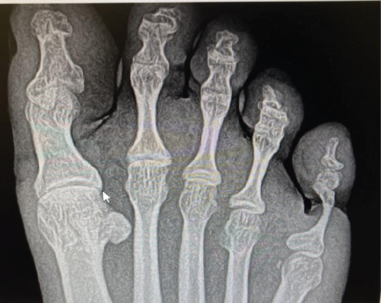 Figure 1: Xrays revealed narrowing of the shaft of the proximal phalanx, marked rotation of the 5th toe, triangular conical appearance of the distal phalanx, with annular indentation in the soft tissues at the base of the 5th toe. Pathognomonic for ainhum Cole stage III . View Figure 1
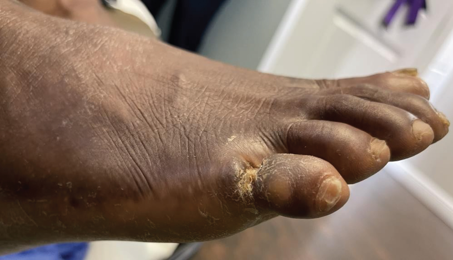 Figure 2: Marked rotation of the 5th toe in 50 year old hiatian patient. View Figure 2
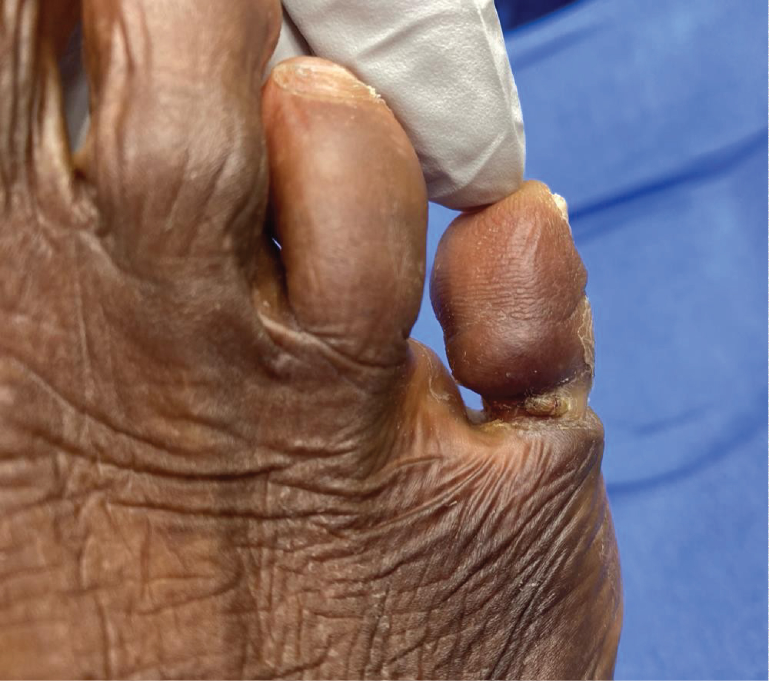 Figure 3: 5th right toe with constricting band encircled the toe at the base with a bulbus appearing distal toe, and external rotation of the nail plate. View Figure 3
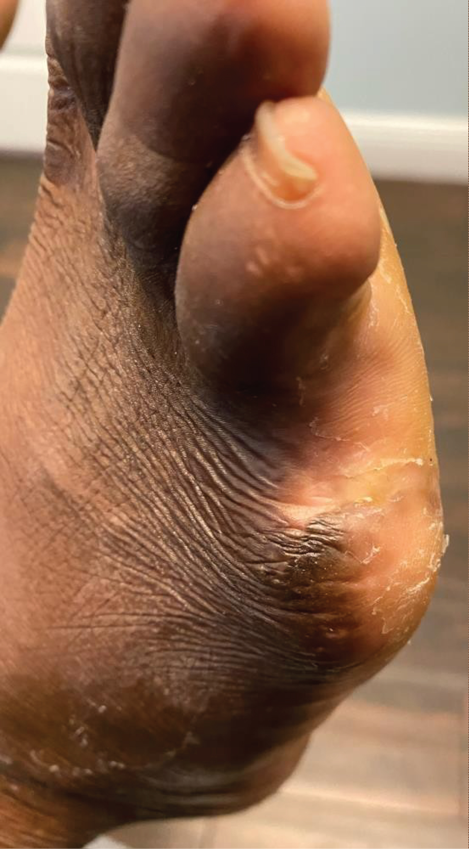 Figure 4: Incision healed without keloid. View Figure 4
Ainhum is rare disease affecting roughly 2% of patients of African descent causing a progressive constricting groove of keratin encircling thee base of the fifth toe due to changes in the prickel cell layer leading to autoamputation and pain as the disease progresses. Cole at a hospital in Nigeria 1965 after examining 1000 African patients that had no foot pain found 2.2% suffered from ainhum [1]. Daccarett in 2002 at a hospital in Chicago with a population mostly of African Americans after review of 6000 random foot radiographs using Cole criteria, noted constriction of soft tissue around the base of the toe and thinning of the proximal phalanx diagnosed 1.7% with ainhum [7]. Browne in a survey of 45,035 patients in central Africa found 0.25% incidence of ainhum [5]. Race seems to be the most predisposing factor as all cases of true ainhum involve patients of African heritage, suggesting ainhum is a genetic disorder. In our case study the patient was of African descent and members of his family also suffered from Ainhum. Support for ainhum caused by a genetic disorder is occurrence in families. Cole found 52 patients were from the yorubas tribe. 5 patients had one parent with the disease, three had a uncle or aunt affected, one patient had 4 members of his family with ainhum [1]. Priya presented a case of familiar ainhum in father and son, the father ainhum affecting the right 4th and 5th toes and left 5th toes, the son at age 8 ainhum affected his right 5th toe that lead to amputation and at age 30 his left 5th toe became affected [3]. Aninhum is painful, and the patient will seek treatment. If ainhum left untreated autoamputation will occur with risk of infection around the fragment of proximal phalanx or even the metatarsal if the stump ulcerates. Longer follow up is needed to see if z plasty is treatment option for early stages of ainhum.
Cole found histologically in 2 very early lesions dermis was normal and stratum germinativum was normal yet lacked melanin pigment and paillae were thickened, with a disorganized prickel cell layer with the absence of nuclei and intercellular bridges in many places. The Statum granulosum was irregular with very large keratohyaline granules, there was no startum lucidum. Gross thickening of the stratum corneum with calcification of the ducts and sweat glands seen. More advanced sections of bands showed chronic inflammation deep to hyperkeratoic epithelium with normal vessel and nerves and sterile absorption of bone [1]. Inflammation and fibrosis are not part of the change seen in ainhum , no relation to sickle cell trait, nor evidence of fibrotic contraction highly specific changes in a constant pattern affecting small number of cells in the prickel cell layer that is responsible for laying down condensed keratin that causes the groove. In our case study the pathology report revealed a right 5th toe with apparent slight proximal constriction with no inflammation or necrosis identified. It is through ainhum is due to trauma from walking barefoot in a patient that is prone to keloiding [1]. In our case study after the amputaiton the incision healed without keloid, supporting the idea that ainhum is a genetic disorder not due to keloiding after trauma.
Cole observed that bone involvement occurred as the groove grows deeper in later stage ainhum, starting on the plantar medial base of the toe encircling the toe as it spreads dorsally [1]. Proximal phalanx are short long bones, long bones main blood supply is the nutrient artery that enters obliquely through the nutrient canal directed away from the growing end. Nutrient foramina in phalanges of the toes are numerous and difficult to identify actual nutrient foramen except in distal phalanx of the great toe, likely that small periosteal vessel entirely responsible for blood supply of the phalanx in the toes [8]. The constricting band as it grows deeper cuts off the nutrient arteries to the proximal phalanx causing the osteolysis distal to the groove.
Complete informed consent was obtained from the patient for the publication of this study and accompanying images
The authors declare that they have no known competing financial interest or personal relationships that could have appeared to influence the work reported in this paper.