We describe the case of a 3-week-old male neonate who developed a large pseudoaneurysm of the left common carotid artery following transcarotid ductal stent placement [1].
The lesion was successfully managed using a stepwise endovascular technique involving embolization with thrombin and coils [2].
The pseudoaneurysm, which caused significant airway compromise, was excluded without surgical intervention [3].
This report emphasis the value of early recognition and individualized endovascular treatment in critically ill neonatal patients [4].
Pseudoaneurysms involving the common carotid artery are infrequently encountered in neonates, and management strategies vary due to their rarity [5]. These lesions are often linked to iatrogenic injury, especially from catheter-based procedures or central line placements [6]. In most published cases, treatment has involved surgical excision and vascular reconstruction [7].
Although effective, surgery can pose considerable risk in neonates, particularly those who are hemodynamically unstable or have underlying congenital heart disease [8]. In selected cases, minimally invasive techniques may offer a safe alternative [9].
A 3-week-old male neonate, diagnosed with pulmonary atresia with an intact ventricular septum and a hypoplastic right ventricle, underwent transcarotid ductal stent placement.
The child had a history of frequent seizures and was on Keppra and inotropic support.
Two days after the procedure, the infant developed a progressively enlarging mass on the left side of the neck, accompanied by stridor and respiratory distress, necessitating intubation.
On examination, the patient was intubated and noted to have a large, pulsatile swelling on the left side of the neck. There was no overlying skin breakdown. Vital signs included a heart rate of 152 bpm, blood pressure of 77/45 mmHg, and oxygen saturation of 82% on mechanical ventilation. The patient weighed 3.3 kg.
Neck ultrasound revealed a large pseudoaneurysm arising from the mid-segment of the left common carotid artery, measuring approximately 6 x 5 cm. (Figure 1). The lesion demonstrated active systolic inflow through a narrow neck. Contrast-enhanced CT showed the pseudoaneurysm causing significant mass effect with tracheal deviation and compression, though airway patency was preserved by the endotracheal tube (Figure 2).
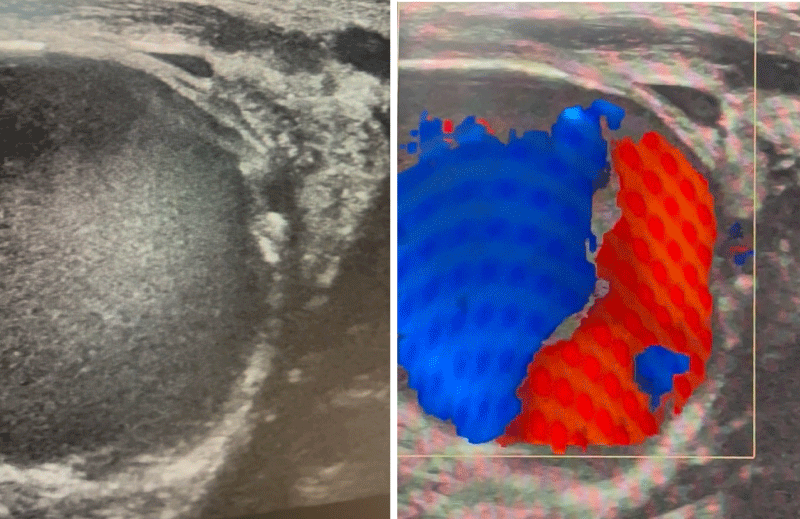 Figure 1: Physical examination
Left: US large pseudo-aneurysm arrises from mid left common carotid artery with small neck opening with sytolic high flow entry site.
Right: Color blood flow shape of pseudo-aneurysm.
View Figure 1
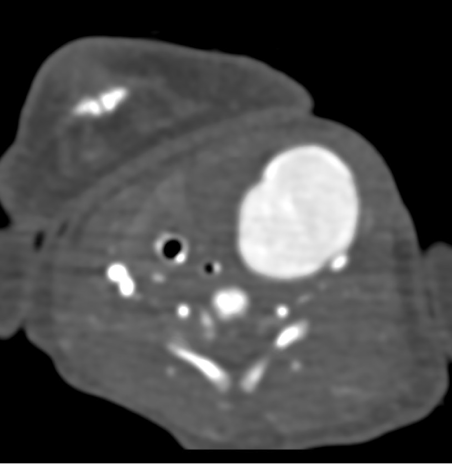 Figure 2: Contrast - enhanced CT showed the pseudoaneurysm.
View Figure 2
After multidisciplinary discussion and consent from the parents, the patient was taken for endovascular intervention. A 4 Fr vascular sheath was inserted into the right femoral artery under ultrasound guidance. Cerebral angiography demonstrated preserved collateral circulation, and neck angiography confirmed a large pseudoaneurysm with a narrow neck (Figure 3).
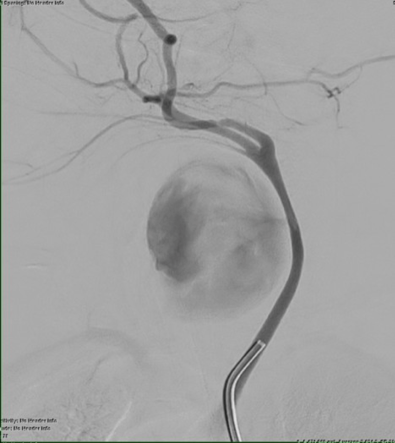 Figure 3: Interventional management.
View Figure 3
An initial attempt at ultrasound-guided compression with a percutaneous 2 Fr sheath insertion into the aneuyrmal neck to the carotid lumen in order to occlude the inflow and acheive hemostasis while applying gentle compression. Although this resulted in 75% thrombosis, residual high inflow required further intervention.
Next, percutaneous embolization with 3 ml of thrombin and 3 ml of Flowseal, and 75 % of saccular thrombosis was achievedwith presence of percutaneous vascular sheath inside the neck and simultaneous gentile compression by ultrasound for 10 minutes (Figure 4, illustrated residual 25% of active high inflow).
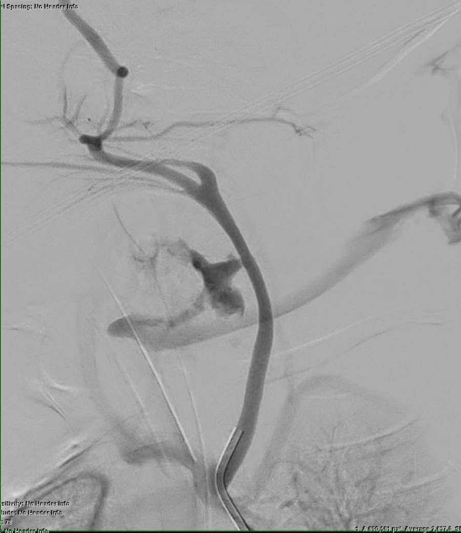 Figure 4: Residual 25% of active high inflow.
View Figure 4
After thrombin injection, A balloon occlusion catheter of proximal carotid in oreder to decrease the high inflow inside the pseudoaneurysm for 3 minutes was unsuccessful (Figure 5).
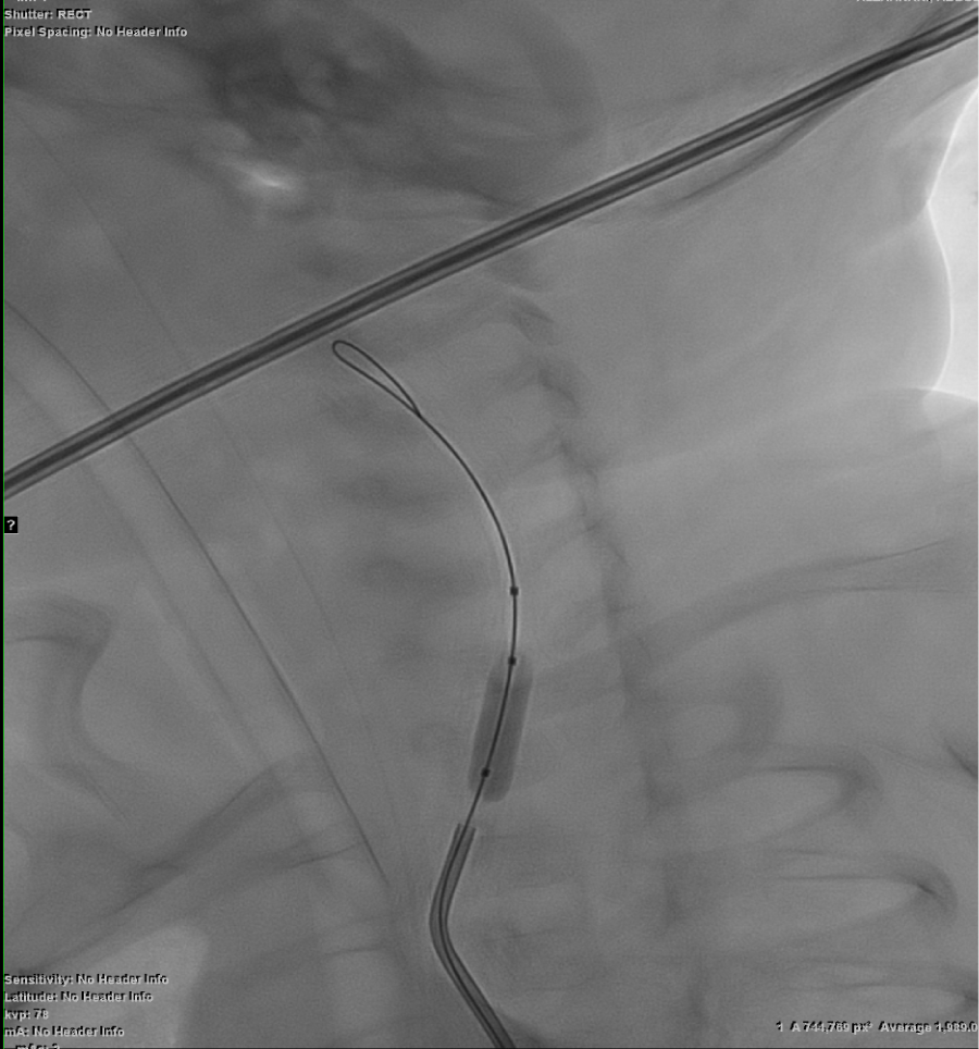 Figure 5: A balloon occlusion catheter of proximal carotid.
View Figure 5
Finally, successful complete thrombosis achieved with multiple coils deployed inside the residual active with exclusion of the pseudo-aneurysm from circulation (Figure 6).
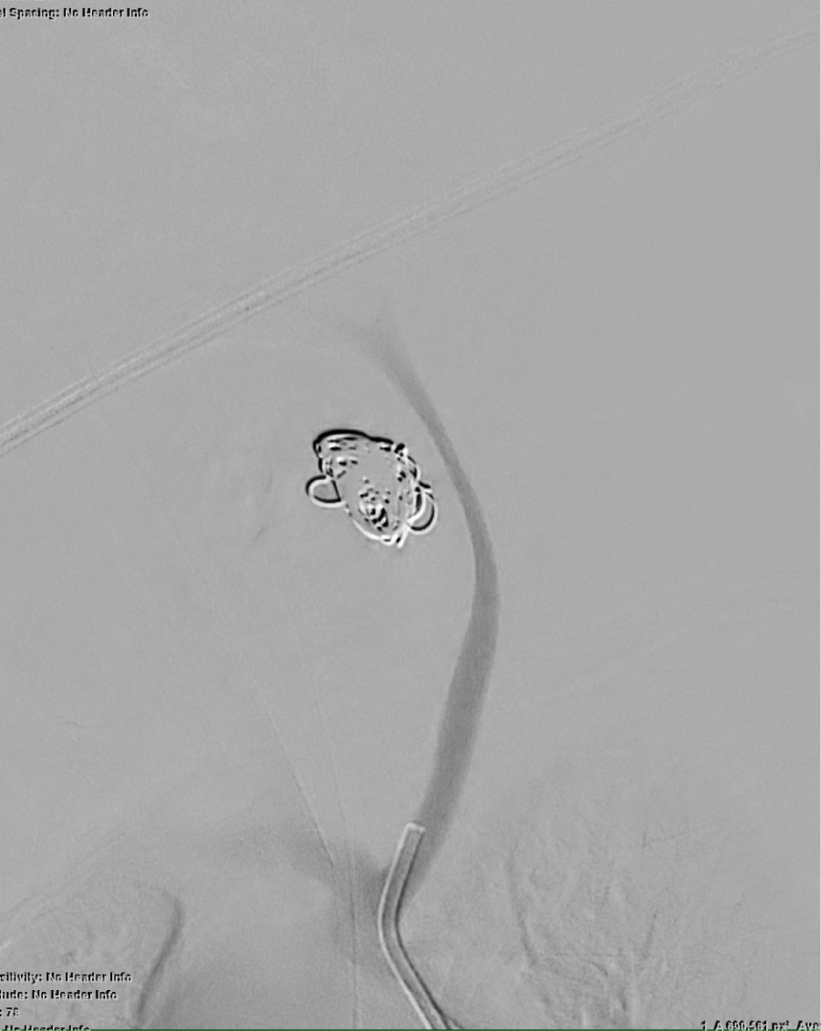 Figure 6: Complete thrombosis and exclusion from the carotid circulation.
View Figure 6
Post-procedure, the neck mass gradually regressed. Follow-up ultrasound confirmed complete thrombosis of the pseudoaneurysm with continued patency of the parent artery. One week later, the residual hematoma was surgically evacuated. The patient was extubated successfully after ensuring airway stability.
Pseudoaneurysms of the carotid artery in neonates are exceedingly rare and pose significant management challenges due to the critical anatomy involved and the delicate physiology of the patient population [1]. Most documented cases in the literature are related to iatrogenic trauma, particularly from catheterizations or central line insertions, and have traditionally been treated surgically through resection and reconstruction [2,3].
While surgery remains the standard approach in many institutions, it is not without considerable risk, especially in neonates with complex cardiac anomalies or respiratory compromise [4]. In recent years, endovascular management has gained attention as a feasible alternative in selected pediatric patients, including the use of thrombin injection, coil embolization, and liquid embolics like glue [5,6]. However, the evidence remains limited, especially in neonates, with only a handful of reports describing successful minimally invasive interventions [7,8].
Our case represents one of the few instances where a large, symptomatic carotid pseudoaneurysm in a neonate was managed entirely by endovascular techniques. A stepwise approach, including initial compression, embolization with thrombin, balloon occlusion, and finally coil embolization, was employed to achieve complete exclusion of the pseudoaneurysm [9]. Notably, brain perfusion was carefully evaluated prior to carotid occlusion attempts, highlighting the importance of pre-procedural imaging and neurovascular planning in such cases [10].
This experience emphasizes the need for early diagnosis and individualized planning. It also adds to the growing body of evidence supporting endovascular approaches as viable, safe, and effective options when performed in specialized centers with multidisciplinary expertise [6,9].
Endovascular treatment should be considered in select neonates with carotid artery pseudoaneurysms, particularly when surgical risk is high. In our patient, the use of thrombin and coils resulted in complete resolution. Tailored approaches and multidisciplinary planning are key to achieving optimal outcomes.