To elucidate age-related changes of the first septal perforator branch (FSB), the authors investigated age-related changes of elements in the FSBs by direct chemical analysis. In addition, the effects of different arterial sizes and genders on element accumulation were investigated in the FSBs. Sixty-two formalin-fixed adult Thai hearts were used and the FSBs were carefully dissected in the hearts. After the arteries were incinerated with nitric acid and perchloric acid, element contents were determined by inductively coupled plasma-atomic emission spectrometry. It was found that both the P and S contents increased significantly in the FSBs with aging, but five element contents such as Ca, Mg, Zn, Fe, and Na did not change significantly with aging. Regarding the relationships among Ca, P, and Mg contents, no significant direct correlations were found in the FSBs, except for an extremely significant direct correlation between Ca and Mg contents. No gender differences and effect of arterial size were found in age-related changes of Ca and P contents in the FSBs. To elucidate whether calcification occurred in the FSBs in old age, both the mass ratios of Ca/P and Mg/Ca were estimated in the FSBs. The mass ratio of Ca/P increased progressively in the FSBs with Ca increase, being not constant. The mass ratio of Mg/Ca decreased gradually in the FSBs with Ca increase, but the average mass ratio of Mg/Ca was moderate, being 8.0 ± 1.3%. These results indicated that calcification scarcely occurred in the FSBs in old age, independently of gender and arterial size.
First septal perforator branch, Coronary artery, Calcium, Phosphorus, Sulfur, Aging
The conduction system of the heart is supplied by the sinoatrial nodal (SAN) artery, the atrioventricular nodal (AVN) artery, the first septal perforator branch (FSB) of the left anterior descending artery (LAD), and the posterior descending branch of the right coronary artery (RCA). These arteries ensure adequate blood supply to maintain the electrical properties in the heart. It is well known that a high accumulation of Ca and P occurs in the proximal sites of the LAD, left coronary artery (LCA), RCA, and left circumflex artery (LCX) with aging [1-3]. For example, Montenegro and Eggen [1] studied the distribution of atherosclerotic lesions along the axis of the coronary artery (topography) and the relationship of topography to concepts of pathogenesis, and reported that a higher prevalence of atherosclerotic lesions in isolated coronary arteries occurred from the first to second centimeter of both the LAD and the RCA, with a decrease in prevalence from 3 cm onward in the LAD and from 5 cm onward in the RCA.
There are a few reports [4-6] on calcification of the FSB. Wasilewski et al. [4] investigated calcification of the LAD including the FSB by multi-slice computed tomography and revealed that calcification was most frequently located in the LAD in proximity to the FSB origin, but it was not located in the FSB. Velican et al. [6] investigated histological changes of four arteries supplying the conduction system of the heart and reported that intimal thickening occurred significantly in the FSB, SAN artery, AVN artery, and posterior descending artery without calcification. However, age-related changes of the FSB had not yet been studied by direct chemical analysis. Therefore, the authors investigated age-related changes of the FSBs using adult Thai hearts from a viewpoint of elements. It was found that calcification scarcely occurred in the FSBs in old age.
The research was carried out on 62 adult Thai hearts (of subjects between 36 and 94 years of age; average age, 70.7 ± 14.5 years) received from the Department of Anatomy, Faculty of Medicine, Chiang Mai University. The perivascular fatty tissue was carefully removed, where necessary, to visualize the epicardial course of the coronary arteries. The FSBs were carefully dissected in the hearts. The FSB arising from the LAD penetrated the myocardium, ran rightwards and downwards, and distributed the interventricular septum. The FSBs were resected from the hearts.
The arterial samples were washed thoroughly with distilled water and were dried at 95 ℃ for 16 h. After 1 ml concentrated nitric acid was added to the dry samples to incinerate, they were heated at 100 ℃ for 2 h. After the addition of 0.5 ml concentrated perchloric acid, they were heated at 100 ℃ for an additional 2 h [7]. The samples were adjusted to a volume of 10 ml by adding ultrapure water and were filtered through filter paper (no. 7; Toyo Roshi, Osaka, Japan). Seven elements of Ca, P, S, Mg, Zn, Fe, and Na were selected for measurement because of the following reasons: Both Ca and P are directly correlated with Mg on calcification [8]; smooth muscles containing S decrease on atherosclerosis [9]; both Zn [10] and Fe [11] are related to atherosclerosis; and Na is an important cation. The resulting filtrates were analysed by inductively coupled plasma-atomic emission spectrometry (iCAP 7400 ICP-OES Duo; Thermo Fisher Scientific Japan Inc., Kanagawa, Japan). The conditions were as follows: 1.15 kW from the radiofrequency forward power, an auxiliary gas flow rate of 0.5 l/min, a nebulizer gas flow rate of 0.55 l/min, a coolant gas flow rate of 12 l/min, a purge gas flow rate of 3.2 l/min, and an exposure time of 10 s. Especially prepared standard solutions of Ca, Mg, Zn, Fe, and Na for atomic absorption spectrometry and phosphate and sulfate ions for ion chromatography were purchased from Wako Pure Chem. Ind. (Osaka, Japan) and were used as standard solutions. The measurement of elements was performed at a fixed wavelength of 588.995 nm for Na, 393.366 nm for Ca, 279.553 nm for Mg, 259.940 nm for Fe, 213.856 nm for Zn, 180.731 nm for S, and 177.495 nm for P. The amount of elements was expressed on a dry weight basis.
Statistical analyses were performed using the GraphPad Prism version 8.0 (GraphPad Software, San Diego, CA, USA). Peason's correlation was used to investigate the association between parameters. It was analysed whether significant differences were found between two slopes and between two intercepts of the regression lines. A p value of less than 0.05 was considered to be significant. Data were expressed as the mean ± standard deviation.
Table 1 indicates ages, sexes, and causes of deaths of the 62 subjects used in the present study. There were eight cases of coronary artery disease in the subjects used in the present study. The incidence of the subjects with coronary artery disease was 12.9% (8/62 cases).
Table 1: Subjects used in the present study. View Table 1
Figure 1 shows age-related changes of seven element contents in the 62 FSBs studied. The correlation coefficients between age and element contents were estimated to be -0.243 (p = 0.057) for Ca, 0.296 (p = 0.019) for P, 0.308 (p = 0.015) for S, -0.157 (p = 0.225) for Mg, -0.233 (p = 0.069) for Zn, 0.213 (p = 0.100) for Fe, and -0.109 (p = 0.398) for Na. Significant direct correlations were found between age and either P or S content in the FSBs, but no significant correlations were found between age and the other element contents such as Ca, Mg, Zn, Fe, and Na. However, both the Ca and Zn contents tended to decrease with aging.
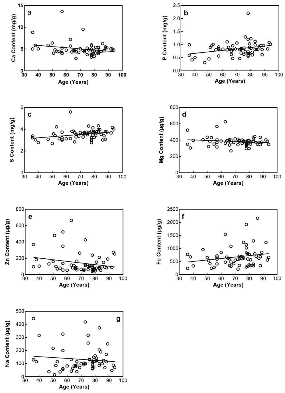 Figure 1: Age-related changes of Ca(a), P(b), S(c), Mg(d), Zn(e), Fe(f), and Na (g) contents in the FSBs.
View Figure 1
Figure 1: Age-related changes of Ca(a), P(b), S(c), Mg(d), Zn(e), Fe(f), and Na (g) contents in the FSBs.
View Figure 1
The average contents of seven elements were 5.029 ± 1.571 mg/g for Ca, 0.841 ± 0.272 mg/g for P, 3.530 ± 0.471 mg/g for S, 384.0 ± 56.09 g/g for Mg, 141.4 ± 125.1 µg/g for Zn, 675.6 ± 379.6 µg/g for Fe, and 131.4 ± 92.65 µg/g for Na. The average content was highest in Ca, and it decreased in the order of S, P, Fe, Mg, Zn, and Na.
Table 2 indicates the incidence of the FSB with the Ca content more than 5 mg/g, which is not contained in a normal artery [12]. The incidence of such an artery was 100% in both the 30s and the 40s of the subjects, and it ranged from 18.2% to 46.7% between the 50s and the 90s of the subjects. The incidence of the FSB with such a Ca content did not increase with aging. The mean incidence was 37.1% in all the FSBs studied.
Table 2: Incidence of an artery with the Ca content more than 5 mg/g. View Table 2
The relationships among seven element contents were examined in the FSBs studied. Table 3 lists the relationships among seven element contents in the FSBs. Extremely significant direct correlations were found both between Ca and Mg contents and between S and Mg contents, and very significant direct correlations were found between Ca and either Zn or Na contents and between Mg and Na contents. However, no significant correlations were found between Ca and P contents and between P and Mg contents. Therefore, no significant direct correlations were found among Ca, P, and Mg contents in the FSBs, except for an extremely significant direct correlation between Ca and Mg contents.
Table 3: Relationships among seven element contents in the FSBs. View Table 3
The used subjects consisted of 36 men and 26 women. The average age of men's subjects was 68.3 ± 14.5 years, whereas it of women's subjects was 74.0 ± 14.1 years. Figure 2 shows age-related changes of Ca and P contents in men and women's FSBs. The correlation coefficients between age and Ca content (Figure 2a) were estimated to be -0.156 (p = 0.363) in men's FSBs and -0.438 (p = 0.025) in women's FSBs. A significant inverse correlation was found between age and Ca content in women's FSBs, but no significant correlation was found in men's FSBs. The correlation coefficients between age and P content (Figure 2b) were estimated to be 0.364 (p = 0.029) in men's FSBs and 0.109 (p = 0.596) in women's FSBs. A significant direct correlation was found between age and P content in men's FSBs, but no significant correlation was found in women's FSBs. Therefore, the Ca content decreased significantly and gradually in women's FSBs with aging, whereas the P content increased significantly and progressively in men's FSBs with aging.
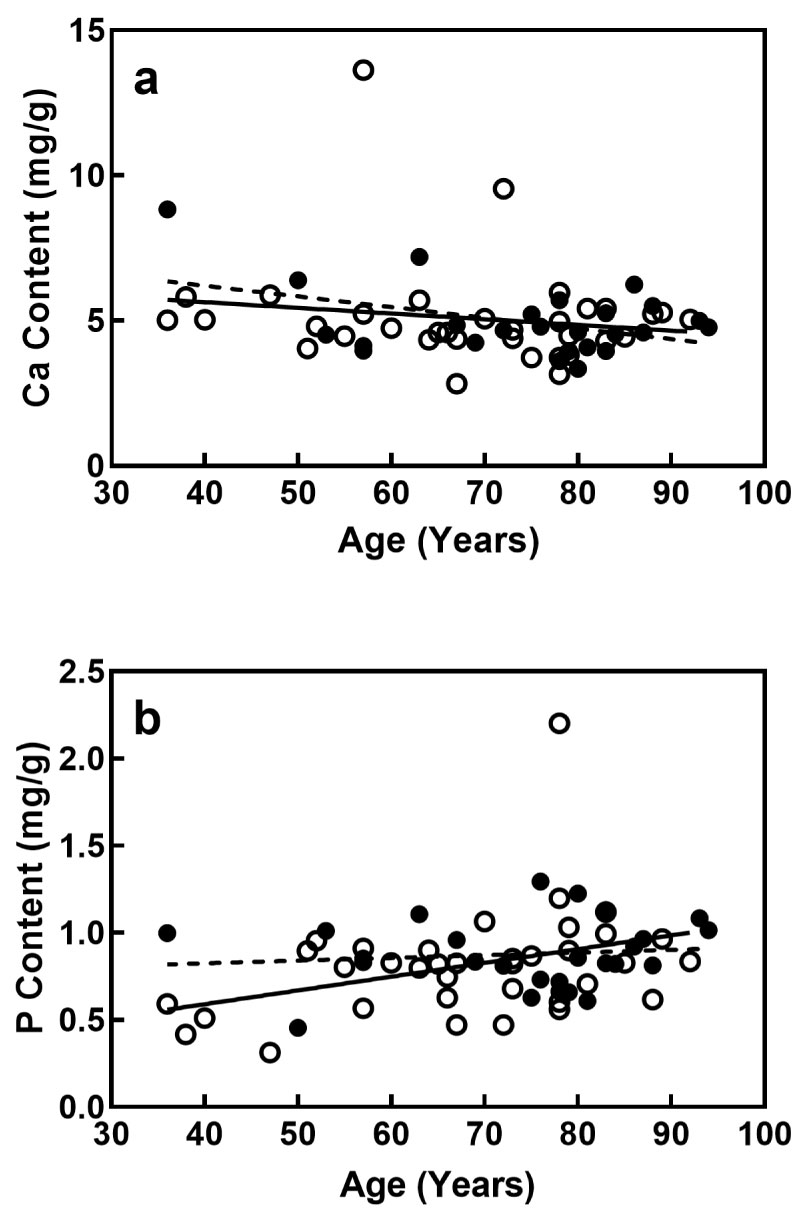 Figure 2: Age-related changes of Ca(a) and P(b) contents in men and women's FSBs. The open and solid circles indicate men and women's FSBs. The straight and dotted lines of trend with age indicate men and women's FSBs, respectively.
View Figure 2
Figure 2: Age-related changes of Ca(a) and P(b) contents in men and women's FSBs. The open and solid circles indicate men and women's FSBs. The straight and dotted lines of trend with age indicate men and women's FSBs, respectively.
View Figure 2
The analysis of the regression lines between age and Ca content in Figure 2a showed that no significant differences were found between the two slopes (p = 0.550) and between the two intercepts (p = 0.962) of the regression lines for men and women's FSBs. The analysis of the regression lines between age and P content in Figure 2b showed that no significant differences were found between the two slopes (p = 0.192) and between the two intercepts (p = 0.634) of the regression lines for men and women's FSBs. Therefore, no gender differences were found in the FSBs with regard to age-related changes of Ca and P contents.
To examine an effect of arterial size on age-related changes of elements, the FSBs were separated into the smaller group (n = 40) less than 1.40 mm of the outer diameter and the larger group (n = 22) more than 1.40 mm of the outer diameter (Figure 3). The smaller group ranged in age from 36 to 94 years (average age, 70.4 ± 14.9 years), whereas the larger group ranged in age from 36 to 88 years (average age, 71.2 ± 14.0 years). The two groups were separately analysed to examine whether age-related changes of Ca and P contents were different between smaller and larger groups.
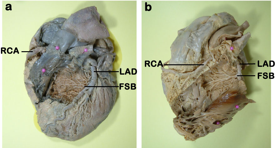 Figure 3: Anterior views of smaller (a) and larger (b) FSBs. FSB, first septal perforator branch; LAD: left anterior descending artery; RCA: right coronary artery.
View Figure 3
Figure 3: Anterior views of smaller (a) and larger (b) FSBs. FSB, first septal perforator branch; LAD: left anterior descending artery; RCA: right coronary artery.
View Figure 3
Figure 4 shows age-related changes of Ca and P contents in both the smaller and larger groups. The correlation coefficients between age and Ca content (Figure 4a) were estimated to be - 0.310 (p = 0.052) in the smaller group and -0.081 (p = 0.719) in the larger group. No significant correlations were found between age and Ca content in two groups. The correlation coefficients between age and P content (Figure 4b) were estimated to be 0.307 (p = 0.054) in the smaller group and 0.304 (p = 0.169) in the larger group. No significant correlations were found between age and P content in two groups.
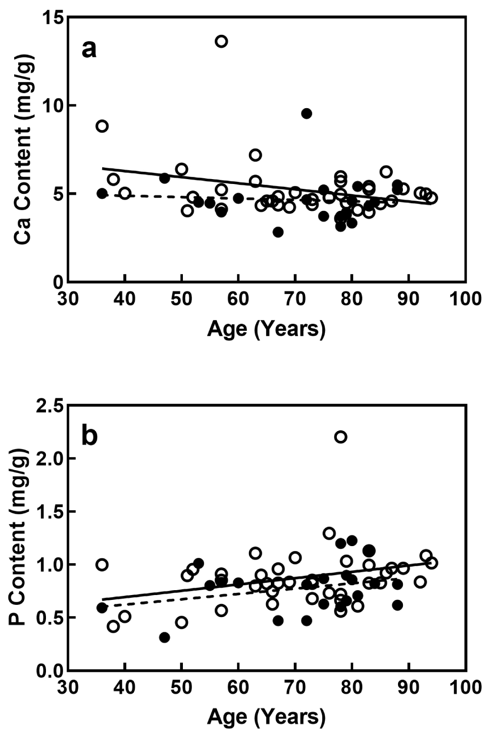 Figure 4: Age-related changes of Ca (a) and P (b) contents in the smaller group less than 1.4 mm of the outer diameter and the larger group more than 1.4 mm of the outer diameter in the FSBs. The open and solid circles indicate smaller and larger groups. The straight and dotted lines of trend with age indicate smaller and larger groups.
View Figure 4
Figure 4: Age-related changes of Ca (a) and P (b) contents in the smaller group less than 1.4 mm of the outer diameter and the larger group more than 1.4 mm of the outer diameter in the FSBs. The open and solid circles indicate smaller and larger groups. The straight and dotted lines of trend with age indicate smaller and larger groups.
View Figure 4
The analysis of the regression lines between age and Ca content in Figure 4a showed that no significant differences were found between the two slopes (p = 0.360) and between the two intercepts (p = 0.162) of the regression lines for the smaller and larger groups. The analysis of the regression lines between age and P content in Figure 4b showed that no significant differences were found between the two slopes (p = 0.850) and between the two intercepts (p = 0.152) of the regression lines for the smaller and larger groups. Therefore, no significant differences were found in age-related changes of Ca and P contents between the smaller and larger groups.
To elucidate whether calcification occurred in the FSBs in old age, both the mass ratios of Ca/P and Mg/Ca were investigated in the 62 FSBs studied. Figure 5 shows changes of both the mass ratios of Ca/P and Mg/Ca in the FSBs as a function of Ca content. The correlation coefficient was estimated to be 0.796 (p < 0.0001) between the mass ratio of Ca/P and Ca content Figure 5a. The mass ratio of Ca/P increased significantly and progressively with Ca increase, being not constant. The average mass ratio (mg/mg) of Ca/P was high in the FSBs studied, being 6.75 ± 3.93.
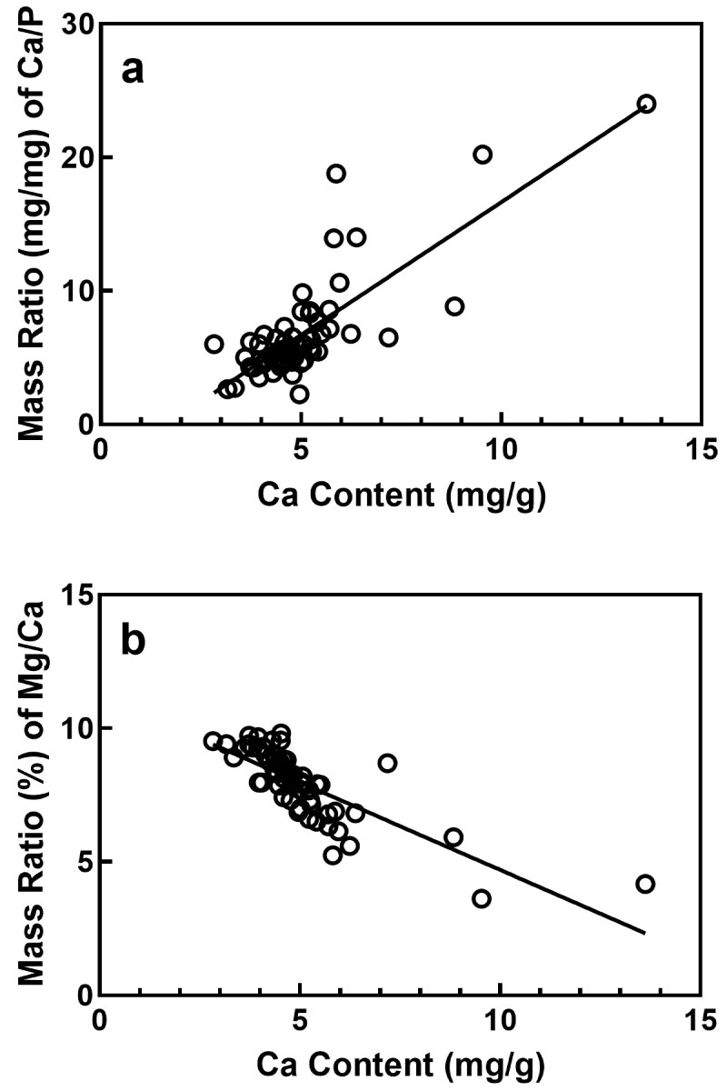 Figure 5: Changes of mass ratios of Ca/P(a) and Mg/Ca(b) in the FSBs. The mass ratios of Ca/P and Mg/Ca are plotted against the Ca content.
View Figure 5
Figure 5: Changes of mass ratios of Ca/P(a) and Mg/Ca(b) in the FSBs. The mass ratios of Ca/P and Mg/Ca are plotted against the Ca content.
View Figure 5
Regarding the mass ratio of Mg/Ca, the correlation coefficient was estimated to be -0.783 (p < 0.0001) between the mass ratio of Mg/Ca and Ca content (Figure 5b). The mass ratio of Mg/Ca decreased significantly and gradually in the FSBs with Ca increase. The average mass ratio (%) of Mg/Ca was moderate in the FSBs studied, being 7.95 ± 1.31%.
There are a few reports [4-6] on calcification of the FSB. Wasilewski et al. [4] investigated the LAD including the FSB in 309 patients with the mean age of 59.9 years by multi-slice computed tomography examination and revealed that calcification was most frequently located in the LAD in proximity to the origin of the FSB, but it was not located in the FSB. Wasilewski et al. [4] proposed that the septal branches generated disturbed flow in the LAD and posterior descending artery in a similar fashion to the myocardial bridge (myocardial bridge effect). Balghith et al. [5] investigated plaque distribution in the LAD by intravascular ultrasound in 50 patients with minimally obstructive coronary artery disease (< 50% angiographic diameter stenosis) and revealed that in the LAD segment adjacent to the origin of the FSB, the mean atheroma area and the mean intimal thickness were substantially greater on the septal side than on the antiseptal side.
To examine age-related changes of the arteries supplying the conduction system of the heart, Velican et al. [6], investigated histological changes of the FSB, SAN artery, AVN artery, and posterior descending artery in the subjects of 6 to 55 years old. They reported that in apparently healthy persons, the incidence of intimal thickening in the FSB increased progressively from 9% at 21-25 years of age to 34% at 51-55 years of age. However, they did not observe calcification in the tunica intima.
Vascular calcification is a hallmark of atherosclerosis. On calcification, deposition of calcium orthophosphate first occurs in the artery. Thereafter, it progresses to amorphous calcium phosphate and then to calcium phosphate crystalline structures such as hydroxyapatite [13]. Calcification occurs in most, but not all, arteries in old age [14].
To elucidate the manner of element accumulation in the arteries with aging, the authors [8,9,15] investigated age-related changes of elements in the arteries and found that when calcification occurred in the arteries, a significant accumulation of Ca, P, and Mg occurred simultaneously in the arteries and both the Ca and P contents were well correlated with the Mg content. In addition, the mass ratio of Ca/P was constant independently of Ca content, and the mass ratio of Mg/Ca was low [16]. In the FSBs studied in the present study, the following results were obtained: Although the P content increased significantly with aging, the Ca and Mg contents did not increase with aging. No significant direct correlations were found among the Ca, P, and Mg contents, except for an extremely significant direct correlation between Ca and Mg contents. The mass ratio of Ca/P increased significantly and progressively with Ca increase, being not constant. The average mass ratio of Mg/Ca was moderate, being 7.95 ± 1.31%. Therefore, it was suggested that calcification scarcely occurred in the FSBs in old age. The present result is compatible with the finding by Wasilewski et al. [4].
The present study revealed that the average content of P was very low in the FSBs studied. The authors [17] previously investigated the Ca, P, S, and Mg contents in 19 kinds of Japanese arteries and found that the average contents of P were very low in both the internal thoracic and the radial arteries, being 0.83 ± 0.25 mg/g in the internal thoracic arteries and 0.85 ± 0.66 mg/g in the radial arteries. These arteries are widely used for coronary artery bypass grafting [18,19]. It is recognized that atherosclerosis scarcely occurs in the internal thoracic artery in old age and occurs rarely in the radial artery [20]. Recently, the authors investigated age-related changes of elements in the SAN artery [21], AVN artery [22], and the posterior intercostal artery [23], and found that the average contents of P were very low in the three arteries. All the internal thoracic artery, radial artery, SAN artery, AVN artery, posterior intercostal artery, and FSB are characterized by a very low average content of P.
In the absence of calcification, the P content of tissue is mostly determined by the nucleic acid content (DNA and RNA) and the phospholipid content of tissue. Nucleic acids in the cell nucleus and the cytosol and phospholipids in the cell membrane are indicators of metabolically active cells [24]. Taking these into consideration, it is reasonable to presume that the P content in the artery indicates the active cell density, namely, the number of active cells per volume. Therefore, it is thought that the active cell density of the FSB increases slightly with aging.
There are two reports [6,25] on the characteristic of the intimal thickening in the coronary arteries. Velican et al. [6] reported that in the arteries supplying the conduction system of the heart, intimal thickening was composed of smooth muscle cells, elastin, and proteoglycans, and that the incidence of intimal thickening in the FSB increased progressively with aging. Likewise, Nakashima et al. [25] reported that major components of intimal thickening in the coronary arteries were smooth muscle cells, elastin, and proteoglycans. The present study revealed that the S content increased significantly in the FSBs with aging. The S content in the artery is mainly contained in the smooth muscle cell, collagen, and elastin [26]. According to the findings by Velican et al. [6] and Nakashima et al. [25], it is speculated that smooth muscle cells, elastin, and proteoglycans may increase mainly in the tunica intima of the FSB with aging.