Introduction: Lipomas of the finger are extremely uncommon, slow-growing benign tumors composed of fibrous adipose tissue, accounting for less than 5% of benign hand tumors. Neurolipoma, fibrolipoma or lipofiromatous Hamartoma is a lipomatous tumoral process involving peripheral nerves and their dividing branches, particularly in the hands and feet.
Case presentation: We report a case of digital neurolipoma of the 3rd finger and discuss the clinical, epidemiological and management aspects of this rare tumor.
Discussion: These tumors represent a diagnostic challenge due to their rarity and neuronal involvement, This tumor has a predilection for the median nerve, but lesions of various nerves have been reported, notably the digital nerve.
Conclusion: Finally, no definitive treatment is available for neurolipoma. Decompression and biopsy for final diagnosis is the current ideal management.
Benign, Finger lipoma, Lipofibromatous hamartoma, Neurolipoma
Finger lipomas are extremely rare, slow-growing benign tumors composed of a mixture of fibrous and fatty tissue, accounting for less than 5% of all benign hand tumors [1]. Neurolipomas are a specific type of lipomas where the fatty tumor grows a surrounding nerve [1]. While most commonly reported in the median and digital nerves of the hand, these lesions can also occur in other nerves like the radial, ulnar, sciatic, cranial, and plantar nerves [2]. Their exact etiology and pathophysiology are unknown, although trauma is suspected to be a potential contributing factor [1,2].
Affected patients usually present with a painless mass that progressively increases in volume, following the area of the affected nerve. Occasionally, in cases of median nerve injury, patients present with symptoms and signs of carpal tunnel syndrome, complaining of numbness and tingling along the palmar aspect of the wrist and hand and, finally, motor deficit indicating an advanced stage [3].
We report a case of digital neurolipoma of the digital nerve of the 3 rd finger in a 36-years-old man.
A 36-year-old-patient, with no significant past medical history, presented with a tumefaction at the base of the left third finger that had been present for over 6 months.
Clinical examination on admission revealed a painless 3-centimeter diameter mass, associated with mild paresthesia, involving the root of the 3 rd left finger, and moved freely relative to both deeper tissues and the skin surface, there were no inflammatory signs. The patient's general examination was unremarkable.
Radiological assessment showed no bone involvement sign, but ultrasound revealed a 4 cm subcutaneous tissue lesion with a lipomatous appearance.
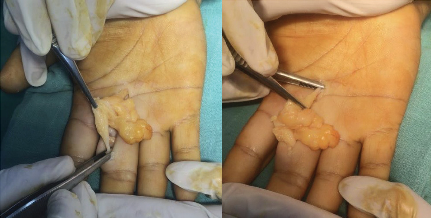
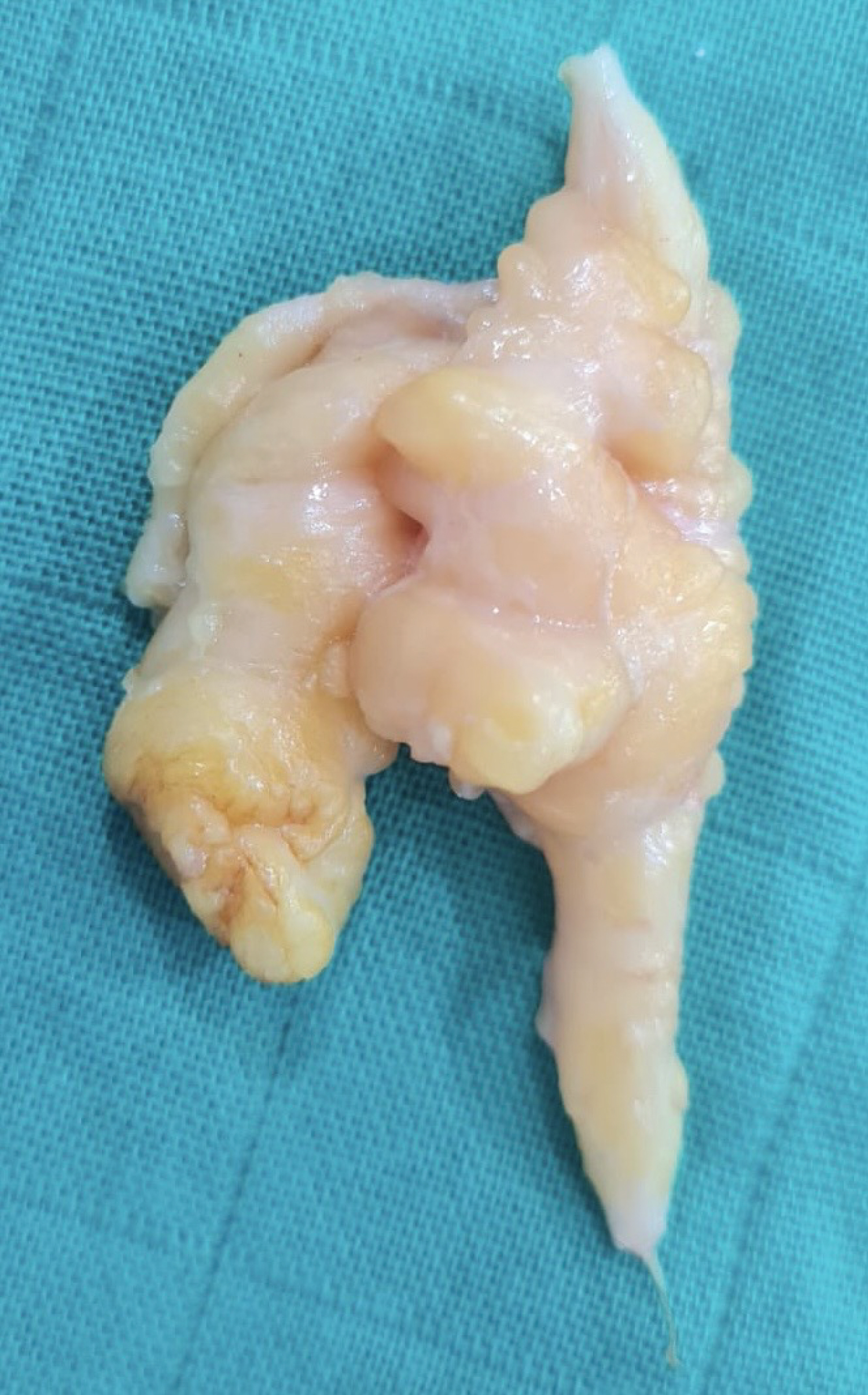
Using a palmar approach to the finger, we found a polylobed formation with a tissue and fat component adhering to the lateral digital nerve of the 3 rd finger (Figure 1). After dissection of the pedicle, the resection was straightforward (Figure 2).
 Figure 1: Intraoperative photograph shows neurolipoma of the digital nerve of the 3rd finger.
View Figure 1
 Figure 2: Soft lobulated mass with a nerve enlarged by fibrofatty tissue.
View Figure 2
Pathological examination was consistent with neurolipoma. The post-operative follow-up was satisfactory, with no local recurrence or sensory-motor disorders.
Neurolipoma is a rare benign tumor of slow evolution and have an unknown origin [2]. While they most commonly affect the median nerve, to which they have a predilection [4,5], cases involving other nerves, particularly the digital nerve, have also been reported in literature.
This condition predominantly affects males in the first three decades of life [6], with a higher prevalence in the left hand [5]. Our patient aligns with this typical presentation: A male in his thirties whose left hand was affected.
The exact cause of neurolipoma formation and its predilection for the median nerve remain unclear. Some authors believe that repetitive microtrauma might trigger the abnormal growth of mature fat cells and fibroblasts in the epineurium [2,7]. Others suggest a potential congenital abnormality affecting adipose tissue development [7].
Patients usually present with a painless mass that has been present for many years, often since childhood, and is discovered in the third or fourth decade of life. Subsequently, patients may experience additional symptoms like paraesthesia, motor deficit and pain due to compression of the affected nerve [8].
Neurolipoma can be misdiagnosed for other nerve tumors like ganglion cysts, hereditary hypertrophic neuritis of Dejerine-Sottas syndrome, vascular malformations, traumatic neuromas, schwannomas, neurofibromas, intraneural lipomas, fibromatosis, and plexiform neurofibroma [2,7]. However, true lipomas are well encapsulated, sharply delineated masses that normally appear on the surface; they originate in the perineurium or epineurium and do not contain neuronal elements, which helps distinguish them from neurolipomas [7].
Lipofibromatous hamartoma is usually diagnosed as a fat-containing soft tissue mass on the CT or MRI [8]. However, a differential diagnosis is crucial to rule out other possibilities [9].
Surgery with microscopic techniques is the mainstay of treatment for suspected neurolipomas. This typically involved an exploratory surgery with excisional biopsy for definitive diagnosis. Surgeons aim to preserve important nerve branches while removing the neurolipoma since radical nerve excision is not recommended [10]. Extensive intraneural dissection carries a high risk of neurological deficit due to ischemic complications [2].
Microscopic examination of the escised tissue is essential for definitive diagnosis of neurolipoma. This reveals characteristic features like fibrous tissue proliferation with infiltration of perineural and epineural tissue [5].
Nerve tumors are rarely found in the hand, and even more rarely in the fingers. Digital neurolipoma is a rare benign tumor of unknown origin that develops from peripheral nerves. Macroscopically, it presents as a lobulated formation with a mobile fat and tissue component, painless and without associated inflammatory signs. The definitive diagnosis is histological after surgical excision, characterized by a proliferation of fibrous tissue with infiltration of perineural and epineural tissue.
This case report follows scare guidelines [11].
No conflict of interest to be disclosed.
Informed consent was obtained from the patient.
This research received no specific grant from any funding agency in the public, commercial, or not-for-profit sectors.
These authors contributed equally.