Metastases in oral cavity possesses a low frequency. They represent approximately 1% of malignant process in mouth, being the mandible the most prevalent and in an even smaller number it affects the temporomandibular joint. We present a 72-year-old male gender patient, who asks for temporomandibular pain initially interpreted as a temporomandibular disorder; after radiographic control an osteolytic lesion is identified in the right mandibular condyle for which is derived to the Maxillofacial Surgery Department. The biopsy evidences an Adenocarcinoma, that by immunohistochemical characteristics suggest a pulmonary primary, which is verified by computed tomography of the thorax. In our literature review we identified 74 cases that involved mandibular condyle. Despite its low frequency, it is important to consider these types of pathologies within the diagnostic hypothesis because its appearance suggest generally a poor survival prognosis.
Condyle, Temporomandibular joint, Maxillary metastasis
Metastasis in oral cavity are rare, they represent approximately 1% of malignant processes in oral [1]. The most affected site is the mandible, affecting principally the molar and premolar region [2]. Those involving mandibular condyle are even less frequent. Usually the primary tumor is known and treated previously to the occurrence of dissemination. However, in a percentage close to 30%, the oral manifestation represents the first finding, which could help detect an undiagnosed primary tumor [3]. The neoplasm that metastasize more frequently in the oral cavity are those that originate in breast, lung, prostate, gastrointestinal system and kidney [4]. In a literature review performed by the authors of this article, 38 cases of mandibular condyle metastasis were identified. Seven cases correspond to metastasis whose primary tumor was located in the lungs.
The first warning signs of possible malignant processes are pain, paresthesia, swelling, tooth mobility and pathological fractures. About 59% of the cases that reported metastasis in the condylar region are presented mimicking symptoms of temporomandibular disorders [2]. In the case of the tumors described in the condylar region, the most frequent symptoms are: disfunction, pain, trismus, limitation and deviation of the mandibular opening, swelling, osteolytic radiographic lesion and pathologic fractures of the condyle neck that may suggest metastatic malignant processes [2].
Due to the non-specific of the clinical signs, the possibility to confuse the diagnose with benign pathologies and the high mortality when is found distant metastasis. The objective of this study is to present a case of a mandibular condyle metastasis that present itself as the only initial manifestation mimicking signs and symptoms of temporomandibular disorders.
71-year-old male patient consulted for painful symptomatology in the right temporomandibular joint (TMJ). A panoramic radiography is requested evidencing a discrete osteolytic lesion in the right mandibular condyle (Figure 1), finding that is interpreted as degenerative disorder of the joint and treated under this idea. After 3 months without resolving, the patient is referred to evaluation by maxillofacial surgeon, who request new panoramic radiography and computed tomography (CT) for bilateral TMJ study, where the important advance of the lesion is manifested in the right TMJ with a full loss of the condyle and part of the mandibular ramus (Figure 2). The patient is admitted to the Oral and Maxillofacial Surgery Service of Hospital del Salvador, Santiago de Chile for evaluation and treatment with presumptive diagnosis of right mandibular condyle osteosarcoma.
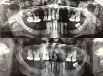 Figure 1: A) Panoramic radiography March 2017. Discrete osteolytic lesion in the right mandibular condyle; B) Panoramic radiography July 2017. The osteolytic lesion involves all the right mandibular condyle. View Figure 1
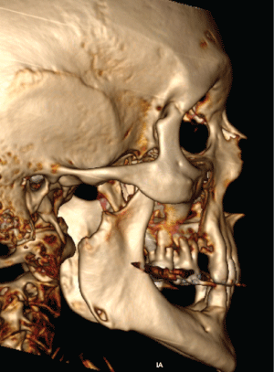 Figure 2: 3D reconstruction. The image shows a complete absence of the right mandibular condyle beacuse the osteolytic lesion. View Figure 2
Within medical history we remark arterial hypertension, benign prostatic hyperplasia, history of chronic smoker and reports a weight loss of 9 kg in the past 6 months. Upon physical examination, the patient presented in the preauricular region a firm consistency, painful to palpation, poorly delimited, 5 cm in diameter swelling with no skin involvement (Figure 3). No joint noises were found, and the patient did not present restrictions in the opening range or in mandibular dynamics. The intraoral examination revealed its condition of superior and inferior partial edentulous, without alterations in oral soft tissues (gingiva, tongue, mouth floor and oropharynx). The patient did not present alterations in the motor function of the facial nerve and sensitive function of the trigeminal nerve, no palpable cervical adenopathy's.
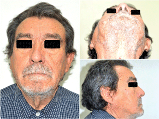 Figure 3: Clinical photography, admission to Hospital del Salvador, July 2017. In the right preauricular region a firm consistency, 5 cm in diameter swelling with no skin involvement. View Figure 3
A new panoramic radiography revealed the extensive advance of the osteolytic process that affects the right mandibular condyle. A craniofacial CT demonstrates the total osteolysis of the right temporomandibular region, occupied by a 3.6-2.8 cm mass in the infra temporal fossa and right pterygomaxillary space (Figure 4). Through preauricular end aural approach the lesion is accessed (Figure 5) obtaining an amorphous, solid, not vascularized tissue sample that is sent to differed biopsy; reporting a moderately differentiated adenocarcinoma, characterized by a solid cellular mass that resemble glandular parenchyma with scarce duct-like structures spread all over the tissue (Figure 6). Immunohistochemical analysis mean to find the primary tumor, reveal positive results to Cytokeratin 7 (CK7+), compatible with an adenocarcinoma. At this point, cannot be ruled out the possibility for a salivary gland adenocarcinoma of parotid origin against a possible metastatic adenocarcinoma, of probably lung origin. The Cluster of differentiation 10 (CD10) for renal origin is also requested because of focal areas of clear cells, with negative response. A computed tomography with contrast of abdomen, chest and pelvis is requested, where a large mass of soft tissue density and irregular borders of 6 × 5.5 cm, with necrotic areas inside is observed in the right pulmonary lobe, widely contacting the pleura at the para mediastinal level, remarking multiple pulmonary nodules and regional adenopathy's (paratracheal, subcardial and supraclavicular) (Figure 7).
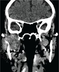 Figure 4: The image show a coronal cut CT. Demonstrates a mass 3.6-2.8 cm that involves the temporomandibular region, the infra temporal fossa and right peterigomaxillary space. The image shows the mass infiltrating in the condylar and ramus bone (osteolytic process). View Figure 4
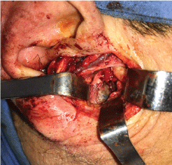 Figure 5: Preauricular approach to the temporomandibular region, it can be observe the total absence of the right mandibular condyle. View Figure 5
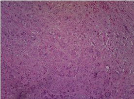 Figure 6: Histology. A solid, cellular mass that resemble glandular acinus was depicted, no dyskeratosis or keratin formation could be seeing, scarse duct-like structures where spread all over the tumor area. A typical cellular architecture with plemorophic and nuclear hypercromatism was common. Multiple biazarre mitoses could be seeing. View Figure 6
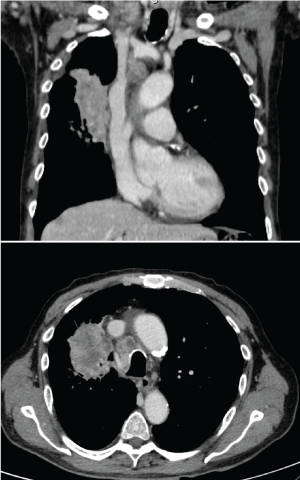 Figure 7: Chest CT coronal and axial cut. The image shows a large mass of soft tissue density and irregular borders of 6 × 5.5 cm, with necrotic areas inside is observed in the right pulmonary lobe, widely contacting the pleura at the paramediastinal level, ramarking multiple pulmonary nodules and regional adenopathies (paratracheal, subcardial and supraclavicular). View Figure 7
With the collected antecedents the case was presented to the oncological committee, before carry on the lung biopsy the patient had a multiple organ dysfunction syndrome, consequently the biopsy was not possible, the patient condition become deteriorated, therefore the committee decided a palliative therapy on this case, within 3 months the patient died.
Metastasis in oral cavity are rare and usually present a poor prognosis, they represent 1% of malignancies in the region. They mainly manifest in the mandible over the maxilla, with the premolar and molar area being the most affected [1,5,6]. Likewise, those that compromise the condylar are even more difficult to find.
The development of metastasis in the maxillofacial region is not always consequent with knowing the primary tumor. Kolokythas, et al. mentions that in 70% of the cases of mandibular condyle metastasis the primary was known and treated. However, in approximately 30% of the remaining cases they are a manifestation of an undiagnosed neoplasm, being its diagnosis and detection a real diagnostic challenge.
Immunohistochemical staining techniques of moderately differentiated adenocarcinomas is commonly asked for origin determination. Because of the clinical findings and the morphological aspect of the tissue specimen at microscope it was decided to made an orientated but not conclusive panel that included CK7 and CD10 both typically used for differentiated renal metastases from pulmonary glandular origin.
Literature review from the authors found a total of 38 metastasis on mandibular condyle. Gender preference was not determined. 18 cases in females (47.4%) and 20 cases in males (52.6%), while the age average was 61-years-old (Table 1). These data were obtained through a computerized review of the literature in Pubmed (MEDLINE) and Clinically search engines, using the search terms: "mandibular condyle", "metastatic" and "temporomandibular joint disorders", finding a total of 49 articles (51 reports). 13 articles (13 cases) were eliminated for being only in "abstract" or "poster" format and those that didn't provide enough information (age, gender, primary tumor, symptoms, etc.), selecting a total of 31 articles for the revision.
Table 1: Clinical findings of 73 mandibular condyle metastasis cases. Results of our revision. View Table 1
In a revision of 390 articles about metastasis in mandibular and maxillary bones, Hirshberg, et al. identified that the most frequent primary site of metastasis for women was breast cancer and in men lung cancer. We identified in our research 9 cases of breast metastasis in females that affected the mandibular condyle and 7 lung metastasis for males [7]. Only 6 prostate cancer, 4 gastrointestinal and 2 uterus cancer were identified (Table 1). We consider, as Hirschberg and Panossian, that the neoplasm that generate metastasis more frequently in the oral cavity are those originated in breast, lung, prostate, gastrointestinal system and kidney, which suggest that different cancers have a different potential to develop metastasis in the maxillofacial region [4].
It has been described that the mandibular body (molar and premolar area) and the angle are the most affected areas, meanwhile the mandibular condyle presents a low frequency to these type of cases. Three theories have been proposed that may explain this behavior. To understand them is important to know that the main route for metastasis are via lymphatic and via hematogenous [8]. Consistent with this, we consider that the main neoplasm that involves the mandible are the carcinomas originated in breast, lung and prostate, which disseminates via hematogenous [9]. The first theory considers that the amount of substantial hematopoietic marrow in the condyle is scarce compared with the one in the mandibular body, which limit the dissemination probabilities [9]. In second place the condyle irrigation is "separated" or "isolated", depending principally from the maxillary and superficial temporal artery, different arteries than those irrigating the medular body. The third explanation gives reason to the existence of a bone plate that limits the propagation to the mandibular condyle space [10-12].
The non-specific clinical presentation of the metastatic pathology makes the diagnosis a great challenge. The first warning signs of possible malignant processes are pain, paresthesia, swelling, tooth mobility and pathological fractures [4]. 50% of the cases that reports metastasis to the condylar region are presented mimicking symptoms of temporomandibular joint disorder [2]. We identified in the literature the principal motives for consultation related to the TMJ were pain with 19 cases (50%) and swelling with 21 cases (55%). Agreeing with [10], other signs and symptoms common at TMJ level were alterations in the opening ranges (6 cases, 15.7%), deviation in mandibular trajectory (5 cases, 13%), numbness (6 cases, 15.7%) and trismus or lockjaw (4 cases, 10.5%). However, is important to consider that in many cases the lesion may be asymptomatic [13].
Our patient presented a preauricular region a firm consistency, painful to palpation, poorly delimited, 5 cm in diameter swelling, no limitation of opening ranges or lateral excursions, no joint noises were found. In the beginning, these symptoms are attributed to TMJ disorders, keeping the patient in interocclusal splint therapy for approximately 4 months. Radiographic appearance is usually an osteolysis [9], despite that there is no pathognomonic image for mandibular metastasis, radiolucency is a common finding [13]. In our report, slight degenerative changes in the condylar cortex were found, which progresses to complete osteolysis of condyle and mandibular ramus in a period of only 6 months. Contrast computed tomography showed a soft tissue density mass replacing condylar and mandibular ramus hard tissues, in addition to the muscles associated to the region. After the results of chest, abdomen and pelvic CT, the condyle biopsy and immunohistochemical markers oriented the lesion present in the condyle was the result of a pulmonary metastasis. We consider critical the chronic smoking history of this patient as a triggering factor. As reported in literature the survival rate is on average a year from the moment of the metastasis finding [14-17]. Similarly, our patient died 4 months after the first and only clinical manifestation at distance of the pulmonary adenocarcinoma at the temporomandibular joint level.
It is reported that approximately 50% of the metastatic lesion that involved the condylar presented with symptoms similar to temporomandibular joint disorders [2]. When presented these type of situations is fundamental the right analysis of imaging test (radiographs, computed tomography, magnetic resonance, etc.), considering benign and malignant pathologies as differential diagnosis. A delay in the diagnosis prolongs start of treatment, time that can be critical when the etiology is unknown and of possible neoplastic origin [3,13]. We identified in our results that 22 patients (57.9% of the cases) did not present a history of previous malignancies, and 16 cases (42.1%) did had a history of cancer. Although the occurrence of these events is usually infrequent, is fundamental to consider them, because as we mentioned their appearance usually suggest a bad prognosis.
The authors have no commercial or financial associations that might create a conflict of interest with the information presented in this manuscript.
This study was approved by ethical board of Hospital del Salvador.